
1. MEN syndromes 2. Granulomatous lesions of lung
MEN syndrome multiple endocrine neoplasia diagram
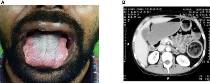
This multi-panel figure illustrates clinical manifestations of Multiple Endocrine Neoplasia (MEN) syndrome, specifically MEN2B. Panel A is a clinical photograph showing the oral cavity of a patient with a mucosal neuroma. The tongue is significantly enlarged (macroglossia) with characteristic nodular, irregular borders and a central whitish-yellow coating, typical of benign nerve sheath tumors associated with this genetic syndrome. Panel B is an axial contrast-enhanced abdominal CT scan demonstrating significant bowel pathology. Several loops of the small and large bowel are severely dilated and distended, consistent with chronic intestinal pseudo-obstruction or megacolon, which can occur due to intestinal ganglioneuromatosis in MEN2B patients. Other visualized anatomy includes the liver, kidneys, and vertebrae. Together, these images serve as educational examples of the extra-endocrine findings in MEN syndromes, highlighting the importance of physical examination of the oral mucosa and radiological assessment of the gastrointestinal tract in suspected cases.
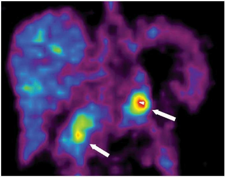
This diagnostic image is a coronal 11C-5-HTP Positron Emission Tomography (PET) scan of the abdomen, specifically illustrating tracer uptake in a patient with Multiple Endocrine Neoplasia type 1 (MEN-1) syndrome. The PET scan utilizes a pseudocolor heatmap where high metabolic activity is indicated by red and yellow, moderate activity by green, and background or low uptake by blue and purple hues. Two distinct focal areas of high radiotracer intensity are identified by white arrows. The arrow on the right points to a localized high-intensity red and yellow signal in the pancreatic head, while the arrow on the left indicates a secondary area of elevated uptake in the pancreatic tail. These findings are characteristic of neuroendocrine tumors (NETs), which typically exhibit high 11C-5-HTP uptake due to increased amine precursor decarboxylation. The liver and surrounding gastrointestinal structures show heterogeneous background tracer distribution. This image serves as a clinical example of PET imaging sensitivity in localizing small pancreatic neuroendocrine tumors that may be occult on traditional CT or MRI.
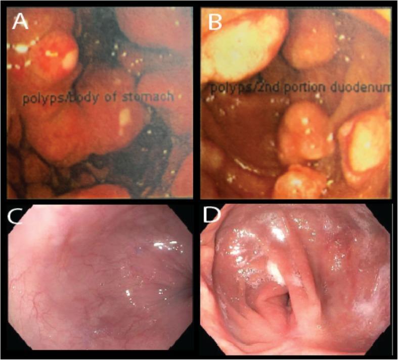
This composite of four endoscopic images illustrates the progression and treatment response of neuroendocrine tumors in a patient with Multiple Endocrine Neoplasia type 1 (MEN-1) and Zollinger-Ellison syndrome (ZES). Panels A and B (dated 2007) show large, multifocal, polypoid-appearing masses. Image A depicts nearly obstructing Enterochromaffin-like (ECL) carcinoid tumors in the gastric body, while Image B shows large gastrinomas in the proximal second portion of the duodenum. These tumors are characterized by a rounded morphology, protruding into the lumen with areas of superficial ulceration. Panels C and D (dated 2018) represent follow-up endoscopy after optimal surgical cytoreduction (near-total gastrectomy and pancreaticoduodenectomy) and medical management with octreotide. Image C shows the proximal stomach, and Image D shows the distal remnant stomach and small bowel anastomosis; both are completely devoid of carcinoid tumors or polyps, displaying smooth, healthy pink mucosa with normal vascular patterns. The comparison highlights the transition from aggressive, multifocal neuroendocrine neoplasia to successful long-term disease control.
granulomatous lung disease histology diagram
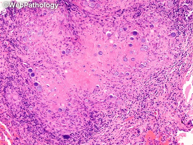
Imaging modality and technique: Histology using light microscopy; routine Hematoxylin and Eosin (H&E) stained paraffin section of lung parenchyma. In this pulmonary coccidioidomycosis specimen, tissue shows granulomatous inflammation with both necrotizing and non-necrotizing granulomas embedded in a background of mixed inflammatory cells, including epithelioid histiocytes, neutrophils, eosinophils, lymphocytes, plasma cells, and multinucleated giant cells. The defining fungal morphology comprises spherules of Coccidioides spp., present as mature and immature forms. Mature spherules are thick-walled, doubly refractile spheres measuring approximately 60–100 μm in diameter and containing numerous endospores 2–5 μm in diameter. Immature spherules are smaller and may be visible as the infection progresses. In rare circumstances, mycelial forms may be observed interspersed with bronchopulmonary fistulas or aerated cavitary lesions. Fungal elements localize within granulomas and interstitial spaces, with occasional minor invasion of adjacent tissue. Clinically, the presence of spherules with endospores in lung tissue is diagnostic of pulmonary coccidioidomycosis and guides antifungal therapy, prognosis, and infection control. Differential considerations include Histoplasma, Aspergillus, Blastomyces, Cryptococcus, and other fungal pathogens; correlation with culture, serology, and imaging is recommended for comprehensive management. This image is educational for pathology training and research on endemic mycoses and granulomatous lung disease, and case discussion for learners everywhere.
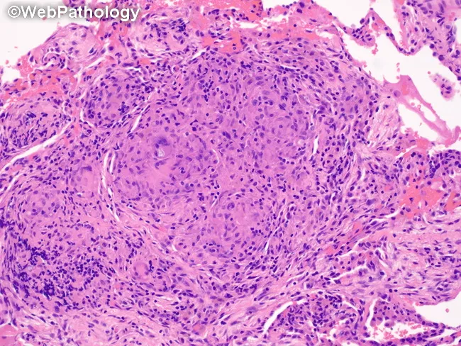
Pulmonary tissue from a wedge resection stained with Hematoxylin and Eosin, analyzed by brightfield light microscopy. The specimen shows robust granulomatous inflammation within the lung parenchyma consistent with a granulomatous infectious process. Nodular aggregates of epithelioid histiocytes are surrounded by a rim of lymphocytes and scattered plasma cells; multinucleated Langhans-type giant cells are present, producing a palisading appearance. The granulomas appear well circumscribed within the interstitium and may encroach upon adjacent alveolar spaces. Central necrosis is variably present and not conspicuously predominant in this field, a feature compatible with coccidioidal granulomatous reaction (coccidioidomycosis) that may display BCG-like granulomas in tissue specimens. The background alveolar architecture is preserved in portions, with mild interstitial inflammation and occasional eosinophils. Although fungal organisms are not definitively visualized in this image, the histology is compatible with fungal infection of the lung in the clinical context of pulmonary coccidioidomycosis, potentially associated with chronic granulomatous response. This image emphasizes characteristic granuloma morphology useful for differential diagnosis from mycobacterial tuberculosis or sarcoidosis, and provides histopathologic confirmation to guide clinical management, including antifungal therapy decisions and serology correlation. It is suitable for education on pulmonary granulomatous disease and wedge resection pathology.
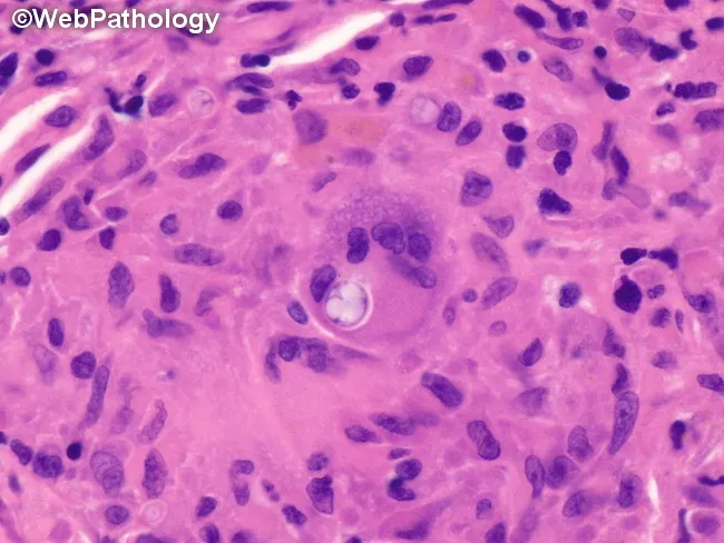
Pulmonary tissue section (lung biopsy) stained with hematoxylin and eosin, examined under brightfield light microscopy at high magnification. The specimen shows a well-formed epithelioid granulomatous reaction within the pulmonary parenchyma, composed of aggregates of epithelioid histiocytes and multinucleated giant cells encircling a developing Coccidioides spherule. An immature, thick-walled spherule is evident within a giant cell, containing sparse endospores. Surrounding lymphocytes and occasional plasma cells contribute to the granulomatous milieu. The background is pink extracellular matrix with scattered alveolar spaces; no overt necrosis is required; note the absence of septate hyphae on H&E. The image epitomizes granulomatous inflammation seen in pulmonary coccidioidomycosis, a fungal disease caused by Coccidioides immitis/posadasii encountered in endemic regions. The spherule's immature stage (BCG-like morphology) is diagnostic in the appropriate clinical context, distinguishing it from other fungal infections such as histoplasmosis or blastomycosis. Clinically, this histology supports a fungal etiology in a patient with respiratory symptoms, fever, and relevant exposure history. Diagnostic significance includes visualizing the fungal spherule within macrophages; differential diagnoses include TB, sarcoidosis, granulomatous pneumonia. Antifungal therapy planning may follow. This image conveys critical features for pathology teaching, endoscopy-guided biopsy interpretation, and research into host-pathogen interactions in endemic mycoses, supporting labeling and targeted antifungal management.
1. MEN Syndromes (Multiple Endocrine Neoplasia)
Overview
- Tumors occur at a younger age
- Tumors arise in multiple endocrine organs, either synchronously (same time) or metachronously (different times)
- Tumors are often multifocal even within one organ
- Tumors are usually preceded by an asymptomatic hyperplastic stage of the cell of origin
- Tumors are generally more aggressive and recur more frequently
MEN-1 (Werner Syndrome)
| Organ | Manifestation | Notes |
|---|---|---|
| Parathyroid | Primary hyperparathyroidism | Most common (80-95%); initial manifestation in most patients; appears by age 40-50; hyperplasias + adenomas |
| Pancreas | Endocrine tumors (NETs) | Leading cause of death; usually aggressive + metastatic; multifocal "microadenomas" throughout pancreas |
| Pituitary | Prolactinoma (most common) | Macroadenoma; some develop acromegaly from somatotropin-secreting tumors |
- Duodenum is the most common site of gastrinomas (more than pancreas)
- Zollinger-Ellison syndrome (gastrinomas) and hypoglycemia (insulinomas) are classic endocrine syndromes
- Carcinoid tumors, thyroid and adrenocortical adenomas, lipomas also occur more frequently
MEN-2A (Sipple Syndrome)
| Organ | Manifestation | Notes |
|---|---|---|
| Thyroid | Medullary carcinoma of thyroid (MCT) | ~100% of patients; multifocal; virtually always with C-cell hyperplasia in adjacent thyroid; secretes calcitonin; clinically aggressive |
| Adrenal medulla | Pheochromocytoma | 40-50%; often bilateral; may arise at extra-adrenal sites; <10% malignant |
| Parathyroid | Hyperplasia | 10-20%; leads to hypercalcemia, renal stones |
MEN-2B (now often called MEN-3)
- Medullary thyroid carcinoma (multifocal, more aggressive than MEN-2A)
- Pheochromocytoma
- Primary hyperparathyroidism does NOT develop
- Mucosal neuromas/ganglioneuromas - lips, tongue, GI tract, respiratory tract, eyes
- Marfanoid habitus - overly long bones of axial skeleton, hyperextensible joints (resembles Marfan syndrome but without cardiovascular defects)
MEN-4 and MEN-5 (Newer Syndromes)
| Syndrome | Gene | Features |
|---|---|---|
| MEN-4 | CDKN1B (inactivating germline mutations) | Phenotypically mimics MEN-1 |
| MEN-5 | MAX tumor suppressor gene | Bilateral pheochromocytomas + other tumors; NO medullary thyroid carcinoma or C-cell hyperplasia (unlike MEN-2) |
Genetic Screening
- MEN-2: Genetic testing is clearly important - all patients with MCT are advised to undergo germline RET mutation testing. All RET mutation carriers should undergo prophylactic thyroidectomy.
- MEN-1: Benefit of early genetic screening is more controversial; however biochemical screening is still practiced.
2. Granulomatous Lesions of the Lung
What is a Granuloma?
Pathogenesis of Granuloma Formation in the Lung
- Antigen deposition in lung parenchyma - either infectious (intracellular organisms) or non-infectious (beryllium, organic dusts)
- Antigen engulfed by antigen-presenting cells (dendritic cells, macrophages) and presented to naive CD4+ T cells in peripheral lymphoid organs
- In the absence of IL-4, exposure of activated CD4+ T cells to IL-12 (from macrophages) directs T cell differentiation toward a Th1 response
- Early IFN-γ up-regulates IL-12 production and IL-12 receptors on Th1 cells
- Chemokines from activated Th1 cells recruit macrophages and other inflammatory cells to the lung
- Accumulation of inflammatory cells within the alveolus (alveolitis) is the initial lesion
- Eventually, activated macrophages and T cells form organized granulomas
Classification of Granulomatous Lung Lesions
A. Infectious Causes
| Organism | Disease | Granuloma Type |
|---|---|---|
| Mycobacterium tuberculosis | Tuberculosis | Caseating (necrotizing) granulomas; Langhans giant cells |
| Mycobacterium leprae | Leprosy | Variable (depends on host immunity) |
| Atypical mycobacteria | MAC disease | Non-caseating or caseating |
| Histoplasma capsulatum | Histoplasmosis | Caseating; calcification common |
| Coccidioides immitis | Coccidioidomycosis | Epithelioid granulomas with spherules + endospores inside giant cells |
| Blastomyces dermatitidis | Blastomycosis | Suppurative + granulomatous |
| Cryptococcus neoformans | Cryptococcosis | Gelatinous, minimal granuloma in immunosuppressed |
| Candida | Disseminated candidiasis | Granulomas in immunocompetent |
| Parasites | Various | Eosinophilic granulomas |
B. Non-Infectious Causes
| Cause | Disease | Key Features |
|---|---|---|
| Unknown antigen | Sarcoidosis | Non-caseating granulomas; CD4+ alveolitis; oligoclonal T cell expansions; unknown inciting antigen |
| Beryllium dust | Chronic beryllium disease (CBD) | HLA-DPB1 alleles (with Glu69 on β-chain) confer susceptibility; beryllium-specific T cell response |
| Organic dusts (mold, bird proteins) | Hypersensitivity pneumonitis (Extrinsic allergic alveolitis) | Including Farmer's lung; known antigenic stimulus |
| Silica particles | Silicosis | Silicotic nodules; can have "eggshell" calcification |
| Granulomatous polyangiitis (Wegener's) | Vasculitis | Necrotizing granulomas + vasculitis; c-ANCA positive |
| Eosinophilic granuloma | Pulmonary Langerhans cell histiocytosis | Langerhans cells (CD1a+, Birbeck granules on EM) |
| Lipids | Granulomatous lipid pneumonia | Lipid-laden macrophages |
| Silicone | Silicone-induced granuloma | Foreign body-type |
C. Causes of Hypercalcemia via Granulomatous Disease (Clinically Important)
Sarcoidosis - Immunopathogenesis (Key)
- Inciting antigen is unknown, but immunopathogenic events are similar to other granulomatous diseases
- Alveolitis is characterized by an accumulation of CD4+ T cells (at BAL)
- These T cells show oligoclonal expansions (identified by TCR Vβ and Vα regions) - indicating a response to conventional peptide antigens
- Different individuals may show T cell clones with related TCRs, suggesting a response to the same (unknown) antigen
- Both the HLA type (MHC class II) of the individual and the stimulating antigen determine which TCRs are used
Histological Features of Granulomas
| Feature | Caseating (e.g., TB) | Non-caseating (e.g., Sarcoidosis) |
|---|---|---|
| Central necrosis | Present ("cheese-like") | Absent |
| Epithelioid histiocytes | Yes | Yes |
| Langhans giant cells | Yes (prominent) | Yes (less prominent) |
| Lymphocyte cuff | Yes | Yes |
| Calcification | Common (healed TB) | Can occur (Schaumann bodies in sarcoid) |
Summary Table
| MEN Syndrome | Gene | Key Organs Affected | Key Mutation Type |
|---|---|---|---|
| MEN-1 (Werner) | MEN1 (menin) | Parathyroid, Pancreas, Pituitary | Loss-of-function (TSG) |
| MEN-2A (Sipple) | RET | Thyroid (MCT), Adrenal (pheo), Parathyroid | Gain-of-function |
| MEN-2B / MEN-3 | RET | Thyroid (MCT), Adrenal (pheo), Neuromas, Marfanoid | Gain-of-function (TK domain) |
| MEN-4 | CDKN1B | Mimics MEN-1 | Loss-of-function (TSG) |
| MEN-5 | MAX | Bilateral pheochromocytomas | Loss-of-function (TSG) |