
Diabetes mellitus
Reading File
Finding Sources
Searching PubMed
"diabetes mellitus" AND "guideline"
Diabetes mellitus is a large topic. Here's the map of what it covers, a brief overview, and then I'll let you pick where to go deep.
Topic breakdown
- Classification & epidemiology - Type 1, Type 2, gestational, monogenic (MODY), secondary causes
- Pathophysiology - autoimmune beta-cell destruction (T1DM) vs insulin resistance/relative deficiency (T2DM)
- Diagnosis - fasting glucose, OGTT, HbA1c criteria, screening
- Acute complications - DKA, hyperosmolar hyperglycemic state (HHS), hypoglycemia
- Chronic microvascular complications - retinopathy, nephropathy, neuropathy
- Chronic macrovascular complications - CAD, stroke, peripheral arterial disease, diabetic foot
- Pharmacologic management - insulin regimens, metformin, GLP-1 agonists, SGLT2 inhibitors, and other oral agents
- Lifestyle management - diet, exercise, weight management
- Special populations - pregnancy (gestational diabetes), pediatric/elderly, renal impairment
- Monitoring & targets - HbA1c goals, glucose monitoring (CGM), follow-up schedules
Brief overview
Diabetes mellitus is a group of metabolic disorders characterized by chronic hyperglycemia resulting from defects in insulin secretion, insulin action, or both.
- Type 1 DM results from autoimmune destruction of pancreatic beta cells, leading to absolute insulin deficiency. It typically presents in childhood/young adulthood, requires insulin from diagnosis, and carries risk of diabetic ketoacidosis.
- Type 2 DM is far more common and involves a combination of insulin resistance in peripheral tissues (muscle, liver, fat) and progressive beta-cell dysfunction. It is strongly linked to obesity, sedentary lifestyle, and genetic predisposition, and often presents insidiously in adults, sometimes discovered only via complications.
- Diagnosis rests on fasting plasma glucose ≥126 mg/dL, 2-hour OGTT glucose ≥200 mg/dL, HbA1c ≥6.5%, or a random glucose ≥200 mg/dL with symptoms.
- Uncontrolled diabetes drives both acute emergencies (DKA in T1DM, HHS in T2DM) and long-term microvascular (retinopathy, nephropathy, neuropathy) and macrovascular (atherosclerotic cardiovascular disease) damage, making it a leading cause of blindness, renal failure, and lower-limb amputation worldwide.
- Management combines lifestyle modification with pharmacotherapy: metformin remains first-line for T2DM, with GLP-1 receptor agonists and SGLT2 inhibitors now favored in patients with cardiovascular or renal disease given demonstrated outcome benefits; insulin is the mainstay for T1DM and advanced T2DM (Textbook of Family Medicine 9e; Brenner and Rector's The Kidney).
- Recent systematic reviews highlight evolving, more individualized guidance - for example, relaxed HbA1c targets and simplified regimens for frail older adults with T2DM, and specific nutritional guideline comparisons for gestational diabetes (PMID: 39572394, PMID: 40732980).
Which area would you like to go deeper into?
Asking for Preferences
Which aspect of diabetes mellitus would you like to explore in detail?
Diabetes mellitus is a large topic. Here's the map of what it covers, a brief overview, and then I'll let you pick where to go deep.
Topic breakdown
- Classification & epidemiology - Type 1, Type 2, gestational, monogenic (MODY), secondary causes
- Pathophysiology - autoimmune beta-cell destruction (T1DM) vs insulin resistance/relative deficiency (T2DM)
- Diagnosis - fasting glucose, OGTT, HbA1c criteria, screening
- Acute complications - DKA, hyperosmolar hyperglycemic state (HHS), hypoglycemia
- Chronic microvascular complications - retinopathy, nephropathy, neuropathy
- Chronic macrovascular complications - CAD, stroke, peripheral arterial disease, diabetic foot
- Pharmacologic management - insulin regimens, metformin, GLP-1 agonists, SGLT2 inhibitors, and other oral agents
- Lifestyle management - diet, exercise, weight management
- Special populations - pregnancy (gestational diabetes), pediatric/elderly, renal impairment
- Monitoring & targets - HbA1c goals, glucose monitoring (CGM), follow-up schedules
Brief overview
Diabetes mellitus is a group of metabolic disorders characterized by chronic hyperglycemia resulting from defects in insulin secretion, insulin action, or both.
- Type 1 DM results from autoimmune destruction of pancreatic beta cells, leading to absolute insulin deficiency. It typically presents in childhood/young adulthood, requires insulin from diagnosis, and carries risk of diabetic ketoacidosis.
- Type 2 DM is far more common and involves a combination of insulin resistance in peripheral tissues (muscle, liver, fat) and progressive beta-cell dysfunction. It is strongly linked to obesity, sedentary lifestyle, and genetic predisposition, and often presents insidiously in adults, sometimes discovered only via complications.
- Diagnosis rests on fasting plasma glucose ≥126 mg/dL, 2-hour OGTT glucose ≥200 mg/dL, HbA1c ≥6.5%, or a random glucose ≥200 mg/dL with symptoms.
- Uncontrolled diabetes drives both acute emergencies (DKA in T1DM, HHS in T2DM) and long-term microvascular (retinopathy, nephropathy, neuropathy) and macrovascular (atherosclerotic cardiovascular disease) damage, making it a leading cause of blindness, renal failure, and lower-limb amputation worldwide.
- Management combines lifestyle modification with pharmacotherapy: metformin remains first-line for T2DM, with GLP-1 receptor agonists and SGLT2 inhibitors now favored in patients with cardiovascular or renal disease given demonstrated outcome benefits; insulin is the mainstay for T1DM and advanced T2DM (Textbook of Family Medicine 9e; Brenner and Rector's The Kidney).
- Recent systematic reviews highlight evolving, more individualized guidance - for example, relaxed HbA1c targets and simplified regimens for frail older adults with T2DM, and specific nutritional guideline comparisons for gestational diabetes (PMID: 39572394, PMID: 40732980).
Which area would you like to go deeper into?
Insulin types
Finding Sources
Finding Sources
Searching PubMed
"insulin analogs" AND "type 1 diabetes"
Reading File
Reading File
Insulin Types
Insulin preparations are classified by their onset, peak, and duration of action, which determines their clinical role as basal (background) or prandial (mealtime/bolus) insulin.
Classification and pharmacokinetics
| Category | Examples | Onset | Peak | Duration | Role |
|---|---|---|---|---|---|
| Rapid-acting analogues | Lispro, aspart, glulisine | 5-20 min | 40-90 min | 3-5 h | Prandial (bolus) - injected just before meals to control postprandial glucose |
| Short-acting ("Regular") | Human Regular (U100 or U500) | ~30 min | 2-4 h | 5-8 h | Prandial, but slower onset than analogues; needs to be given 30 min before meals |
| Intermediate-acting | NPH (human) | 2-4 h | 4-10 h | 10-20 h | Basal, but has a distinct peak so can cause mid-interval hypoglycemia |
| Long-acting analogues | Glargine (U100/U300), detemir | ~1.5-2 h | Minimal/no peak | 16-24 h | Basal - steadier, flatter profile than NPH |
| Ultra-long-acting | Degludec | ~2 h | No peak | >40 h | Basal - very flat, allows flexible dosing time |
| Premixed | 70/30 (NPH/regular), 75/25 (NPL/lispro), 70/30 (NPH/aspart) | <15-60 min | 1-10 h (dual peak) | 10-18 h | Combines basal + prandial coverage in one injection, fewer daily injections but less flexible |
(Goldman-Cecil Medicine, Table 210-4, p. 2474; Symptom to Diagnosis: An Evidence-Based Guide, Table 12-9)
Clinical use
- Basal-bolus regimen (the standard for type 1 DM and advanced type 2 DM): long-acting insulin (glargine, detemir, or degludec) supplies basal coverage, while rapid-acting analogues (lispro, aspart, or glulisine) are given before meals for prandial coverage. Rapid-acting analogues should be injected within about 10 minutes of eating - Harrison's Principles of Internal Medicine, 22nd ed.
- NPH is older and cheaper but has a defined peak, creating more risk of hypoglycemia between meals or overnight compared with long-acting analogues.
- Total daily insulin requirement in adults with type 1 DM is typically 0.3-1.0 units/kg/day, split roughly 50% basal / 50% prandial, with prandial dosing further adjusted using carbohydrate counting (e.g., 1 unit per 10-15 g carbohydrate) plus a correction factor for elevated pre-meal glucose - Goldman-Cecil Medicine, p. 2474.
- Rapid- and long-acting analogues produce less hypoglycemia and less weight gain than older human insulins (regular, NPH), but cost more.
- Premixed insulins reduce the number of daily injections (useful for patients who struggle with multiple injections) but offer less flexibility in timing and dose adjustment.
- Insulin pumps (CSII) use only rapid-acting insulin, with the pump's basal rate programming substituting for a separate long-acting basal insulin.
Recent evidence note
A 2026 systematic review and meta-analysis of RCTs found that biosimilar insulins are therapeutically equivalent to reference (originator) insulins in glycemic control and safety, including when patients are switched between the two (PMID: 41311227) - relevant if you encounter biosimilar insulin products, which are increasingly used to reduce cost. This doesn't change core mechanism/classification teaching but is worth knowing for current prescribing practice.
Want me to go deeper into insulin dosing algorithms, sliding-scale vs. basal-bolus protocols in the hospital, or insulin pump/CGM technology?
Pathophysiology of insulin in bodu
Pathophysiology of Insulin in the Body
1. Insulin secretion
Insulin is produced by pancreatic beta cells in the islets of Langerhans. Glucose entering the beta cell via GLUT2 transporters is metabolized through glycolysis, raising intracellular ATP. This ATP rise closes ATP-sensitive K+ (K_ATP) channels, causing membrane depolarization. Depolarization opens voltage-gated Ca2+ channels, and the resulting Ca2+ influx triggers exocytosis of insulin-containing secretory granules (Guyton and Hall Textbook of Medical Physiology; Katzung's Basic and Clinical Pharmacology).
Secretion is biphasic: a rapid first-phase release from pre-formed granules, followed by a sustained second phase reflecting ongoing insulin synthesis. Secretion is amplified by incretin hormones (GLP-1, GIP) released from the gut after a meal, and modulated by amino acids, fatty acids, and autonomic input (Harrison's Principles of Internal Medicine, 22nd ed).
2. Insulin receptor and signal transduction
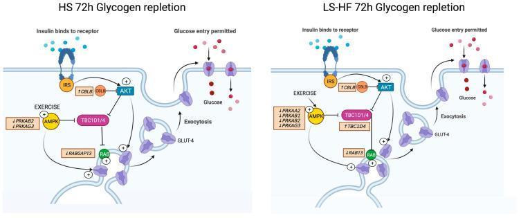
The insulin receptor is a transmembrane glycoprotein made of two extracellular alpha subunits (the insulin-binding/recognition site) and two transmembrane beta subunits, linked as an alpha2-beta2 heterotetramer. The beta subunit has intrinsic tyrosine kinase activity - the insulin receptor is classified as a receptor tyrosine kinase (Lippincott Illustrated Reviews Biochemistry; Katzung's Basic and Clinical Pharmacology).
Mechanism:
- Insulin binds the alpha subunits, causing a conformational change.
- This activates the beta subunit's tyrosine kinase, which autophosphorylates the receptor and phosphorylates downstream substrates, chiefly insulin receptor substrates (IRS-1, IRS-2).
- Phosphorylated IRS proteins recruit and activate PI3-kinase, which generates PIP3 and activates Akt (protein kinase B).
- Akt drives translocation of GLUT4 glucose transporters from intracellular vesicles to the plasma membrane in muscle and adipose tissue, allowing glucose uptake.
- A parallel Ras/MAP kinase pathway mediates insulin's growth-promoting and gene-expression effects (Costanzo Physiology, 7th ed; Guyton and Hall Textbook of Medical Physiology).
3. Metabolic actions by target tissue
Liver
- Promotes glycogen synthesis (activates glycogen synthase) and inhibits glycogenolysis and gluconeogenesis, suppressing hepatic glucose output.
- Stimulates lipogenesis (fatty acid and triglyceride synthesis) and inhibits ketogenesis.
Skeletal muscle
- Stimulates GLUT4-mediated glucose uptake.
- Promotes glycogen synthesis and protein synthesis (via mTOR activation); inhibits proteolysis.
Adipose tissue
- Stimulates glucose uptake and triglyceride synthesis.
- Inhibits hormone-sensitive lipase, suppressing lipolysis and free fatty acid release - a key reason why insulin deficiency leads to unchecked lipolysis and ketogenesis (as seen in DKA).
Overall systemic effect: insulin is the primary anabolic, fuel-storage hormone - it lowers blood glucose by promoting cellular uptake and storage (glycogen, triglycerides, protein) and by suppressing catabolic pathways (glycogenolysis, gluconeogenesis, lipolysis, proteolysis) (Robbins, Cotran & Kumar Pathologic Basis of Disease - "Insulin Action and Insulin-Signaling Pathways").
4. Relevance to disease states
- Type 1 DM: autoimmune beta-cell destruction eliminates insulin secretion, leading to unopposed lipolysis/ketogenesis and hepatic glucose overproduction (DKA risk).
- Type 2 DM: post-receptor signaling defects (impaired IRS-1/PI3K/Akt activation) cause insulin resistance in liver, muscle, and fat, combined with progressive beta-cell secretory failure - the two-hit pathophysiology of hyperglycemia in type 2 diabetes.
- Insulin receptor mutations/autoantibodies (rare) cause severe insulin resistance syndromes (Goldman-Cecil Medicine, "Insulin-Receptor Mutations").
Want me to go deeper into the molecular basis of insulin resistance in type 2 DM, or the counter-regulatory hormone system (glucagon, catecholamines, cortisol, GH) that opposes insulin action?
This is a shared conversation. Sign in to Orris to start your own chat.