

What is this Without pain Skin is protruding i seem but i dont know exactly what this is
plantar wart verruca plantaris sole of foot
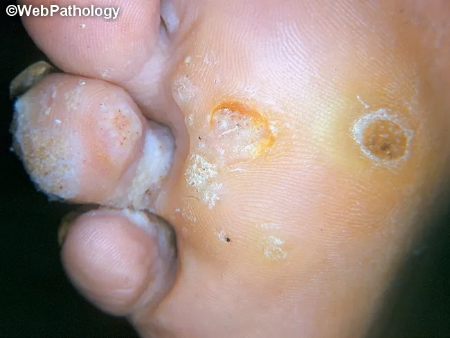
Clinical photography of a plantar foot displaying a verruca plantaris (plantar wart). Modality is a high‑quality gross image used in dermatology documentation. Precise location is the plantar surface of the foot (sole), with a focal, endophytic lesion that is rough and hyperkeratotic. Visual features include a circular to oval papule with a central crater or indent, irregular surface texture, and surrounding callus‑like thickening. Punctate black dots representing thrombosed capillaries may be seen within the lesion, while surrounding epidermis shows white scaling and fissuring. The lesion is typically mildly elevated above the surrounding skin and may be tender with pressure; it often coexists with other warts on the same sole. The clinical impression is verruca plantaris caused by human papillomavirus infection, with characteristic epidermal hyperplasia, papillomatosis, and hyperkeratosis evident on gross inspection. Differential diagnoses include callus/corn, plantar keratoderma, and foreign body reaction; histology would show koilocytosis and viral cytopathic effects if examined. This image supports clinical decision making, differential diagnosis, and treatment planning (topical salicylic acid, cryotherapy, or laser), and serves as an educational reference for students, residents, and clinicians evaluating plantar lesions. Clinical correlation includes assessing for additional lesions and patient history of HPV exposure; follow-up imaging is not required.
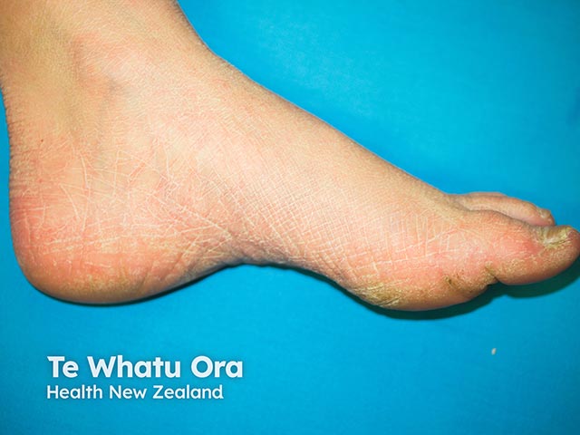
This is a macroscopic clinical photograph of the plantar aspect of the foot, captured for dermatology/foot-health assessment. Imaging modality: Clinical photography; technique: high-resolution color image of the sole, direct view, with standard diffuse lighting and no magnification. Anatomical location: Plantar surface of the hindfoot and midfoot, primarily the heel region, extending to the medial plantar arch. Visual features: a red-orange to pink-tan hyperkeratotic plaque with a thick, rough surface and fissuring; surrounding hypochromic to xerotic skin; margins are relatively ill-defined but the lesion remains clearly localized to the plantar skin. The lesion appears as a diffuse keratotic patch rather than a single discrete nodule, with variable thickness across the plantar surface. Pathology (clinically): evidence of epidermal hyperplasia with thickened stratum corneum (hyperkeratosis) and surface fissures, typical of friction-induced plantar keratopathy; no signs of vesiculation, purulence, or acute inflammation evident. Diagnostic significance: characteristic of friction-related plantar hyperkeratosis or callus; differential considerations include plantar corn, plantar wart (verruca plantaris), and diffuse plantar keratoderma; correlate with symptoms and history. Clinical correlation: evaluate footwear, gait, pressure points, activity level; screen for diabetes risk; may respond to keratolytics, debridement, padding, and orthotic optimization; useful for monitoring response to therapy and for educational and research material.
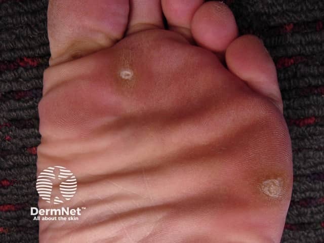
Clinical photography of the plantar surface of the foot demonstrating a classic hard corn (clavus) due to repetitive pressure. The lesion is a small, well‑circumscribed, hyperkeratotic papule with a central keratin plug, surrounded by peripheral epidermal thickening (callus) on the weight‑bearing sole. The surrounding skin is erythematous to slightly inflamed under pressure. The core appears pale yellow to white and slightly translucent; the lesion projects above the adjacent skin, producing a painful sensation with direct compression. This appearance distinguishes clavus from a plantar wart or simple callus by the presence of a dense keratinized core and a more pointed cone shape toward the dermis. The lesion is typically located at pressure points near the metatarsal heads or along the distal toe margins, often due to ill‑fitting footwear or abnormal gait. Etiology is chronic friction and pressure; pathophysiology involves localized epidermal hyperplasia and keratinocyte hypertrophy. Management focuses on debulking the hyperkeratosis (paring), application of keratolytics (salicylic acid), protective footwear, and orthotic correction; addressing footwear fit reduces recurrence risk. Differential diagnoses include plantar callus, plantar wart (verruca plantaris), and foreign body granuloma. Clinically correlates with pain on palpation and history of footwear‑related irritation. This image is educational for dermatology, podiatry, primary care, and medical education in skin pathology.
What Is It?
Key Features Visible in Your Image
| Feature | What I See |
|---|---|
| Location | Plantar surface (sole) of the foot |
| Lesion | Small, round, raised, rough/hyperkeratotic papule |
| Skin ridges | Skin lines distort around the lesion (classic wart sign) |
| No black dots visible | Can be hidden under thick overlying callus |
| No pain | Consistent - warts at non-pressure spots are often painless |
Why No Pain?
How It Spreads
- HPV enters through tiny cuts or breaks in the skin
- Picked up from floors, pool areas, shared shoes/socks
- Can spread to other parts of your foot if scratched
What You Should Do
- Salicylic acid (over-the-counter or prescription strength) - applied daily to gradually dissolve the wart
- Cryotherapy (liquid nitrogen freezing) - done in a clinic, very effective
- Laser treatment or minor surgery - for stubborn warts
Disclaimer: This is general information based on image analysis and is not a medical diagnosis. Please have it examined by a healthcare professional to confirm and get appropriate treatment.