
psoriasis from rooks and fitzpatrick
psoriasis
psoriasis from rooks and fitzpatric
psoriasis plaques skin silvery scales
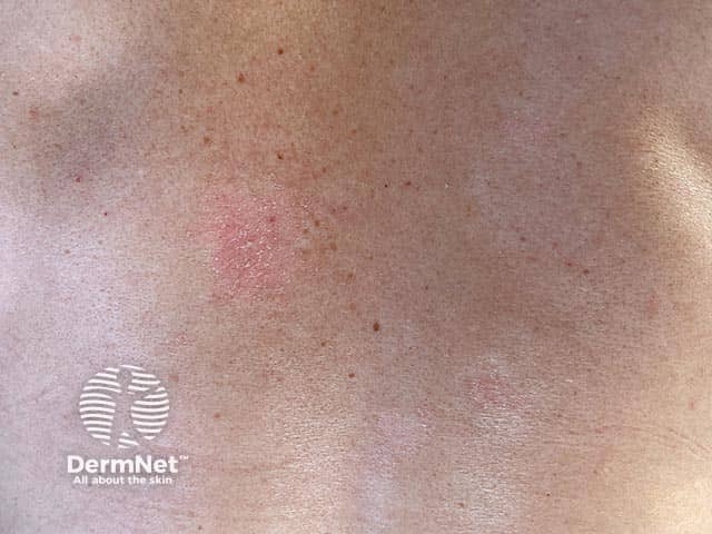
Clinical photography of early superficial plaque psoriasis on the back (posterior trunk). The image depicts well-demarcated, erythematous plaques with overlying loosely adherent silvery scales. On gentle manipulation, the scale becomes more prominent, and scratching accentuates the shedding of scales, compatible with the classic Auspitz phenomenon. The plaques appear epidermal in location, with discrete to coalescing patches centered on the dorsal torso and sparing unaffected skin. The skin surface shows slight hyperkeratosis, mild epidermal thickening (acanthosis), and minimal surrounding edema. There is no purulence or crusting; the lesions are non-ulcerated and without vesiculation. This appearance is characteristic of plaque psoriasis, a chronic, immune-mediated dermatosis with rapid keratinocyte turnover and inflammatory infiltrates. The clinical relevance includes differentiation from eczema and tinea corporis; a dermatitis with silvery scale is more psoriasis-like, whereas eczema typically exhibits lichenification and oozing, and tinea shows ring-like margins with central clearing. The image highlights features important for diagnosis: plaque morphology, scaling pattern, and trunk distribution. In educational and research contexts, this image supports teaching psoriasis pathophysiology, differential diagnosis, and treatment planning, including topical corticosteroids, vitamin D analogs, and phototherapy. Clinically, the finding warrants evaluation for associated comorbidities such as psoriatic arthritis and metabolic syndrome for comprehensive patient care.
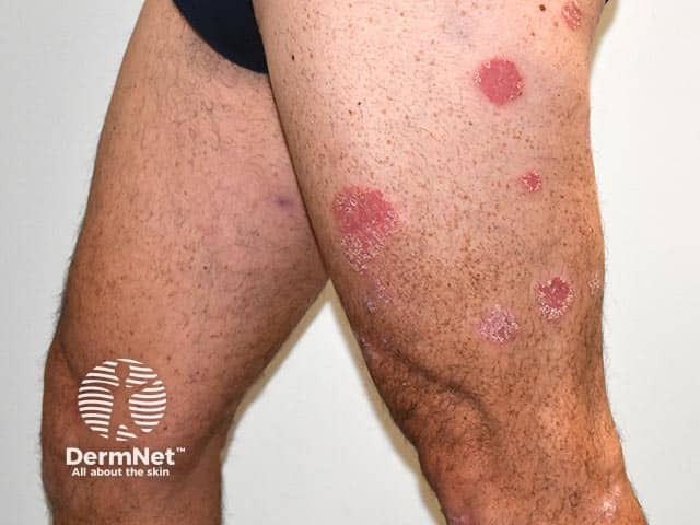
Clinical photography of cutaneous psoriasis on the lower legs, captured as a frontal close-up of extensor surfaces. This dermatologic image depicts multiple well-circumscribed, erythematous plaques with characteristic silvery white scale, distributed on the anterolateral aspects of the shins and extending to surrounding skin. Plaques range from several centimeters in diameter to smaller lesions, with clearly defined borders and a slightly raised, plaque-like morphology. There is mild to moderate surface scaling and partial crusting in several plaques, with surrounding islands of normal-appearing skin. The photograph emphasizes the chronic, relapsing nature of plaque psoriasis, a T-cell mediated, inflammatory dermatosis affecting the integumentary system. The regional localization corresponds to typical extensor surfaces of the limbs; however, lesions may be symmetrical and bilateral. Differential considerations include eczema/atopic dermatitis, tinea corporis, pityriasis rosea, or nummular dermatitis, though the combination of well-demarcated plaques and fine silvery scales strongly supports psoriasis. Clinical relevance: diagnosis is usually clinical, supported by history and, if needed, dermoscopic or histopathologic correlation. This image is useful for educational demonstrations of plaque morphology, treatment monitoring, and illustrative references for dermatology training, telemedicine review, and psoriasis severity assessment in clinical trials. Documented features include scaling pattern, plaque size variability, and potential Koebnerization in susceptible individuals.
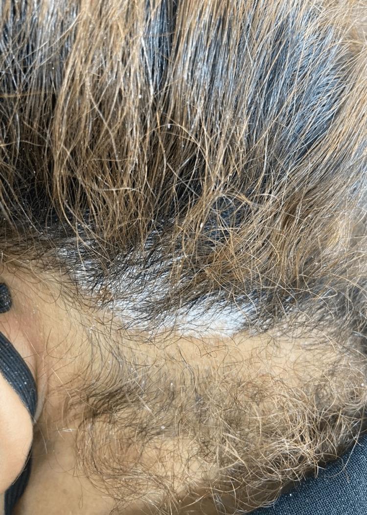
This clinical photograph shows a close-up view of a patient's scalp, demonstrating hallmark signs of scalp psoriasis. The image reveals multiple well-circumscribed, erythematous plaques covered by thick, micaceous (silvery-white) scales. These scales are non-uniformly distributed across the scalp and appear heavily concentrated at the base of the hair follicles. Many scales have detached from the skin surface and are visible clinging to individual hair shafts, creating a characteristic flaky or 'powdery' texture throughout the surrounding hair. The presentation is typical for chronic plaque psoriasis affecting the scalp, specifically in the context of paradoxical reactions or drug-induced dermatoses. Key educational features include the contrast between the silvery scales and the underlying skin, the focal density of the plaques, and the typical involvement of the hair-bearing areas without resulting in primary scarring alopecia.
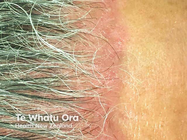
Clinical photography of the frontal scalp and anterior hairline demonstrates plaque psoriasis involvement. The image shows erythematous, well-demarcated plaques along the hairline and extending onto the forehead with fine silvery scales. Lesions are located on sun-exposed facial/forehead skin around the temples and brow region, with involvement of hair-bearing skin and adjacent non-hair-bearing skin. The plaques display classic psoriatic features: erythema, prominent scaling, and mild desquamation, with edge accentuation at hair follicle openings. The skin between plaques remains largely uninvolved, and there is no evident crusting or oozing in this view. The appearance is consistent with plaque psoriasis (psoriasis vulgaris) rather than seborrheic dermatitis, tinea faciei, or atopic dermatitis; however, atypical mimickers exist. The image is captured in a frontal/anterior view using standard color photography, suitable for documenting disease extent on the hairline and forehead. These findings have diagnostic significance in supporting a psoriasis diagnosis and in guiding topical therapies and subsequent phototherapy planning. Clinically, recognizing localized facial/forehead psoriasis impacts management decisions and prognosis, as facial involvement may require gentler formulations and careful irritation minimization. Potential clinical use cases include dermatology education, teledermatology assessment, monitoring treatment response, and research on facial psoriasis phenotypes.
psoriasis histology acanthosis parakeratosis neutrophils Munro
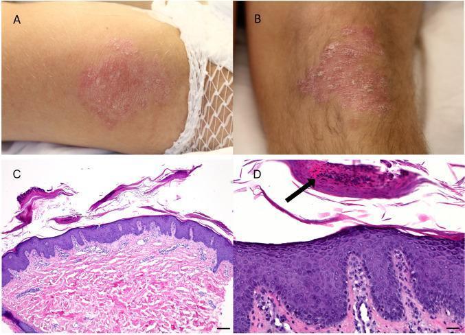
A multi-panel medical image illustrating the clinical and histological features of psoriasis. Panels A and B are clinical photographs showing well-demarcated, erythematous, scaly plaques located on the extensor surfaces of the elbows and knees. Panels C and D are photomicrographs of a skin biopsy stained with Hematoxylin and Eosin (H&E). Low-power magnification (C) demonstrates classic psoriasiform acanthosis, characterized by regular epidermal hyperplasia with elongated rete ridges and perivascular lymphocytic infiltration in the upper dermis. High-power magnification (D) reveals parakeratosis (retention of nuclei in the stratum corneum), dilated and tortuous capillary vessels within the papillary dermis, and a Munro microabscess—a distinct collection of neutrophils within the stratum corneum (indicated by a black arrow). The combination of clinical distribution on extensor surfaces and hallmark histological findings supports a diagnosis of psoriasis vulgaris, presented here in the context of a paraneoplastic manifestation of thymic carcinoma.
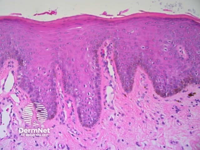
Light microscopy examination of a cutaneous punch biopsy stained with Hematoxylin and Eosin reveals epidermal hyperplasia characterized by acanthosis with elongation of the rete ridges. The stratum corneum shows varying thickness consistent with hyperkeratosis. The epidermal architecture remains orderly without described dysplasia or malignant features. The epidermal-dermal junction is preserved. In the superficial dermis there is a mild perivascular inflammatory infiltrate composed predominantly of lymphocytes and occasional histiocytes, without conspicuous neutrophilic microabscesses. The overall pattern is compatible with a chronic inflammatory or hyperplastic skin process rather than an acute infection. The combination of thickened epidermis, elongated rete pegs, and modest dermal inflammation can be seen in conditions such as chronic dermatitis, atopic dermatitis, or friction-related hyperkeratosis; psoriasis-like acanthosis should be considered if parakeratosis and neutrophils in the stratum corneum are present, which is not definitively demonstrated here. The findings are non-specific and require clinical correlation with lesion morphology, distribution, duration, and patient history. Clinically this image provides educational representation of basic cutaneous histology: epidermal hyperplasia, hyperkeratosis, dermal inflammatory infiltrate, and intact dermal connective tissue. Potential uses include teaching dermatopathology, validating histologic criteria for chronic inflammatory dermatoses, and aiding differential diagnosis discussions in dermatology.
psoriasis treatment biologics IL-17 IL-23
From Rook's Dermatology (5e) & Fitzpatrick's Dermatology
Definition & Overview
Clinical Features
Chronic Plaque Psoriasis (Psoriasis Vulgaris)
- Relatively symmetric distribution of sharply defined, erythematous, scaly plaques
- Sites of predilection: scalp, elbows, knees, lumbosacral area, hands, feet
- Genitalia involved in up to 45% of patients
- Plaques can persist for months to years at the same locations
- Remissions lasting at least 5 years occur in ~15% of patients
- Rook's Dermatology 5e, Ch. 8
Guttate Psoriasis
- More common in children and adolescents
- Frequently preceded by upper respiratory tract infection (Group A Streptococcal pharyngitis)
- Rapid onset of small, erythematous, drop-like (guttate) papules - can be generalized
- Elevated antistreptolysin O (ASOT) in over half of patients
- Superantigen-mediated T-cell activation is the proposed mechanism; SPEC-expressing Streptococcus has been identified in lesional skin
- Estimated to be the initial presentation of psoriasis in up to 20% of patients
- Fitzpatrick's Dermatology, Vol. 1-2; Rook's 5e
Pustular Psoriasis
- Characterized by sterile neutrophilic pustules
- Two main types:
- Generalized (von Zumbusch): acute fever, widespread erythema, lakes of pus, can be life-threatening
- Localized (palmoplantar pustulosis): confined to palms and soles
Inverse (Flexural) Psoriasis
- Affects intertriginous areas (axillae, groin, submammary folds, intergluteal cleft)
- Typical silvery scale is absent due to moisture
- Presents as shiny, smooth, well-demarcated erythematous plaques
- Differential diagnosis includes seborrheic dermatitis, tinea cruris, erythrasma
- Rook's Dermatology 5e, Ch. 73
Erythrodermic Psoriasis
- Involves >90% of body surface area
- Can arise from unstable chronic plaque psoriasis or abrupt withdrawal of systemic therapy
- Complications: thermoregulatory failure, high-output cardiac failure, sepsis
Pruritus in Psoriasis
Histopathology
- Neutrophils accumulate between keratinocytes forming spongiform pustule of Kogoj (in upper stratum spinosum)
- Accumulation of neutrophils and remnants in stratum corneum = microabscess of Munro
- These two are pathognomonic for psoriasis (and AGEP)
- Acanthosis with elongated, "squared-off" rete ridges (some coalescing at their bases)
- Parakeratosis with absence of stratum granulosum (hypogranulosis)
- Dilated, tortuous capillaries extending into elongated club-shaped dermal papillae
- Thin suprapapillary plate - explains the Auspitz sign (pinpoint bleeding on scale removal)
- Modest perivascular infiltrate of lymphocytes and histiocytes
- Rook's Dermatology 5e, Ch. 8
Pathogenesis
- Dermal dendritic cells produce TNF-alpha + IL-23
- IL-23 (IL-23p19 + IL-12/23p40) drives differentiation of Th17 cells
- Th17 cells produce IL-17, which promotes neutrophil-predominant inflammation
- IL-17 is now the key therapeutic target in modern biologics
- Chemokines attract neutrophils (forming Munro abscesses) and lymphocytes (via epidermotropism)
- CTACK, GRO-alpha, TARC, and other chemokines are upregulated
- Dendritic cells also secrete chemokines, attracting T cells and stimulating conjugate formation
- Fitzpatrick's Dermatology, Vol. 1-2
- Marked reduction in basal keratinocyte transit times
- Abnormal differentiation with loss of granular layer
- Anti-metabolites (methotrexate, UVB) and immunomodulators both address this
Nail Psoriasis
| Finding | Mechanism |
|---|---|
| Pitting (irregular, large, deep) | Parakeratotic foci in proximal nail matrix |
| Oil drop / Salmon patch | Exocytosis of leukocytes beneath nail plate (nail bed involvement) |
| Onycholysis with erythematous border | Distal nail bed parakeratosis |
| Subungual hyperkeratosis | Nail bed/hyponychium involvement |
| Splinter hemorrhages | Increased capillary fragility |
| Leukonychia | Midportion matrix involvement |
- Nail psoriasis is strongly associated with psoriatic arthritis and enthesitis
- In toenails, often clinically indistinguishable from onychomycosis (requires mycology/histology)
- Koebner phenomenon worsens nail symptoms
- Rook's Dermatology 5e, Ch. 71
Psoriatic Arthritis
| Type | Features |
|---|---|
| Mono/oligoarthritis (most common) | DIP, PIP joints; "sausage digit" (dactylitis) |
| Symmetric polyarthritis | Resembles RA but RF-negative |
| DIP-predominant | Nail psoriasis almost universally present |
| Spondyloarthropathy | Sacroiliitis, spinal involvement |
| Arthritis mutilans | Severe destructive, "opera-glass" deformity |
Severity Assessment
- Based on body surface area in 4 regions (head, upper limbs, trunk, lower limbs) plus grading of erythema, induration, and scaling
- Cumbersome; more used in clinical trials than routine practice
- Other indices: Scalp Psoriasis Severity Index, Nail Psoriasis Severity Index, Static Physician Global Assessment
Treatment
Topical Treatments
- Corticosteroids: mainstay; risk of atrophy in flexural/occluded sites
- Vitamin D analogues (calcipotriol/calcitriol): first-line for mild-moderate
- Coal tar: anti-proliferative, anti-inflammatory
- Anthralin (dithranol): effective but staining/irritation limits use
- Topical retinoids (tazarotene)
- Calcineurin inhibitors (tacrolimus, pimecrolimus): useful in flexural/facial psoriasis
- Roflumilast (newer PDE4 inhibitor): useful in intertriginous sites
Phototherapy
- NB-UVB (311-313 nm): preferred for moderate disease, safe in pregnancy
- PUVA (psoralen + UVA): effective but increases risk of skin cancer long-term
- UV also has immunomodulatory properties beyond anti-proliferative effects
Systemic Treatments (Conventional)
| Agent | Mechanism | Key Notes |
|---|---|---|
| Methotrexate | Anti-folate; anti-proliferative + immunomodulatory | Hepatotoxicity risk; gold standard for decades |
| Cyclosporine | Calcineurin inhibitor; blocks T-cell activation | Rapid response; nephrotoxicity limits long-term use |
| Acitretin | Retinoid | Teratogenic; especially useful for pustular/erythrodermic psoriasis |
| Hydroxyurea | Anti-proliferative | Second-line; cytopenias |
Biologics (Targeted Therapy)
- Infliximab, etanercept, adalimumab, certolizumab
- First-generation biologics; highly effective but lower than IL-17/IL-23 agents
- Ustekinumab: targets both IL-12 and IL-23 via shared p40 subunit
- Guselkumab, risankizumab, tildrakizumab
- Highly selective; excellent long-term safety data
- Secukinumab, ixekizumab (IL-17A antagonists)
- Bimekizumab (anti-IL-17A/F dual blocker)
- Fastest onset; especially effective for nail and scalp psoriasis
- Brodalumab: blocks IL-17RA; very high efficacy
Triggering / Exacerbating Factors
- Streptococcal infection (especially guttate psoriasis; superantigen mechanism)
- Drugs: beta-blockers, lithium, antimalarials, ACE inhibitors, NSAIDs, abrupt corticosteroid withdrawal
- Stress (psychological)
- Koebner phenomenon (trauma to skin)
- Alcohol and smoking
- HIV infection: worsens psoriasis paradoxically despite T-cell depletion; erythrodermic variant most common in sub-Saharan Africa
Special Situations
Anogenital Psoriasis
- Silvery scale is absent in flexural sites
- Women: labia majora and mons pubis (labia minora usually spared)
- Men: glans and shaft of penis
- Differential includes tinea cruris, erythrasma, seborrheic dermatitis, Reiter disease
- Treatment: moderately potent corticosteroid-antibiotic-antifungal combinations; tacrolimus/pimecrolimus; systemic agents for widespread disease
- Rook's Dermatology 5e, Ch. 73
HIV-Associated Psoriasis
- Plaque psoriasis remains most prevalent variant in US studies
- Red flags for HIV: rupioid psoriasis, keratoderma blennorrhagica, acute severe onset, multiple morphologies simultaneously
- Pathogenesis paradoxical: T-cell-destructive disease worsening a T-cell-mediated condition
- Treatment: ART is first-line for moderate-severe; TNF inhibitors used with caution (risk of worsening immunosuppression, PML from JC virus reactivation)
- Fitzpatrick's Dermatology, Vol. 1-2
Scalp Psoriasis
- Very common; plaques at hairline extend onto forehead
- Thick, micaceous scaling concentrated at follicular bases
- Does not cause scarring alopecia
Key Clinical Signs (Summary)
| Sign | Description |
|---|---|
| Auspitz sign | Pinpoint bleeding on removal of scale (dilated papillary capillaries) |
| Koebner phenomenon | New psoriatic lesions at sites of skin trauma/injury |
| Woronoff ring | Blanched halo around resolving plaques |
| Oil drop sign | Yellow-orange subungual discoloration (nail psoriasis) |
| Spongiform pustule of Kogoj | Neutrophils in upper stratum spinosum (active disease) |
| Munro microabscess | Neutrophil remnants in stratum corneum (pathognomonic) |
What's the latest on JAK inhibitors and other emerging psoriasis therapies?
JAK inhibitor psoriasis deucravacitinib TYK2
psoriasis emerging therapy novel treatment 2024 2025
JAK inhibitors psoriasis 2025 deucravacitinib new approvals emerging therapies
PMID: 38899421
PMID: 40078858
https://www.hcplive.com/view/psoriasis-in-2025-year-review
PMID: 41013260
spesolimab IL-36 receptor psoriasis pustular
JAK Inhibitors & Emerging Psoriasis Therapies (2024-2026)
1. TYK2 Inhibitors - The Star Class
Deucravacitinib (Sotyktu) - FDA Approved
- Mechanism: Allosteric TYK2 inhibitor - binds the regulatory pseudokinase (JH2) domain, NOT the conserved ATP-binding (JH1) domain shared by all JAKs. This gives it extraordinary selectivity for TYK2 over JAK1/2/3.
- Targets: Downstream signaling of IL-23, IL-12, and type I interferons
- Approvals:
- 2022: FDA-approved for moderate-to-severe plaque psoriasis
- March 2025: FDA-approved for active psoriatic arthritis - first and only TYK2 inhibitor for PsA
- Efficacy: A network meta-analysis of 20 RCTs (7,564 patients, PMID 38899421) ranked deucravacitinib 3mg QD and 3mg BID as the best oral agent when combining efficacy (PGA 0/1, PASI-75) and safety
- 5-year data (POETYK PSO-1/PSO-2 LTE): Consistent durable response rates with maintained safety - no prominent signals for MACE, thrombosis, or malignancy that concern pan-JAK inhibitors. Published 2025 in JEADV.
- Safety advantage over pan-JAK inhibitors: The FDA's boxed warning for pan-JAK inhibitors (risk of MACE, malignancy, DVT) does NOT apply to deucravacitinib because of its allosteric selectivity
Zasocitinib (TAK-279) - Phase 3 Complete
- Next-generation, highly selective oral TYK2 inhibitor by Takeda
- Latitude Phase 3 trials (Dec 2025): Positive topline data - superiority over placebo AND apremilast for sPGA 0/1 and PASI-75 at Week 16. All 44 ranked secondary endpoints met. PASI-75 response evident as early as Week 4
- Compared favorably to deucravacitinib in earlier Phase 2 data (Armstrong et al., JAMA Dermatol. 2024)
ESK-001 (IMMpulse trial)
- Another investigational selective TYK2 inhibitor
- Being evaluated in the IMMpulse trial; discussed at expert panels alongside zasocitinib as a potential next entrant in the class
2. Icotrokinra (JNJ-77242113) - A Landmark Agent
- Mechanism: First-in-class oral peptide IL-23 receptor antagonist - selectively blocks the IL-23 receptor intracellularly, functioning like a biologic but taken by mouth
- ICONIC-ADVANCE 1 & 2 (Sept 2025, presented at EADV 2025 Paris): Icotrokinra was superior to deucravacitinib for skin clearance at both Week 16 and Week 24 in moderate-to-severe plaque psoriasis
- ICONIC-LEAD (52-week): Sustained clearance with adverse event rates comparable to placebo
- ICONIC-TOTAL (Oct 2025): Efficacy in difficult-to-treat areas - scalp, hands, genitals, feet - in adults and adolescents ≥12 years
- This agent blurs the line between oral drugs and biologics, bringing IL-23 pathway precision to oral delivery
3. Pan-JAK Inhibitors in Psoriasis-Adjacent Indications
| Agent | JAK Selectivity | Status in Psoriasis Disease |
|---|---|---|
| Tofacitinib (Xeljanz) | JAK1/3 | Used off-label in psoriasis; less favored for skin due to boxed warning |
| Upadacitinib (Rinvoq) | JAK1-selective | FDA-approved for PsA (not skin psoriasis); expanding in pediatric settings (Elevate-Derm, July 2026) |
| Brepocitinib | TYK2 + JAK1 dual | Under investigation for PsA and skin psoriasis |
| Filgotinib | JAK1-selective | Being studied in PsA |
| Ivarmacitinib | JAK1-selective | Phase 2 data in PsA |
4. IL-36 Receptor Inhibition - Spesolimab (Spevigo)
- Target: IL-36 receptor (a novel pathway distinct from IL-17/IL-23)
- Indication: FDA-approved for generalized pustular psoriasis (GPP) flares - a rare, life-threatening variant
- Mechanism: Blocks IL-36 signaling, which is central to the neutrophilic cascade in GPP. A 2025 JCI study (PMID 40591420) showed spesolimab mitigates residual inflammation AND prevents future GPP flares
- IV formulation for acute flares; subcutaneous formulation under study for maintenance
- Also showing activity in other IL-36-mediated dermatoses (palmoplantar pustulosis, DITRA)
5. New Approvals & Formulations (2025)
- Roflumilast foam (PDE4 inhibitor): FDA approval expanded for specific psoriasis indications including intertriginous/flexural sites - relevant for areas where corticosteroids cause atrophy
- Guselkumab (Tremfya): Additional approvals for specific indications and age groups in 2025
- Biosimilars: Interchangeable biosimilars for TNF inhibitors (adalimumab, etanercept) reducing financial barriers - now covered under expanded Medicare criteria per the Masterclasses in Dermatology 2024 review (PMID 40078858)
6. Gut Microbiota-Targeted Therapy
7. Comorbidity Prevention - A New Goal
Summary Table: Oral Agent Pipeline
| Agent | Class | Status (2026) |
|---|---|---|
| Deucravacitinib | TYK2 inhibitor (allosteric) | FDA-approved (psoriasis + PsA) |
| Zasocitinib | TYK2 inhibitor | Phase 3 complete - filing expected |
| ESK-001 | TYK2 inhibitor | Phase 2 |
| Icotrokinra | Oral IL-23R antagonist (peptide) | Phase 3 complete - filing expected |
| Apremilast | PDE4 inhibitor | Approved (moderate disease) |
| Roflumilast | PDE4 inhibitor (topical/foam) | Approved (topical) |