
Ewings sarcoma
Ewing sarcoma histology small round blue cells
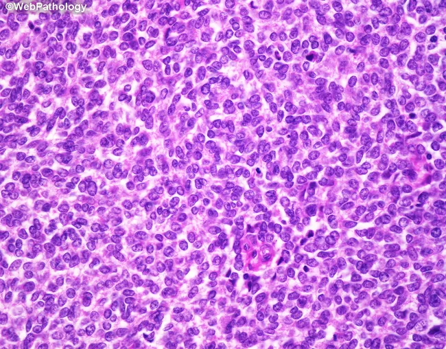
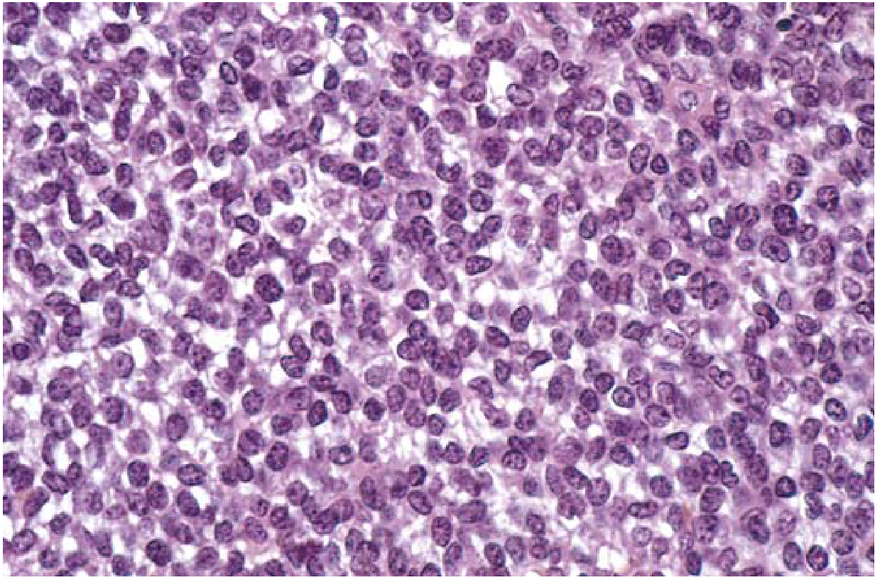
This is a light microscopy image of a bone tumor biopsy stained with Hematoxylin and Eosin (H&E). The image demonstrates the classic small round blue cell morphology of Ewing sarcoma. The tumor is composed of uniform, small, primitive-appearing cells with scant pale eosinophilic or clear cytoplasm and rounded to oval nuclei with evenly dispersed chromatin and small or inconspicuous nucleoli. Cytoplasmic borders are indistinct, producing a syncytial appearance. Mitotic activity is variable and may be increased. Intracytoplasmic glycogen is common and can be highlighted with periodic acid–Schiff (PAS) staining; this feature supports the diagnosis of this entity. The tumor cell nests may arrange around delicate vasculature to form pseudorosettes; true Homer-Wright rosettes may be present in some cases. The stroma is a delicate vascular network; necrosis is frequent, appearing as large geographic areas or smaller foci. Together these features produce a dense, sheets-and-nests pattern with a high nuclear-to-cytoplasmic ratio typical of small round blue cell tumors. The image is diagnostic when integrated with immunohistochemical and molecular studies (e.g., CD99 positivity, EWSR1 rearrangement) but the morphology shown is highly characteristic of Ewing sarcoma. This histology is pathognomonic in the appropriate clinical context and is essential for prognosis and treatment planning.
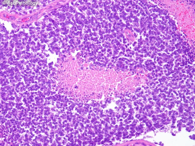
Imaging modality: Histopathology using light microscopy of formalin-fixed, paraffin-embedded bone tissue stained with Hematoxylin and Eosin (H&E). The slide displays a skeletally derived lesion with features classic for Ewing sarcoma. At high cellular density, there are sheets of small round blue cells with scant cytoplasm and round to oval nuclei showing hyperchromasia and brisk mitotic activity. The tumor cells are arranged in uniform clusters, separated by delicate fibrous strands, creating a highly cellular, often undulating landscape. Areas of geographic necrosis are frequently seen within the tumor, producing pink, acellular zones that disrupt cohesive cell architecture. Cytoplasmic scantiness and high nuclear-to-cytoplasmic ratio are typical; occasional rosette-like formations (Homer-Wright) may be present but are not required for diagnosis. The overall histologic pattern corresponds to a high-grade small round blue cell neoplasm in bone. Although not shown in this image, CD99 membrane positivity is commonly observed in Ewing sarcoma and, together with molecular testing for EWSR1-FLI1 fusion (t(11;22)) or related rearrangements, strengthens the diagnosis. Clinically, this histology correlates with aggressive bone lesions in children and adolescents and has important differential considerations including lymphoma, rhabdomyosarcoma, neuroblastoma, and osteosarcoma. Correlation with radiology and immunohistochemistry is essential for definitive diagnosis and prognostic assessment in clinical practice overall.
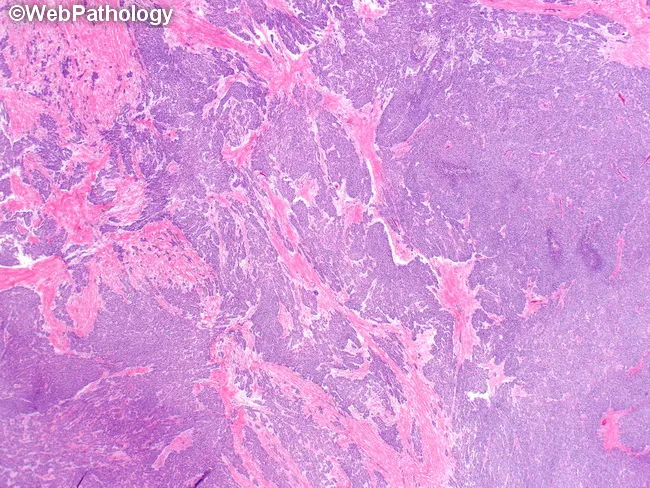
This light-microscopy histology slide shows Desmoplastic small round cell tumor (DSRCT) morphology. Hematoxylin and eosin (H&E) staining reveals sharply demarcated nests and anastomosing trabeculae of small round blue cells with scant cytoplasm and hyperchromatic nuclei, embedded in a densely desmoplastic fibrous stroma. The tumor cells exhibit brisk mitotic activity and occasional rosette-like patterns. The desmoplastic background contains abundant collagen with prominent vascularity, contributing to the fibrous, hyalinized appearance. While not visible by this stain alone, typical immunophenotype includes coexpression of epithelial and mesenchymal markers (cytokeratins, EMA) and desmin in a dot-like perinuclear pattern; WT1 positivity supports the diagnosis. Molecular confirmation commonly shows EWS-WT1 fusion from t(11;22)(p13;q12). Clinically, DSRCT is an aggressive small round cell sarcoma arising primarily in the abdomen or pelvis with potential for peritoneal spread and multicentric involvement; prognosis is poor despite multimodal therapy. This image is valuable for educational purposes, illustrating the characteristic nest-desmoplastic matrix contrast and serving as a reference in differential diagnosis against Ewing sarcoma/PNET, neuroblastoma, and rhabdomyosarcoma. It underscores the need for ancillary studies to confirm diagnosis in pathology practice. This image emphasizes classic DSCRT morphology and the essential role of molecular testing in confirming the EWS-WT1 translocation. This makes it valuable for research and education.
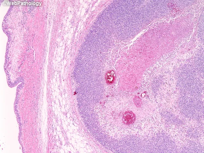
This histopathology image represents a small round blue cell tumor of the renal pelvis, consistent with primitive neuroectodermal tumor (PNET)/Ewing family tumor. Acquired from a renal pelvis specimen showing invasion into the urothelial lining, the section is stained with hematoxylin and eosin and viewed under light microscopy at low to intermediate magnification. The tumor is organized in broad sheets of uniform, round cells with scant cytoplasm and hyperchromatic nuclei, yielding a classic high nuclear-to-cytoplasmic ratio. Mitotic figures are frequent and apoptotic activity may be evident; the surrounding renal parenchyma shows desmoplastic response and focal invasion of the mucosa, with the urothelium on the left preserved in outline. The lesion’s morphology mirrors other Ewing sarcoma family tumors and underlines the close morphologic and genetic kinship to EWSR1-associated neoplasms. Immunophenotyping (where performed) would typically show CD99 (MIC2) expression and may reveal EWSR1 gene rearrangement (e.g., EWSR1-FLI1) by molecular testing. Clinically, such tumors are aggressive and require multidisciplinary management. This image highlights the diagnostic significance of recognizing small round blue cell morphology in the renal pelvis, differentiating from Wilms tumor, lymphoma, rhabdomyosarcoma, and neuroblastoma, and underscores the role of histology complemented by molecular diagnostics for accurate classification and staging.
"Ewing sarcoma"[MeSH Terms] AND treatment
Ewing Sarcoma
Overview
- Robbins & Kumar Basic Pathology, p. 786
- Campbell's Operative Orthopaedics 15th Ed 2026, p. 1221
Epidemiology
| Feature | Detail |
|---|---|
| Peak age | 5-25 years (80% under 20) |
| Sex | Slight male predominance |
| Race | Predominantly White/European origin; exceedingly rare in Black individuals |
| Incidence | <1 per 1 million per year |
| Annual US cases | ~200 |
Pathogenesis / Molecular Genetics
- t(11;22)(q24;q12) - EWSR1-FLI1 fusion: ~85% of cases (most common)
- t(21;22)(q22;q12) - EWSR1-ERG fusion
- t(7;22)(p22;q12) - EWSR1-ETV1 fusion
- Robbins & Kumar Basic Pathology, p. 786
Sites of Involvement
- Long bones: Commonly originates in the metaphysis but typically extends extensively into the diaphysis; often a large portion or the entire bone is involved
- Flat bones: Pelvis (most common flat bone), ribs, scapula
- Spine: Primary vertebral Ewing sarcoma is rare (3.3-15%); sacrum involved in up to 50% of spinal cases
- Common metastatic sites: Lungs (most common), then bone
Clinical Features
-
Pain - the most universal symptom; often insidious onset
-
Painful, tender, warm, erythematous, swollen mass - can mimic osteomyelitis (a classic mimicker)
-
Systemic symptoms: intermittent low-grade fever, leukocytosis, elevated ESR/CRP, anemia
-
A needle aspirate may resemble pus - always send biopsy specimens for both culture AND pathology
-
Average delay from symptom onset to diagnosis: 34 weeks (15 weeks patient delay + 19 weeks physician delay)
-
Campbell's Operative Orthopaedics 15th Ed 2026, p. 1222
Imaging
Plain Radiograph
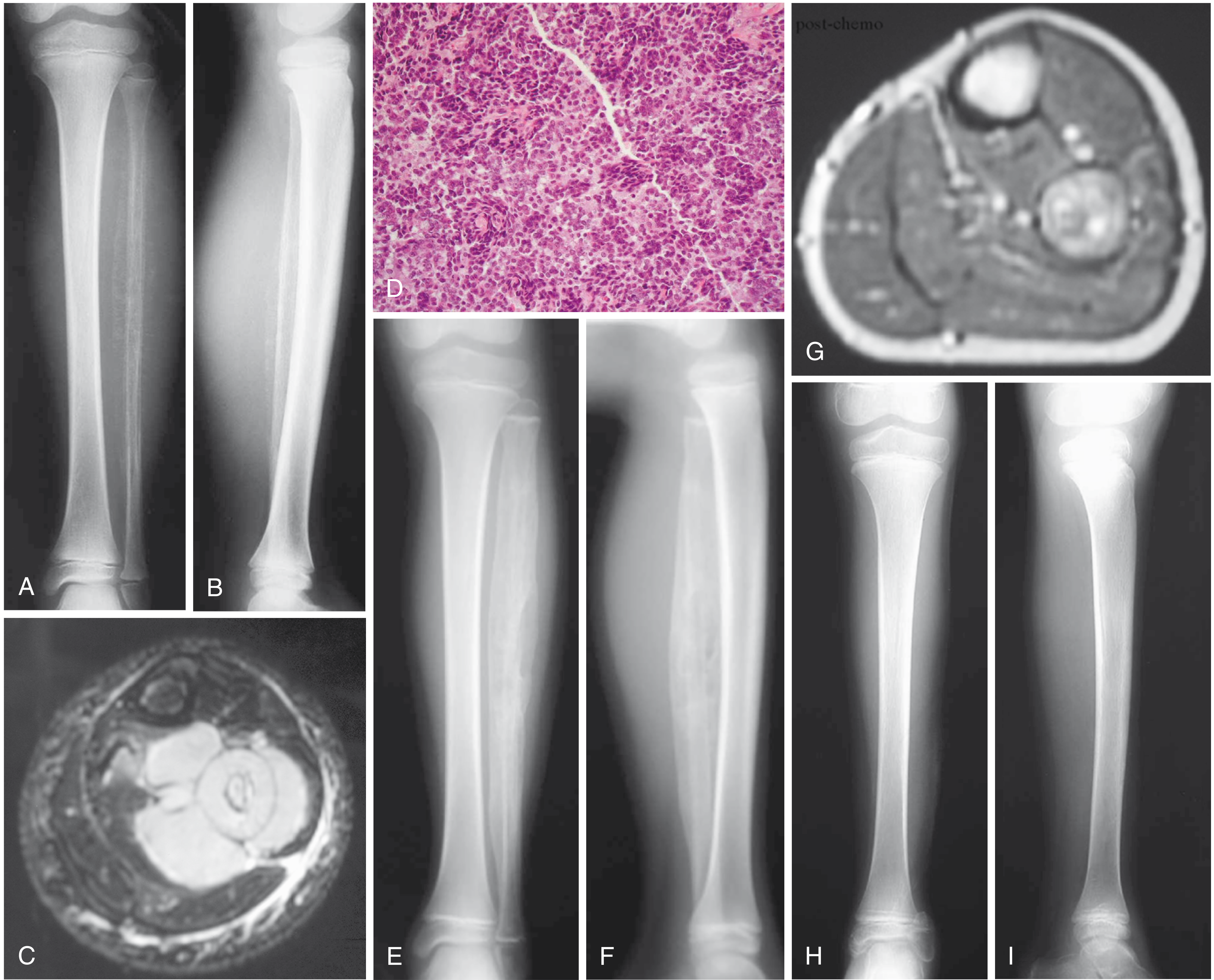
- Destructive, permeative ("moth-eaten") lytic lesion
- Classic "onion-skin" periosteal reaction (multilayered periosteal bone deposition from the advancing tumor)
- In flat bones: nonspecific destructive appearance
MRI
- Gold standard for local staging; defines full extent of intramedullary and soft-tissue involvement (typically extends well beyond plain film abnormality)
- Vertebral Ewing: isointense on T1, iso- to hyperintense on T2, intense gadolinium enhancement; "curtain sign" - invasion of spinal canal with paraspinal soft-tissue extension
- Entire bone must be imaged
CT
- Greater sensitivity than plain film for permeative destruction, especially in spine
- CT chest mandatory (lungs = most common metastatic site)
Nuclear Medicine
-
Bone scan: bone is second most common metastatic site
-
FDG PET/CT: now the new standard for initial staging, detecting recurrence, and evaluating metastatic disease; high sensitivity and accuracy; initial SUV of primary tumor correlates with prognosis
-
Grainger & Allison's Diagnostic Radiology, p. 1285-1286
-
Campbell's Operative Orthopaedics 15th Ed 2026
Pathology / Histology
- Homer-Wright rosettes may be present (circular groupings of cells with central fibrillary core)
- Geographic necrosis common
Differential Diagnosis of Small Round Blue Cell Tumors
| Tumor | Distinguishing Feature |
|---|---|
| Lymphoma | PAS negative, reticulin positive; LCA, CD20/CD3 positive |
| Neuroblastoma | Urinary catecholamines elevated; N-MYC amplification |
| Rhabdomyosarcoma | Desmin, myoglobin, muscle-specific actin positive |
| Osteosarcoma | Produces osteoid matrix |
| Small cell carcinoma | Cytokeratin positive |
| Wilms tumor | Renal location, WT1 |
| DSRCT | EWS-WT1 fusion t(11;22)(p13;q12); desmoplastic stroma |
Immunohistochemistry for Ewing Sarcoma
- CD99 (MIC-2 gene product): strong membranous positivity - most specific marker
- PAS positive (intracellular glycogen)
- Reticulin negative
- EWSR1 rearrangement by FISH: confirmatory
Staging Workup
| Investigation | Purpose |
|---|---|
| MRI of entire involved bone | Local staging, soft-tissue extent |
| CT chest | Pulmonary metastases (most common) |
| Bone scan | Bone metastases (2nd most common) |
| FDG PET/CT | Systemic staging + baseline for response monitoring |
| Bone marrow aspirate | Rule out diffuse marrow involvement |
Treatment
1. Neoadjuvant Chemotherapy (first)
- VDC: Vincristine + Doxorubicin + Cyclophosphamide
- IE: Ifosfamide + Etoposide
2. Local Control (surgery and/or radiation)
- Wide surgical resection is preferred for resectable tumors - avoids long-term complications of radiation (especially growth disturbance in children)
- Radiation for unresectable tumors (e.g., large pelvic tumors), positive margins, or high-risk sites (spine)
- After neoadjuvant chemo, repeat MRI typically shows marked reduction in soft-tissue mass and increased ossification on X-ray
3. Adjuvant Chemotherapy (consolidation)
Salvage Therapy (metastatic/recurrent disease)
-
Cyclophosphamide + Topotecan
-
Irinotecan + Temozolomide
-
Gemcitabine + Docetaxel
-
Goldman-Cecil Medicine, p. 1441-1443
Prognosis
| Scenario | 5-Year Event-Free Survival |
|---|---|
| Localized disease (multiagent chemo) | ~70% |
| Clinically detectable metastatic disease | Poor |
- Large tumor volume
- Pelvic/axial location
- Presence of metastases at diagnosis
- Poor histologic response to neoadjuvant chemotherapy
- Elevated LDH
Key Memory Points
- t(11;22)(q24;q12) - EWSR1-FLI1 - present in >90%
- "Onion-skin" periosteal reaction - classic X-ray sign
- Small round blue cells + PAS positive (glycogen) + CD99 positive
- Diaphysis of long bones + flat bones (pelvis most common flat bone site)
- Mimics osteomyelitis clinically (fever, leukocytosis, warmth, swelling)
- Peak age: 10-20 years; rare in Black individuals
- Treatment: VDC/IE chemotherapy + surgery ± radiation
- Does NOT produce bone or cartilage (unlike osteosarcoma)