
Malunion - Cubitus, varus valgus long answer question for orthopaedic exam mbbs
Reading File
Now let me search the medical library for content on cubitus varus and valgus malunion:
Finding Sources
Finding Sources
Excellent results. Now let me read the most relevant sections from multiple books simultaneously:
Reading File
Reading File
Reading File
Let me read more content and fetch the clinical images:
Reading File
Reading File
Loading Image
Loading Image
Now let me get the S Das surgical textbook content and fetch the osteotomy diagram:
Reading File
Loading Image
Finding Sources
Excellent. Now let me get the tardy ulnar nerve palsy section and the carrying angle/clinical details:
Reading File
Reading File
I now have comprehensive content from multiple authoritative textbooks. Let me compile the complete long-answer question response:
Malunion - Cubitus Varus and Cubitus Valgus
Long Answer - Orthopaedic MBBS Exam
Definition
Malunion refers to the healing of a fracture in a faulty position - with angulation, rotation, or shortening that results in deformity or functional loss.
Cubitus varus and cubitus valgus are the two major angular deformities at the elbow resulting from malunion, most classically following supracondylar fractures of the humerus in children.
Normal Carrying Angle
The elbow in full extension shows a physiological valgus alignment between the upper arm and forearm - the carrying angle - which is normally 9-14° (slightly greater in women, 2-3° more). This allows the arm to clear the hip during carrying. It is measured with the forearm in full supination and the elbow fully extended.
- Bailey and Love's Short Practice of Surgery, p. 505
CUBITUS VARUS
Definition and Deformity
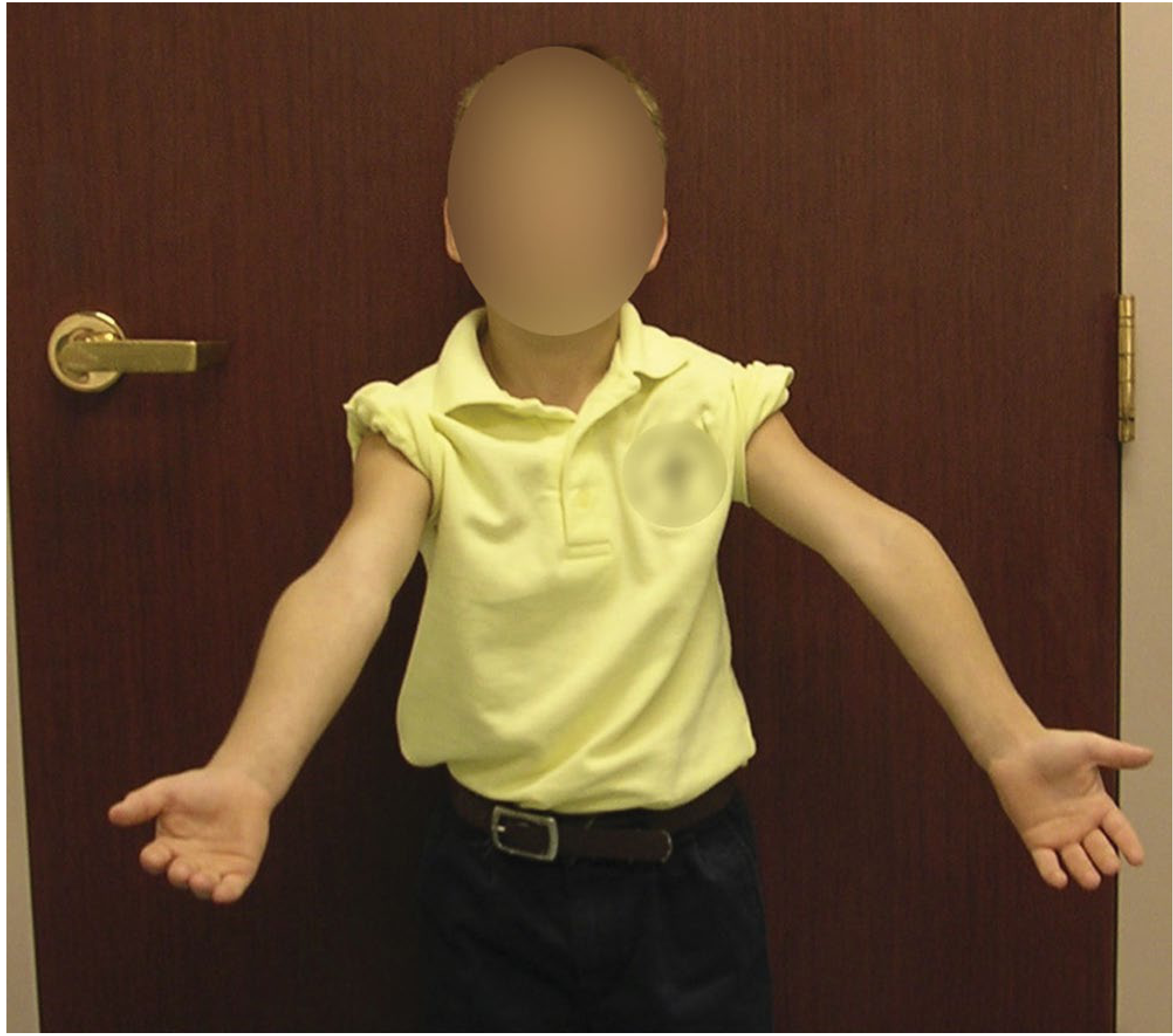
Cubitus varus is a reversal of the normal carrying angle, so the forearm angles inward (medially). The colloquial name is "gun-stock deformity" because the profile of the arm resembles the stock of a rifle.
Aetiology
- Most commonly follows a malunited supracondylar fracture of the humerus in children
- It is the most common angular deformity after supracondylar fracture
- Not primarily due to growth arrest - it is the result of malunion of the distal fragment in varus tilt
- Varus tilting of the distal fragment (medial tilt/collapse) is the single most important cause
- Medial comminution, medial column instability, and inadequate reduction/pinning lead to this collapse
Mechanism of Coronal Tilting
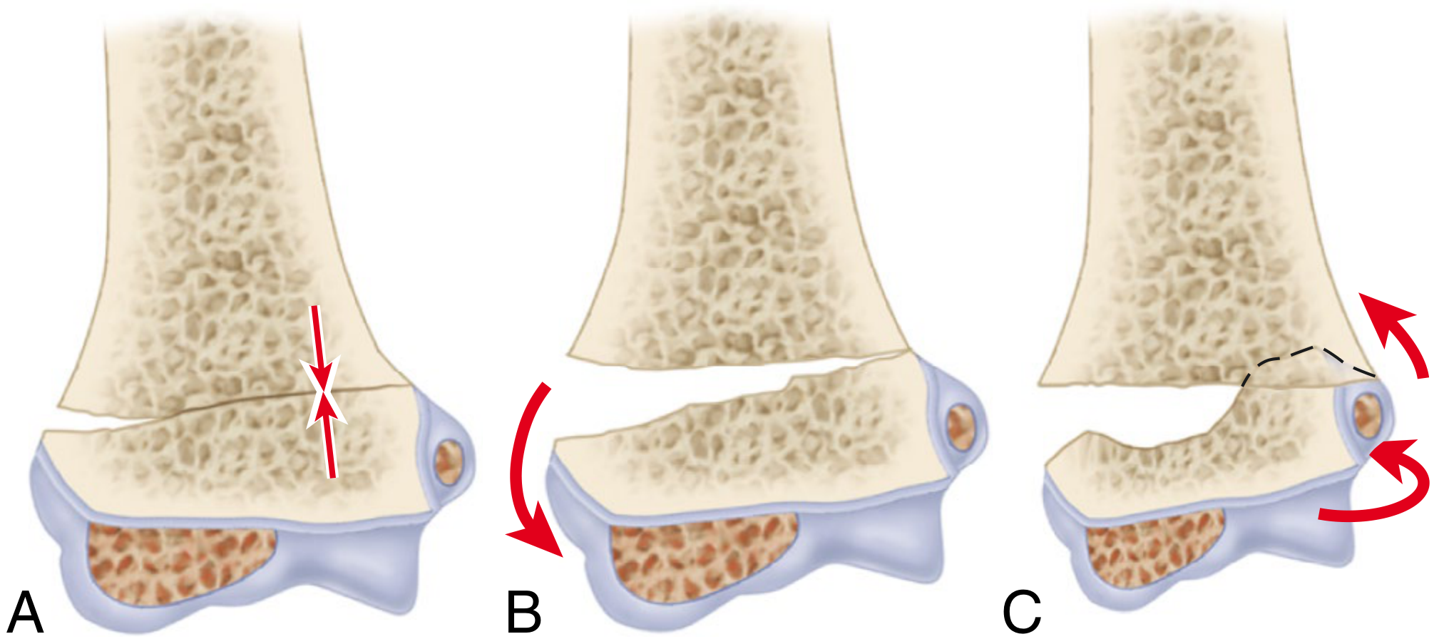
Type II fractures with medial column instability can collapse into varus when treated with cast alone without pinning. Osteonecrosis and delayed growth of the trochlea is an extremely rare additional cause.
Clinical Features
- Obvious medial angulation of the forearm (gun-stock deformity)
- Most noticeable in full extension
- Tenderness over the lateral collateral ligament complex
- Prominent tendon of the medial head of triceps
- Usually good cosmesis is the primary concern - function is generally NOT significantly impaired in uncomplicated cases
Complications of Cubitus Varus
- Lateral elbow instability / Posterolateral rotatory instability (PLRI) - the mechanical axis, olecranon, and triceps line of pull are all displaced medially; the repetitive external rotation torque stretches the lateral collateral ligament complex
- Tardy ulnar nerve palsy - cubitus varus deformities can also rarely be associated with this
- Snapping of the medial head of triceps over the medial epicondyle
- The link to elbow instability in adulthood has been documented up to 51 years after the original childhood injury
- Signs: positive posterolateral rotatory apprehension test, positive lateral pivot shift, posterolateral rotatory drawer test
- Campbell's Operative Orthopaedics, 15th Ed 2026, p. 1776
Remodelling
- 100% remodelling has been shown in children younger than 5 years
- Only minimal remodelling occurs after age 8 years - surgical correction becomes necessary
Treatment
Indications for surgery: Cosmetic deformity causing psychosocial distress; functional limitations; associated PLRI; deformity >15° in older children/adolescents
Types of supracondylar osteotomy:
Three basic types have been described:
- Lateral closing wedge osteotomy - easiest, safest, most stable, most commonly used; corrects coronal and (if placed obliquely) sagittal plane deformity; also allows axial rotational correction
- Medial opening wedge osteotomy with bone graft
- Oblique osteotomy with derotation
Uchida three-dimensional osteotomy - corrects medial and posterior tilt and rotational deformity simultaneously.
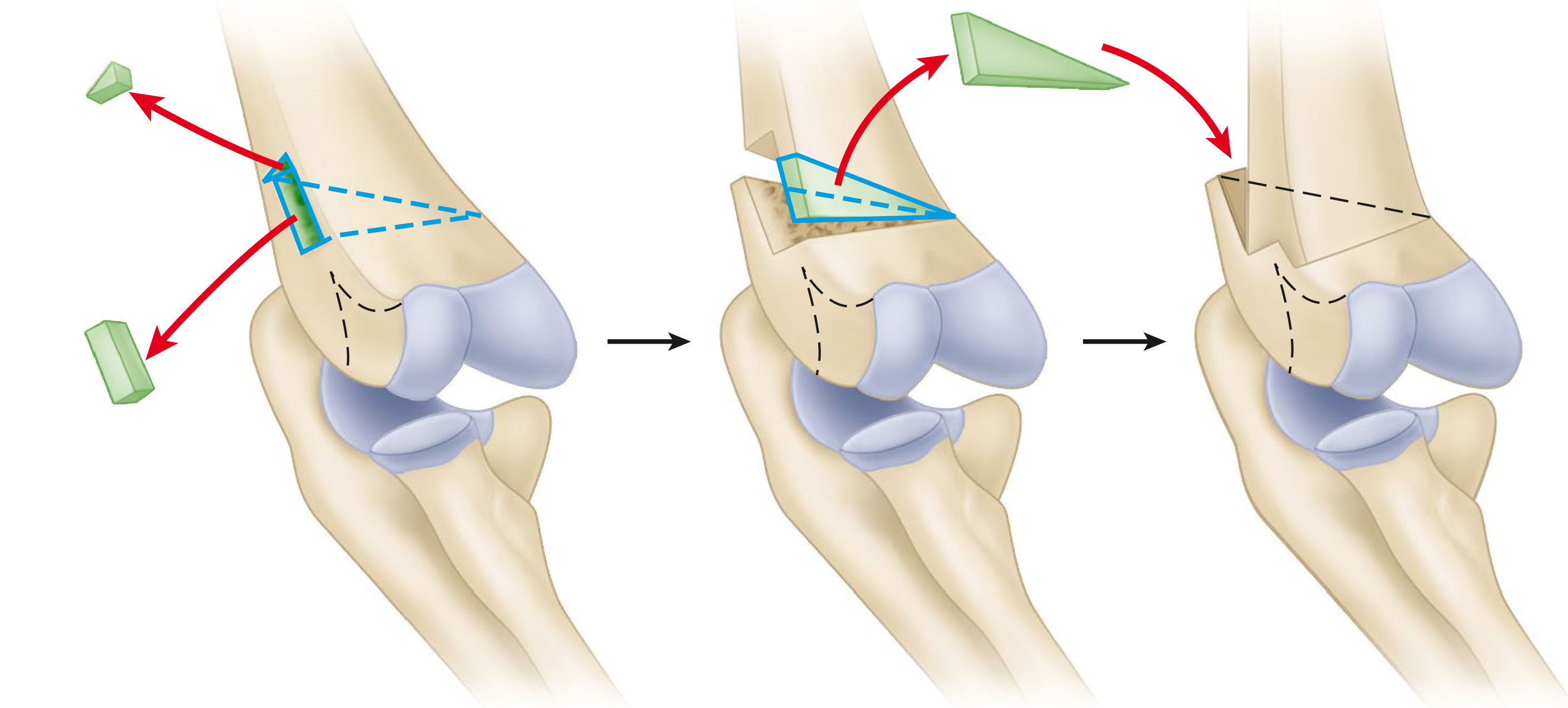
Lateral Closing Wedge Osteotomy - Key Steps:
- Lateral incision; tourniquet inflated
- Insert 2 K-wires into lateral condyle before osteotomy (just distal to planned distal cut)
- Make closing wedge osteotomy laterally, leaving medial cortex intact initially; cuts angled in sagittal plane if sagittal correction needed
- Weaken medial cortex with drill holes; apply valgus stress with forearm in pronation and elbow flexed to complete osteotomy
- Advance K-wires from lateral condyle into medial cortex of proximal fragment
- Fixation tailored to age: K-wires + screws for younger children; plate-and-screw for adolescents
Improved elbow flexion from 101° to 126° has been reported with this technique.
For severe deformity (>15°) with high functional demands or PLRI: osteotomy combined with lateral collateral ligament reconstruction is recommended.
- Campbell's Operative Orthopaedics, 15th Ed 2026
CUBITUS VALGUS
Definition
Cubitus valgus is an abnormal increase in the normal carrying angle (>15-20°), so the forearm deviates laterally beyond normal.
Aetiology
| Cause | Mechanism |
|---|---|
| Malunion of lateral condyle fracture | Most common cause of progressive cubitus valgus |
| Malunited distal humeral fracture | Less common |
| Physeal arrest (lateral side) | Growth disturbance after lateral condyle injury |
| Nonunion of lateral condyle fracture | Progressive deformity as lateral side fails to heal |
- Nonunion of lateral condylar fractures is actually more often responsible for cubitus valgus than simple malunion of supracondylar fractures
- Because the normal carrying angle naturally increases from childhood to adulthood, an increase in valgus is less cosmetically noticeable than varus reversal
Clinical Features
- Increased carrying angle on inspection
- May be well tolerated for years
- The critical late complication is tardy ulnar nerve palsy
Tardy Ulnar Nerve Palsy - The Classical Complication
Definition: A delayed, progressive ulnar nerve palsy occurring years after the original injury.
Mechanism: In cubitus valgus, the ulnar nerve (which runs in the cubital tunnel posterior to the medial epicondyle) is gradually stretched as the valgus angle increases over time. The nerve becomes elongated with each flexion-extension cycle, leading to traction neuropathy.
Features:
-
Develops years (5-20 years) after the original fracture
-
Progressive weakness of ulnar-innervated intrinsic muscles (interossei, hypothenar, medial two lumbricals)
-
Clawing of the ring and little finger (claw hand - more pronounced in ring and little fingers)
-
Loss of sensation on the medial 1.5 fingers and medial palm
-
Wasting of hypothenar eminence and dorsal interossei
-
Positive Froment's sign (flexion of IP joint of thumb on paper-holding test)
-
Positive Wartenberg's sign (little finger abduction)
-
Campbell's Operative Orthopaedics, 15th Ed 2026; S Das Manual on Clinical Surgery, 13th Ed
Treatment
Conservative: Splinting, activity modification, padding of the elbow - limited role in established cases
Surgical options for tardy ulnar nerve palsy:
- Anterior transposition of the ulnar nerve (subcutaneous, intramuscular, or submuscular) - relocates the nerve to prevent further stretching
- Medial epicondylectomy - partial or complete removal of the medial epicondyle
- In situ decompression - for mild/early cases
For the underlying cubitus valgus deformity:
- Medial opening wedge osteotomy or lateral closing wedge osteotomy of the distal humerus to restore normal carrying angle
- Often combined with nerve transposition
Comparison Table: Cubitus Varus vs Cubitus Valgus
| Feature | Cubitus Varus | Cubitus Valgus |
|---|---|---|
| Carrying angle | Reversed (negative) | Increased (>15-20°) |
| Common name | Gun-stock deformity | - |
| Most common cause | Malunited supracondylar fracture | Malunited / nonunited lateral condyle fracture |
| Primary concern | Cosmesis (poor) | Tardy ulnar nerve palsy |
| Functional impairment | Usually minimal initially | Progressive neurological deficit |
| Late complication | PLRI, snapping triceps | Tardy ulnar nerve palsy |
| Treatment | Lateral closing wedge osteotomy | Ulnar nerve transposition ± osteotomy |
Key Exam Points (High-Yield)
- Cubitus varus = gun-stock deformity - result of malunion of supracondylar fracture (medial tilt of distal fragment); primarily a cosmetic problem but late PLRI can occur
- Cubitus valgus = tardy ulnar nerve palsy - the feared late complication; caused most often by malunion/nonunion of lateral condyle fracture, not supracondylar
- Cubitus varus is NOT primarily due to growth arrest - it is malunion
- The carrying angle is normally 9-14°; measured with elbow extended and forearm supinated
- Remodelling of varus: complete below age 5, minimal above age 8
- Osteotomy of choice for cubitus varus: lateral closing wedge osteotomy (most stable, safe, commonly used)
- Tardy ulnar nerve palsy: traction neuropathy from stretching of the ulnar nerve over years in cubitus valgus; treatment is anterior transposition ± correction of deformity
Sources: Campbell's Operative Orthopaedics 15th Ed 2026 | Miller's Review of Orthopaedics 9th Ed | Bailey and Love's Short Practice of Surgery 28th Ed | S Das Manual on Clinical Surgery 13th Ed
This is a shared conversation. Sign in to Orris to start your own chat.