
Give me the name of the four ana- anastomoses of the heart with a small description. And also... The dedicated photos op is on.
- Right and left coronary arteries - anastomose at the apex and posterior interventricular groove
- Atrial anastomoses - connections between atrial branches
- Septal anastomoses - between anterior and posterior interventricular branches
- Marginal branch anastomoses - around the right heart border
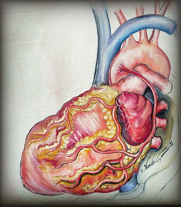
coronary circulation blood supply heart anatomy
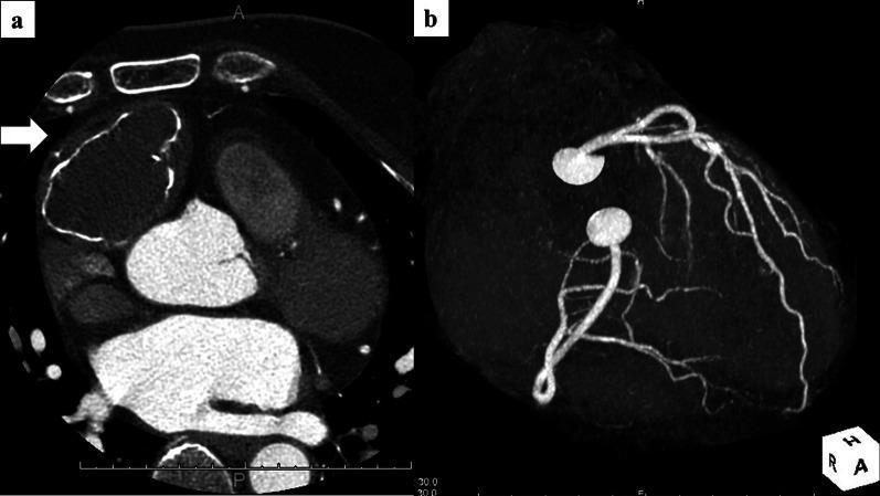
This dual-panel image displays diagnostic cardiovascular imaging. Panel (a) is an axial chest CT scan showing a large, well-circumscribed mass (white arrow) measuring approximately 50 x 45 x 40 mm in the anterior mediastinum, adjacent to the heart. The mass exhibits central intermediate attenuation and prominent, irregular marginal calcification along its periphery. Panel (b) shows a 3D reconstruction from a Coronary CT Angiography (CCTA) visualizing the coronary artery tree. The reconstruction demonstrates normal coronary anatomy without evidence of coronary aneurysms, stenosis, or abnormal feeding arteries originating from the coronary circulation to supply the mediastinal mass. These findings are clinically significant in the differential diagnosis of anterior mediastinal tumors, such as mature cystic teratomas, and in preoperative planning to exclude vascular involvement or parasitized blood supply from the coronary arteries.
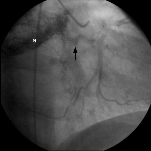
This diagnostic image is a coronary angiogram obtained during cardiac catheterization, showing the left coronary artery system. The visual focus is on the left circumflex (LCx) artery, indicated by a black arrow. Distinct from normal coronary anatomy, there is evidence of pathologically significant neovascularization, characterized by multiple small, tortuous, and irregular vessels branching from the LCx. These vessels lead to a discrete area of intense contrast enhancement, or 'tumor blush' (labeled 'a'), located in the proximity of the heart. This finding is indicative of a hypervascular mass, such as a cardiac paraganglioma, which is recruiting blood supply from the coronary circulation. The image demonstrates the use of angiography in identifying extrinsic or intrinsic cardiac tumors based on their vascular signatures and feeding vessel origins. It serves as an educational example of how coronary neovascularization and tumor blushes appear in clinical imaging for cardiac oncology and cardiology diagnostics.
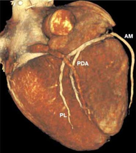
This diagnostic image is a 3D surface-rendered reconstruction of a human heart, specifically focused on the inferior surface to demonstrate coronary arterial anatomy. The rendering shows a reddish-brown textured myocardium with prominent vasculature. The image illustrates a right-dominant coronary circulation system, highlighting the distal branches of the right coronary artery (RCA). Three key arterial branches are annotated: the Acute Marginal (AM) artery curving along the right ventricular margin, the Posterior Descending Artery (PDA) coursing within the posterior interventricular sulcus toward the apex, and a Posterolateral (PL) branch extending toward the diaphragmatic surface of the left ventricle. This visualization is characteristic of Cardiac Computed Tomography Angiography (CCTA) and is used in clinical cardiology to assess coronary artery patency, anatomical variations, and the presence of atherosclerotic disease. The educational focus is on identifying posterior cardiac landmarks and understanding the distribution of blood supply in a dominant right coronary system.
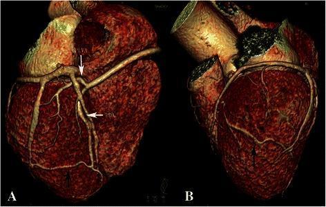
This medical image displays a 3D volume-rendered reconstruction of the heart and coronary arteries derived from cardiac Computed Tomography Angiography (CTA). Panels A and B illustrate the vascular anatomy of a rare congenital anomaly: a single right coronary artery (RCA) with the complete absence of the left coronary artery. In Panel A (posterior-lateral view), the RCA is shown bifurcating into the posterior left ventricular artery (PLVA) and the posterior descending artery (PDA). A black arrow identifies a small, aberrant branch originating from the distal PDA near the posterior cardiac apex. Panel B (anterior-lateral view) demonstrates the distal course of this small branch as it wraps around the apex to terminate on the anterior wall of the left ventricle. This visualization is critical for understanding compensatory collateral circulation and coronary anomalies, specifically how a dominant RCA can provide blood supply to myocardial territories typically served by the left coronary system. The image highlights essential anatomical landmarks, including the cardiac apex and the major posterior coronary branches.
heart coronary arteries anterior posterior surface anatomy labeled
four anastomoses of heart coronary arteries anatomy classical description
https://www.kenhub.com/en/library/anatomy/blood-supply-of-th…
coronary artery anastomosis apex interventricular posterior
The Four Anastomoses of the Heart
1. At the Apex of the Heart
2. In the Posterior Interventricular (Inferior) Groove
3. Around the Right Ventricular Outflow Tract (Conus/Infundibular Anastomosis)
4. In the Atrial Wall (Atrial Anastomoses)
Summary Table
| # | Location | Vessels Involved |
|---|---|---|
| 1 | Apex of the heart | LAD (anterior interventricular) ↔ PDA (posterior interventricular) |
| 2 | Posterior (diaphragmatic) AV groove / crux | Right coronary ↔ Left circumflex |
| 3 | Right ventricular outflow tract | Conus branch of RCA ↔ Infundibular branch of LAD (Circle of Vieussens) |
| 4 | Posterior atrial wall | Atrial branches of RCA ↔ Atrial branches of LCx |
Clinical Note