
Pneumonia x ray
pneumonia chest X-ray radiograph consolidation
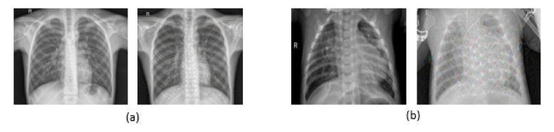
This diagnostic image comparison features four posterior-anterior (PA) chest X-ray radiographs organized into two groups for educational analysis of pneumonia. Group (a) displays normal chest X-rays, characterized by clear lung fields, distinct costophrenic angles, and visible bronchovascular markings. Group (b) illustrates pneumonia-affected lungs, demonstrating hallmark pathological signs including increased haziness, pulmonary infiltrates, and areas of consolidation that obscure normal lung parenchymal detail and cardiac borders. Each group includes one original radiograph and one version processed with a 'Deep Dream' filter, a computer vision technique used here to accentuate texture and pattern recognition for deep learning model training. The filtered pneumonia image in group (b) highlights abnormal mesh-like opacities and parenchymal irregularities. This comparison serves as educational material for medical imaging informatics and radiology, demonstrating the visual differences between healthy pulmonary tissue and inflammatory consolidation associated with infectious pneumonia in both raw and processed data formats.
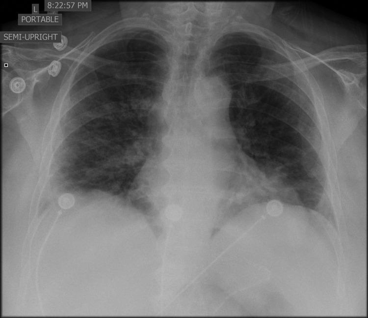
This diagnostic image is a portable, semi-upright anteroposterior (AP) chest X-ray of an adult patient. The radiograph demonstrates diffuse, bilateral pulmonary opacities and areas of consolidation. The increased density is most prominent in the middle and lower lung zones, obscuring the normal bronchovascular markings and suggesting multifocal pneumonia. External medical hardware is visible, including multiple electrocardiogram (ECG) leads positioned on the chest wall. The cardiac silhouette and mediastinal contours appear within normal limits for this projection. The trachea is midline. The visible portions of the diaphragm and costophrenic angles are partially obscured by the parenchymal opacification. This imaging presentation is clinically significant for the assessment of viral or atypical pneumonias, such as COVID-19 or influenza, which frequently present with such bilateral, diffuse infiltrates.
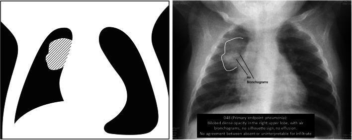
This educational composite contains a pediatric chest radiograph and a corresponding simplified anatomical diagram illustrating the WHO criteria for primary endpoint pneumonia. The right-sided image is an anteroposterior (AP) diagnostic X-ray of a child's chest. It demonstrates a focal, bilobed dense opacity in the right upper lobe, highlighted by a white outline. Significant radiographic features within this consolidated region include air bronchograms—linear lucencies representing air-filled bronchi against fluid-filled alveoli. There is no evidence of a silhouette sign at the heart border, and no pleural effusion is visible. The left-sided image is a high-contrast schematic diagram serving as a teaching tool; it uses a diagonal hatched pattern to map the exact anatomical location and morphology of the consolidation described in the radiograph. This material is designed for epidemiological training to standardize the identification of radiologically confirmed pneumonia, focusing on dense consolidation and distinguishing features like air bronchograms and the presence or absence of pleural fluid.
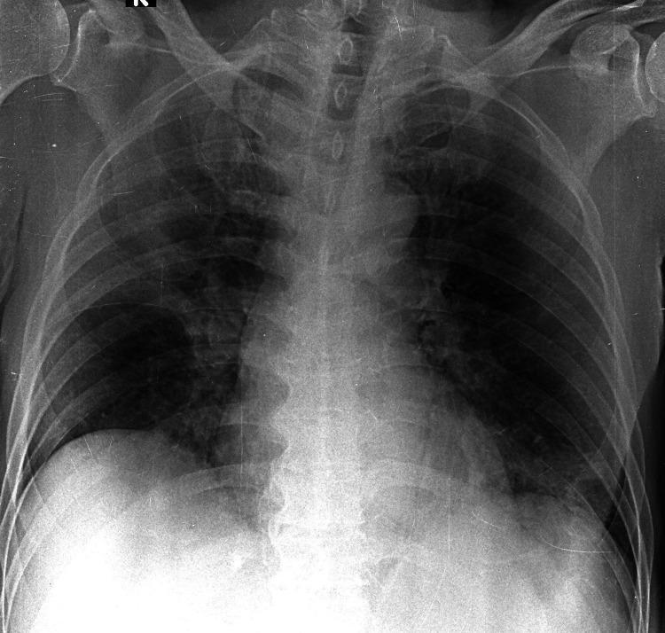
This diagnostic image is an anteroposterior (AP) chest X-ray of a patient with moderate SARS-CoV-2 (COVID-19) pneumonia. The radiograph demonstrates multi-focal areas of increased pulmonary opacity with an atypical, non-uniform distribution. Specifically, there is focal consolidation visible in the right upper lobe and the left basal zone. The right peripheral lower zone exhibits ground-glass opacities (GGO), characterized by hazy increases in lung density that do not obscure the underlying bronchovascular structures. The cardiac silhouette and mediastinal contours appear within normal limits. Clinically, this imaging illustrates moderate disease severity based on standardized scoring systems (Brixia, RALE, and mCXR), where the bilateral involvement of different lung zones reflects typical viral pneumonia patterns but with specific regional consolidation. The educational focus is on identifying diverse parenchymal infiltrates associated with COVID-19, ranging from ground-glass patterns to dense consolidations.
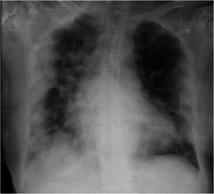
This diagnostic image is an anteroposterior (AP) chest X-ray demonstrating a case of viral pneumonia complicated by Acute Respiratory Distress Syndrome (ARDS). The radiograph reveals diffuse, bilateral, patchy alveolar opacities and ground-glass infiltrates. The abnormalities are primarily concentrated in the peripheral and basal regions of both lung fields, a hallmark distribution often seen in COVID-19 pneumonia. There is evidence of more confluent consolidation in the left lung compared to the right, particularly involving the lower lobe. The cardiac silhouette appears moderately enlarged, and the mediastinal contours are visible despite the overlapping pulmonary infiltrates. This image serves as an educational example of progressive pulmonary parenchymal involvement in severe respiratory infections, illustrating the typical radiographic progression from localized opacities to multi-lobar consolidation characteristic of clinical ARDS.
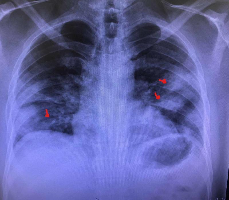
This diagnostic image is a posteroanterior (PA) chest X-ray demonstrating multifocal pneumonia. The radiograph reveals diffuse, bilateral, increased opacities throughout both lung fields. Notable findings include heterogeneous, ill-defined patchy opacities and areas of consolidation, predominantly in the mid and lower lung zones, as indicated by red arrows. These findings are consistent with alveolar filling and interstitial thickening, which obscure normal vascular markings. The cardiac silhouette and mediastinal contours appear within normal limits, though partially obscured by the adjacent pulmonary infiltrates. This image serves as a clinical example of multifocal infectious processes, commonly seen in community-acquired or viral pneumonia, illustrating the typical radiographic presentation of patchy air-space disease and its distribution across multiple lobes.
Pneumonia - Chest X-Ray Findings
Key Radiographic Signs
1. Lobar / Segmental Consolidation
- The classic finding - an area of homogeneous opacity replacing normal aerated lung
- Does not cross fissures (lobar distribution)
- Strongly suggests bacterial pneumonia (sensitivity 54%, specificity 90%, LR+ 5.6)
- Air bronchograms are often visible within the consolidation - dark linear streaks of air-filled bronchi against opacified alveoli
- Streptococcus pneumoniae (the most common cause of CAP) produces asymmetric lobar consolidation with air bronchograms
2. Multifocal / Patchy Infiltrates
- Bilateral or unilateral patchy opacities in multiple lobes
- Typical of atypical pneumonia (Mycoplasma, Chlamydophila, Legionella), viral pneumonia, or aspiration
- Opacities are ill-defined and may coalesce
3. Ground-Glass Opacity (GGO)
- Hazy increase in lung density that does not obscure bronchovascular markings
- Seen in early/mild disease or viral pneumonia (e.g. COVID-19, influenza, varicella)
- May progress to frank consolidation as disease worsens
4. Air Bronchograms
- Linear lucencies (dark lines) representing air-filled bronchi surrounded by consolidated (fluid-filled) alveoli
- A hallmark sign confirming true consolidation rather than collapse or pleural effusion
- Seen in bacterial and organizing pneumonia, ARDS, pulmonary edema
5. Other Associated Findings
| Finding | Implication |
|---|---|
| Pleural effusion | Parapneumonic effusion or empyema |
| Cavitation | Anaerobes, Klebsiella, S. aureus, TB |
| Hilar lymphadenopathy | TB, fungal, atypical infections |
| Interstitial pattern | Viral, Mycoplasma, PCP (P. jirovecii) |
| "Crazy paving" pattern (CT) | COVID-19 organizing pneumonia, PAP |
Radiographic Patterns by Organism
| Organism | Typical CXR Pattern |
|---|---|
| S. pneumoniae | Lobar consolidation, air bronchograms, asymmetric |
| Klebsiella | Upper lobe consolidation, "bulging fissure" sign, cavitation |
| S. aureus | Bilateral nodular/patchy, cavitation, pneumatoceles (especially in children) |
| Mycoplasma | Interstitial / reticulonodular, lower lobe peribronchial thickening |
| Legionella | Unilateral then bilateral rapidly spreading consolidation |
| Varicella (VZV) | Multiple acinar nodules with tendency to coalesce |
| COVID-19 | Peripheral bilateral GGO and consolidation, lower lobe predominant |
| PCP (P. jirovecii) | Bilateral perihilar interstitial pattern; lobar consolidation rare |
Normal vs. Pneumonia - Comparison
Limitations of CXR
- CXR cannot distinguish the causative organism with certainty - radiographic patterns overlap
- According to Symptom to Diagnosis (4th ed.), lobar consolidation in fever < 1 week strongly suggests bacterial pneumonia (LR+ 8.0), but pattern alone cannot differentiate S. pneumoniae from P. aeruginosa or Legionella
- In HIV-positive patients, 82% have CT abnormalities (effusions, cavities, abscesses) not visible on plain CXR - HRCT should be considered in non-responders
- CT chest is more sensitive and should be used when CXR is negative but clinical suspicion is high, or when the patient fails to respond to treatment