
Acute arterial occlusion
acute arterial occlusion limb ischemia angiogram
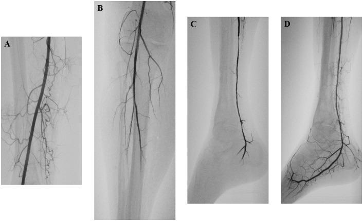
This set of diagnostic angiograms illustrates a case of acute limb ischemia and the subsequent management using embolectomy and intra-arterial thrombolysis. (A) Angiogram of the thigh shows a patent superficial femoral artery with no signs of atherosclerosis. (B) Image of the knee and upper calf shows a patent popliteal artery, but reveals acute distal occlusion of the posterior tibial artery and chronic occlusion of the anterior tibial and peroneal arteries. (C) Follow-up angiogram of the ankle/foot after mechanical embolectomy with a Fogarty catheter shows restored flow in the posterior tibial artery, yet persistent distal occlusion with no contrast filling in the plantar arch. (D) Final angiogram 24 hours after adjunctive intra-arterial administration of recombinant tissue plasminogen activator (rtPA) demonstrates complete restoration of distal perfusion, with excellent filling of both the posterior tibial artery and the plantar arch. This progression highlights the combined efficacy of surgical and pharmacological interventions in treating peripheral arterial embolism and restoring microvascular patency.
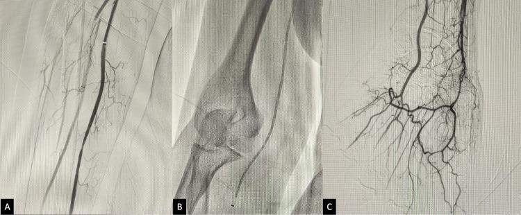
This composite diagnostic image consists of three digital subtraction angiogram (DSA) frames (A, B, and C) illustrating the endovascular revascularization of a right upper limb following acute thrombotic occlusion. Frame A shows the brachial artery post-thrombolysis with restored patency in the main vessel and visible collateral branching, though distal opacification remains incomplete. Frame B is an intra-procedural fluoroscopic image showing the elbow joint (distal humerus and proximal radius/ulna) with a distal access catheter (DAC) positioned for thromboaspiration. Frame C demonstrates the successful final result, showing complete recanalization and robust contrast filling of the distal arterial network, including the radial and ulnar arteries and the deep and superficial palmar arches in the hand. The series illustrates key steps in managing acute limb ischemia: the transition from initial pharmacological thrombolysis to mechanical thromboaspiration, ending with definitive restoration of distal perfusion. The visualization of the intricate palmar arterial anatomy in Frame C serves as the clinical endpoint for successful recanalization.
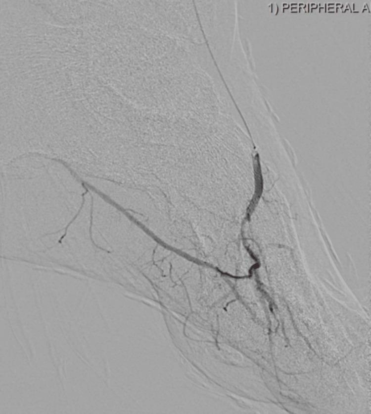
This diagnostic image is a peripheral digital subtraction angiogram (DSA) of the left foot, demonstrating post-intervention revascularization in a patient with peripheral artery disease (PAD) and critical limb ischemia. The image shows the distal anterior tibial artery and the dorsalis pedis artery with clear contrast opacification. A guidewire and an interventional balloon catheter are visible within the arterial lumen, positioned for balloon angioplasty to resolve a previous occlusion. The angiogram illustrates the restoration of flow into the plantar arch and smaller pedal branches. The vessel diameter appears relatively smooth and patent following the atherectomy and angioplasty procedure, with no evidence of residual high-grade stenosis or acute occlusion in the visualized field. This educational material highlights the interventional management of infrapopliteal arterial disease and the technical execution of pedal revascularization to improve distal perfusion.
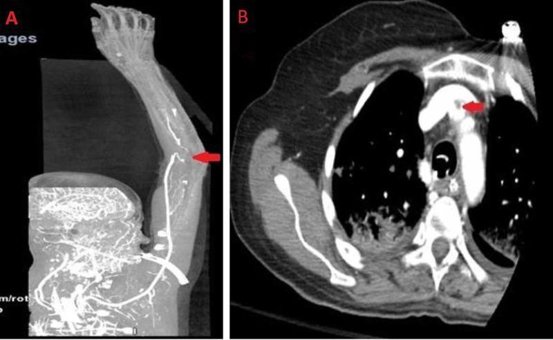
This figure consists of two computed tomography (CT) angiogram images highlighting vascular pathology in the right upper extremity and mediastinum. Image A is a maximum intensity projection (MIP) reconstruction showing the right upper limb arterial supply. A red arrow points to an occlusion at the distal bifurcation of the brachial artery. There is a conspicuous absence of contrast enhancement in the radial and ulnar arteries, indicating distal ischemia and lack of flow. Image B is an axial CT scan of the chest at the level of the aortic arch. A red arrow identifies a localized filling defect within the origin of the right innominate (brachiocephalic) artery, consistent with a small mural thrombus. The surrounding vessel walls and mediastinal structures appear otherwise unremarkable. These images serve as educational examples of arterial embolism or thrombosis leading to acute limb ischemia, emphasizing the importance of recognizing filling defects and abrupt vascular cut-offs in cross-sectional imaging.
"acute limb ischemia" AND management
Acute Arterial Occlusion
Etiology / Causes
1. Arterial Embolism (most dramatic)
- An embolus is a body carried in the bloodstream and lodged distally to obstruct flow.
- Cardioarterial embolism accounts for the majority; sources include:
- Atrial fibrillation (left atrial thrombus)
- Myocardial infarction (left ventricular mural thrombus)
- Valvular disease (mitral stenosis, infective endocarditis vegetations)
- Cardiac aneurysm
- Arterioarterial embolism: from ulcerated atherosclerotic plaques or aortic/peripheral aneurysms
- Common lodgment sites (lower limb) in order of frequency:
- Common femoral artery bifurcation
- Popliteal artery bifurcation
- Common iliac bifurcation
- Aortic bifurcation ("saddle embolus")
- Upper limb: brachial artery bifurcation > axillary artery
2. Acute Arterial Thrombosis
- Often occurs on a background of atherosclerotic stenosis (acute-on-chronic)
- Most common site: lower end of femoral artery at the adductor (subsartorial) canal
- Presentation is often less dramatic because collateral circulation has developed over time
- Differentiation from embolism is essential as management differs
3. Arterial Trauma
- Penetrating wounds disrupting arterial walls
- Pressure from angulated bone fragments
- Intimal rupture following fracture/dislocation
- Fractures/dislocations commonly associated:
- Supracondylar fracture of humerus
- Supracondylar fracture of femur
- Dislocated knee, elbow, or shoulder
Clinical Features: The 6 "P"s
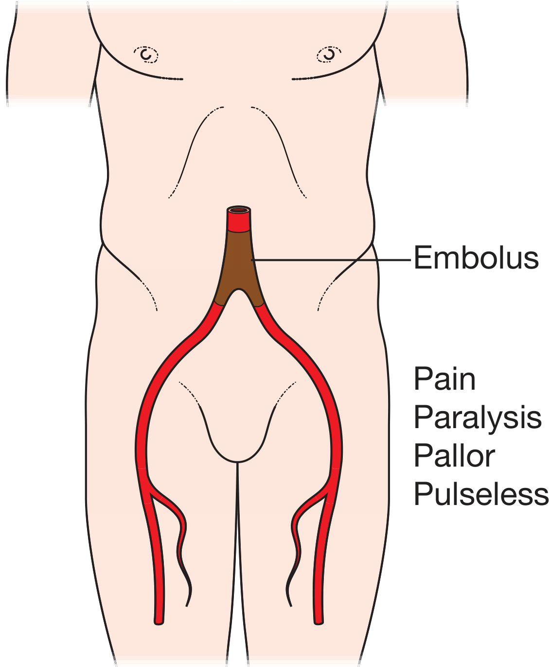
| Sign | Detail |
|---|---|
| Pain | Sudden, severe; first and most prominent symptom; distal to occlusion |
| Pallor | "Waxy" cadaveric pallor; progresses to cyanosis within ~1 hour; guttered veins |
| Pulselessness | Absent pulses distal to obstruction |
| Paraesthesia | Numbness/tingling; sensory nerves are highly ischemia-sensitive |
| Paralysis | Motor deficit indicates advanced ischemia; carries prognostic significance |
| Poikilothermia | Cold limb; appreciable temperature change at level of occlusion |
Neurologic symptoms have the greatest prognostic value. Intact motor + sensory function = limb will survive. Motor deficit = imminent irreversible ischemia.
Level-specific Presentations
| Level | Pain | Motor Loss | Color Change |
|---|---|---|---|
| Aortic bifurcation | Both lower limbs, hips | Hip/knee movements | Below hip/midthigh |
| Femoral | Leg and foot | Leg | Below knee |
| Popliteal | Lower leg and foot | Toes | Foot only |
| Axillary | Whole upper arm | Wrist/fingers | Hand and distal forearm |
| Brachial | Distal forearm/hand | - | Fingers only |
Rutherford Classification (Staging)
| Category | Status | Sensory Loss | Muscle Weakness | Arterial Doppler | Venous Doppler |
|---|---|---|---|---|---|
| I - Viable | Not immediately threatened | None | None | Audible | Audible |
| IIa - Marginally threatened | Salvageable if promptly treated | Minimal (toes) or none | None | Inaudible | Audible |
| IIb - Immediately threatened | Requires immediate revascularization | More than toes + rest pain | Mild-moderate | Inaudible | Audible |
| III - Irreversible | Amputation; major tissue loss | Profound, anesthetic | Profound paralysis/rigor | Inaudible | Inaudible |
Embolism vs. Thrombosis: Key Distinction
| Feature | Embolism | Thrombosis |
|---|---|---|
| Onset | Sudden, dramatic | Gradual/progressive |
| Background | Normal vessels | Atherosclerotic disease |
| Collaterals | Absent | Present (less severe ischemia) |
| Cardiac history | Often present (AF, MI) | PAD history |
| Contralateral pulses | Normal | Often reduced |
| Treatment priority | Surgical embolectomy | Thrombolysis or bypass |
Investigations
- Clinical diagnosis first - Do not delay surgery for investigations in severe ischemia
- ECG and cardiac exam - Identify source (AF, MI)
- Doppler ultrasound - Non-invasive; assesses flow; operator-dependent
- CTA / MRA - Excellent for localizing occlusion; CTA preferred if renal function normal
- Digital Subtraction Angiography (DSA) - Gold standard; allows simultaneous endovascular treatment; most anatomically detailed
Management
Immediate (all patients):
- IV fluids + supplemental oxygen
- Aspirin 81 mg
- IV Heparin: 100 U/kg bolus then infusion (target aPTT 2.0x normal) - prevents thrombus propagation
Rutherford Class I (Viable):
- Anticoagulation + observation
- Urgent workup and elective intervention planning
Rutherford Class IIa-IIb (Threatened):
- Surgical embolectomy with Fogarty balloon catheter - mainstay for embolic occlusion
- Catheter-Directed Thrombolysis (CDT) with rtPA - preferred for thrombotic occlusion, bypass graft occlusion; allows slower lysis without open surgery
- Mechanical thrombectomy - endovascular; useful when thrombolysis is contraindicated
Rutherford Class III (Irreversible):
- Primary amputation - revascularization will not salvage the limb and carries risk of reperfusion injury/systemic toxicity
Fogarty Embolectomy (surgical):
- Balloon catheter passed beyond the clot, inflated, then withdrawn to extract the thrombus
- Performed via groin (common femoral) or arm incision under local/general anesthesia
- Followed by intraoperative angiography to confirm adequate clearance
Post-revascularization:
- Monitor for reperfusion injury and compartment syndrome (fasciotomy may be needed)
- Long-term anticoagulation to prevent recurrence (especially if source is cardiac)
- Treat underlying cause (rate control/anticoagulation for AF, statins, antiplatelet therapy)
Angiographic Appearance
Recent Evidence (2025-2026)
- A 2025 meta-analysis (PMID 40428785) comparing thrombolytic therapy vs. open surgical intervention in ALI found comparable limb salvage rates; thrombolysis may offer a lower early complication profile in selected patients.
- A 2026 meta-analysis (PMID 41232816) comparing endovascular vs. open surgical approach found no significant difference in 30-day mortality or amputation rates, supporting individualized treatment selection based on ischemia severity and operator expertise.