
Ligament of treitz
Reading File
The Ligament of Treitz (suspensory muscle/ligament of the duodenum) is a fibromuscular band that marks the duodenojejunal flexure, traditionally used as the anatomical landmark separating the upper from lower GI tract.
Searching sources...
Finding Sources
Finding Sources
Reading File
Reading File
Reading File
Loading Image
Finding Sources
Reading File
Reading File
Loading Image
Here is a complete overview of the Ligament of Treitz from authoritative surgical and anatomical textbooks:
Ligament of Treitz (Suspensory Muscle of the Duodenum)
Definition and Structure
The ligament of Treitz - formally called the suspensory muscle of the duodenum - is a fibromuscular band composed of smooth muscle fibers that connects the ascending (4th) part of the duodenum to the trunk of the superior mesenteric artery (SMA) and the right crus of the diaphragm. It is located at the duodenojejunal flexure, at approximately the level of L2, to the left of the aorta and anterior to the left renal vein.
(Color Atlas of Human Anatomy Vol. 2, Internal Organs)
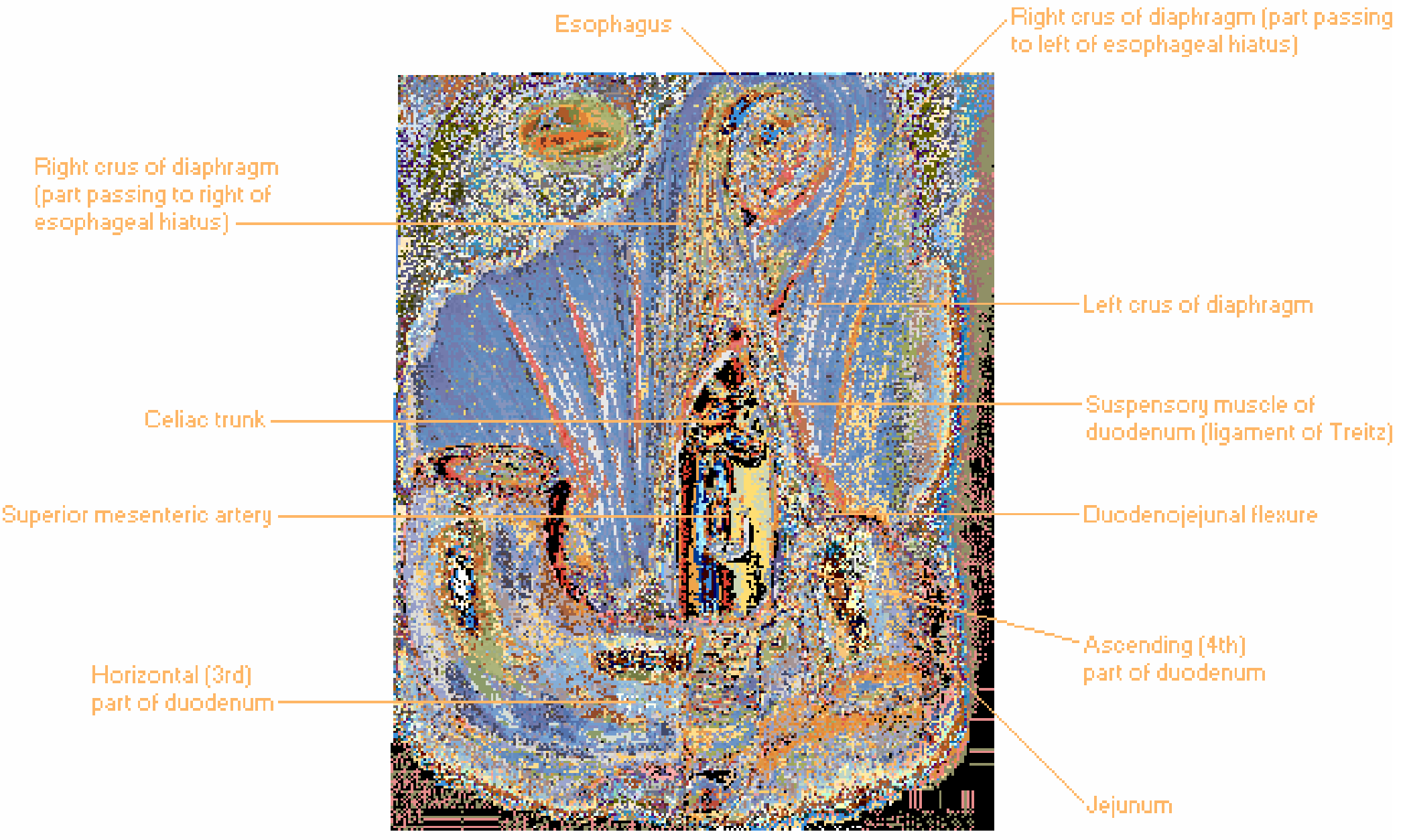
Position and Relations
- Lies at the duodenojejunal flexure, where the retroperitoneal duodenum transitions back into the intraperitoneal jejunum
- Located to the left of the spine (left of the aorta), posterior to the transverse mesocolon
- Anterior to the left renal vein
- Below the transverse colon; the jejunum emerges from the base of the posterior aspect of the transverse mesocolon at this point
(Mulholland and Greenfield's Surgery, 7th ed.)
Embryological Significance
The ligament of Treitz is one of the two normal points of intestinal fixation established during fetal gut development:
- The cecum in the right iliac fossa
- The duodenojejunal junction at the ligament of Treitz
During midgut rotation (weeks 10-12 of gestation), the duodenojejunal junction passes posterior to the SMA and fixes at this location. The normal intestinal mesentery runs in a broad base from the ligament of Treitz to the cecum - this broad-based attachment is what prevents volvulus.
In intestinal malrotation, this fixation fails: the mesenteric base is neither broad nor fixed, placing the entire midgut at risk for volvulus.
(Mulholland and Greenfield's Surgery, 7th ed.)
Clinical Importance
1. Upper vs. Lower GI Bleeding Landmark
The ligament of Treitz is the anatomical boundary between upper and lower GI bleeding:
- Upper GI bleeding (UGIB): source proximal to the ligament of Treitz
- Lower GI bleeding (LGIB): source distal to the ligament of Treitz
(Fischer's Mastery of Surgery, 8th ed.)
2. Small Bowel Measurement Reference
After massive small bowel resection, the remaining length is measured from the ligament of Treitz. Less than 100 cm of small bowel beyond the ligament of Treitz is associated with permanent short gut syndrome.
(Fischer's Mastery of Surgery, 8th ed.)
3. Enteral Feeding Landmark
Post-pyloric feeding tubes are ideally placed distal to the ligament of Treitz (i.e., into the jejunum). Studies show jejunal feeding beyond this landmark reduces the incidence of aspiration pneumonia in hospitalized patients.
(Yamada's Textbook of Gastroenterology, 7th ed.)
4. Surgical Landmark in Major Operations
- Whipple procedure (pancreaticoduodenectomy): Division of the jejunum and mobilization of the ligament of Treitz is a key step. The loose attachments are taken down carefully to avoid injury to the inferior mesenteric vein (IMV).
- Gastric bypass / bariatric surgery: The jejunum is divided with a GIA stapler ~20 cm distal to the ligament of Treitz to create the Roux limb.
- Left colectomy (medial-to-lateral approach): The IMV is identified lateral to the ligament of Treitz, which is divided for exposure.
(Fischer's Mastery of Surgery, 8th ed.; Current Surgical Therapy, 14th ed.)
5. Division of the Ligament (Surgical Technique)
When dividing the ligament of Treitz (open or laparoscopic):
- With the patient in reverse Trendelenburg, the omentum and transverse mesocolon are elevated cephalad
- The peritoneum along the left lateral aspect of the proximal jejunum is incised
- The suspensory muscle is divided along the anterior and cranial aspect of the 3rd and 4th portions of the duodenum
- This lowers the 3rd/4th duodenum from the aortomesenteric angle - the space between the duodenum and SMA origin should allow passage of two fingers once complete
(Fischer's Mastery of Surgery, 8th ed.)
6. Treitz Hernias
Internal herniation of small intestinal loops into the paraduodenal fossae (the superior and inferior duodenal fossae near the ligament of Treitz) is called a Treitz hernia - a potentially life-threatening cause of intestinal obstruction and necrosis.
(Color Atlas of Human Anatomy, Vol. 2)
Summary Table
| Feature | Detail |
|---|---|
| Official name | Suspensory muscle of the duodenum |
| Composition | Smooth muscle fibers |
| Location | Duodenojejunal flexure, ~L2 |
| Connects | Ascending duodenum → SMA trunk / right diaphragmatic crus |
| GI boundary | Upper vs. lower GI tract |
| Embryological role | Fixed point of midgut mesentery; prevents volvulus |
| Key surgical relevance | Whipple, gastric bypass, left colectomy, UGIB workup |
This is a shared conversation. Sign in to Orris to start your own chat.