
Dental plaque hypothesis for 5 marks bds final year exam notes
Reading File
The dental plaque hypothesis covers the role of plaque bacteria in initiating periodontal disease. Let me search the textbook library for the most relevant content.
Searching sources...
Finding Sources
Finding Sources
Finding Sources
Finding Sources
Finding Sources
Finding Sources
Finding Sources
Finding Sources
Reading File
Searching the Web
dental plaque hypothesis nonspecific specific ecological BDS final year notes
Reading Web Page
https://periobasics.com/critical-analysis-of-various-plaque-…
Loading Image
I now have comprehensive content from the medical library and authoritative web sources. Here are complete 5-mark BDS final year exam notes on the dental plaque hypotheses:
Dental Plaque Hypothesis - BDS Final Year Exam Notes (5 Marks)
Definition of Dental Plaque
Dental plaque is an adherent deposit forming on the tooth surface composed almost entirely of bacteria derived from the normal oral flora. It is the most prevalent and densest of human biofilms. The biofilm forms in relation to the acquired enamel pellicle (a thin organic film of salivary proteins and glycoproteins) rather than the mineralized tooth itself. Mature plaque contains 300-400 bacterial species.
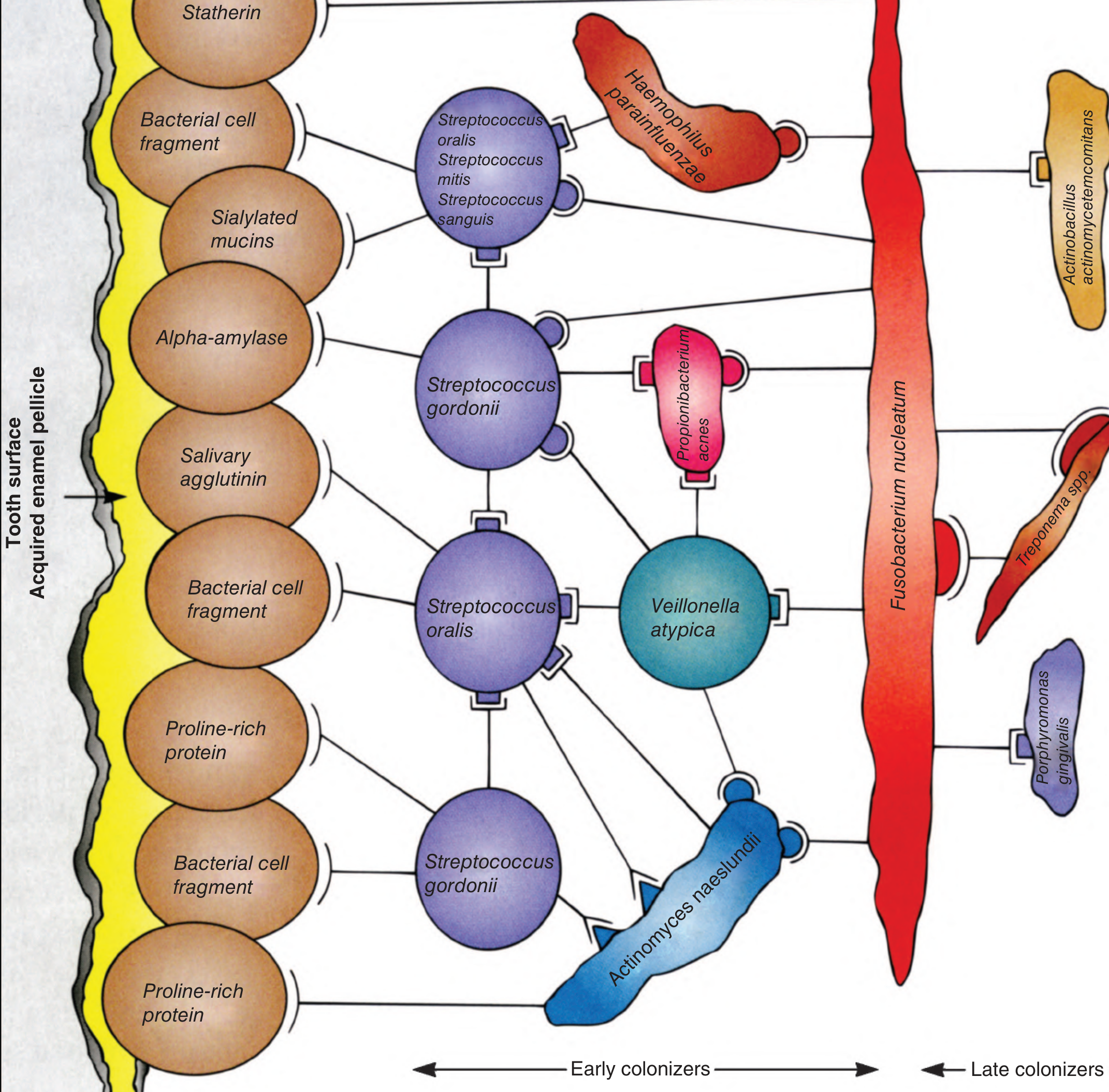
Figure: Dental plaque biofilm. Early colonizers (Streptococci, Actinomyces) bind to the pellicle; late colonizers (P. gingivalis, Treponema, Fusobacterium) bind to earlier bacteria.
Stages of Plaque Formation
- Pellicle formation - Salivary proteins coat the tooth surface
- Early colonizers (0-2 days) - Gram-positive aerobes: Streptococcus sanguis, S. mutans, S. mitis, S. gordonii, Actinomyces species; adhere via ionic, hydrophobic, and lectin-like interactions
- Late colonizers (2-4 days onward) - Gram-negative anaerobes: Porphyromonas gingivalis, Prevotella, Fusobacterium nucleatum, Treponema denticola, Aggregatibacter actinomycetemcomitans
- Maturation - High-molecular-weight glucan polymers synthesized (mainly by S. mutans) act as cement, binding the biofilm together
The Four Major Plaque Hypotheses
1. Non-Specific Plaque Hypothesis (1930s - Loesche, 1976)
- Concept: The sheer accumulation of dental plaque, regardless of bacterial composition, causes periodontal disease
- Mechanism: All plaque bacteria collectively produce metabolic by-products (acids, toxins, enzymes) - disease severity is proportional to the quantity of plaque
- Treatment implication: Focused entirely on mechanical plaque removal (scaling, oral hygiene)
- Limitation: Does not explain why some individuals with large amounts of plaque remain disease-free while others with minimal plaque develop severe periodontitis
2. Specific Plaque Hypothesis (Loesche, 1976)
- Concept: Only specific pathogenic bacteria within the plaque are responsible for periodontal disease
- Key pathogens identified:
- Porphyromonas gingivalis
- Treponema denticola
- Aggregatibacter actinomycetemcomitans (especially in aggressive periodontitis)
- These form Socransky's "Red Complex" - the most pathogenic group
- Mechanism: Shift from gram-positive aerobes to gram-negative anaerobes; specific bacteria initiate inflammatory response leading to tissue destruction and alveolar bone loss
- Treatment implication: Targeted antimicrobial therapy against specific pathogens
- Limitation: Does not fully explain why these bacteria can sometimes be present in the absence of disease; doesn't account for host immune variation
3. Ecological Plaque Hypothesis (Marsh, 1994)
- Concept: Disease results from an ecological imbalance (dysbiosis) in the microbial community driven by environmental changes, rather than the simple presence or absence of specific bacteria
- Mechanism:
- Environmental stresses (altered pH, reduced oxygen, changes in gingival crevicular fluid, immune status) shift the microbial balance
- This promotes the overgrowth of pathogenic species over harmless commensal flora
- Periodontal disease = disruption of the ecological equilibrium
- Key principle: A pathogen need not be introduced from outside - it can emerge from within the existing microflora when conditions change
- Treatment implication: Restore ecological balance (e.g., controlling inflammation, pH modulation) rather than just eliminating bacteria
- Significance: Introduced the concept of dysbiosis now central to periodontal literature
4. Keystone Pathogen Hypothesis (Hajishengallis et al., 2012)
- Concept: Certain bacteria, though present in low numbers, exert a disproportionately large effect on the microbial community
- Key example: Porphyromonas gingivalis
- Subverts host innate immune responses (complement system, TLR signaling)
- Alters the biofilm environment to favor growth of other pathogenic bacteria
- Creates a state of immune dysregulation leading to chronic inflammation
- Distinction from earlier models: Disease is NOT caused by the keystone pathogen directly but by the dysbiotic community it enables
- Limitation: Keystone pathogens can be any species, and some are not necessarily pathogenic in all contexts
Socransky's Microbial Complexes (Related Concept)
Socransky (1998) classified periodontal bacteria into color-coded complexes:
| Complex | Bacteria | Association |
|---|---|---|
| Red | P. gingivalis, T. denticola, T. forsythia | Strongly disease-associated |
| Orange | Fusobacterium, Prevotella intermedia | Bridge between early and red complex |
| Yellow/Green/Purple | S. sanguis, Capnocytophaga | Early colonizers, health-associated |
Summary Table for Quick Revision
| Hypothesis | Key Concept | Treatment Focus | Limitation |
|---|---|---|---|
| Non-specific | All plaque is harmful | Mechanical removal | Ignores individual susceptibility |
| Specific | Only select bacteria cause disease | Targeted antibiotics | Bacteria present without disease |
| Ecological | Dysbiosis / environmental shift | Restore balance | Difficult to quantify ecological shifts |
| Keystone pathogen | Low-abundance bacteria drive dysbiosis | Disrupt keystone | Any species can be a keystone |
Exam Tip
For a 5-mark answer, cover:
- Definition and composition of dental plaque (1 mark)
- Non-specific plaque hypothesis with limitation (1 mark)
- Specific plaque hypothesis with key bacteria and limitation (1 mark)
- Ecological plaque hypothesis - Marsh 1994, dysbiosis concept (1 mark)
- Keystone pathogen hypothesis - P. gingivalis example (1 mark)
Sources: Jawetz Melnick & Adelberg's Medical Microbiology 28th Ed; periobasics.com; dentalcare.com CE Course
Generate a comparison table (PPT) of all four hypotheses easy to understand
Reading File
Reading File
Reading File
Reading File
Good, I have enough to build the PPT. Let me create it now.
Running Command
mkdir -p /home/daytona/workspace/dental-plaque-ppt && ls /home/daytona/workspace/dental-plaque-ppt
Writing File
~/dental-plaque-ppt/build.js
const pptxgen = require("pptxgenjs");
const pres = new pptxgen();
pres.layout = "LAYOUT_WIDE"; // 13.3" x 7.5"
pres.title = "Dental Plaque Hypotheses";
pres.author = "BDS Final Year";

// ─── PALETTE ───────────────────────────────────────────────────────────────
const C = {
  navy:    "0F2D4A",   // dominant dark
  teal:    "0D7377",   // accent 1
  sky:     "14A9AD",   // accent 2
  mint:    "32E0C4",   // highlight
  cream:   "F5F5F0",   // slide bg
  white:   "FFFFFF",
  gray:    "64748B",
  lgray:   "E2E8F0",
  // hypothesis colours
  h1:      "E74C3C",   // Non-specific  – red
  h2:      "2980B9",   // Specific      – blue
  h3:      "27AE60",   // Ecological    – green
  h4:      "8E44AD",   // Keystone      – purple
  h1l:     "FADBD8",
  h2l:     "D6EAF8",
  h3l:     "D5F5E3",
  h4l:     "E8DAEF",
};

// ─── HELPERS ────────────────────────────────────────────────────────────────
function titleSlide(pres, C) {
  const s = pres.addSlide();
  // full dark background
  s.background = { color: C.navy };

  // top accent bar
  s.addShape(pres.shapes.RECTANGLE, { x: 0, y: 0, w: 13.3, h: 0.18, fill: { color: C.mint }, line: { color: C.mint } });

  // big title
  s.addText("DENTAL PLAQUE\nHYPOTHESES", {
    x: 0.8, y: 1.2, w: 11.7, h: 2.4,
    fontSize: 52, bold: true, color: C.white,
    fontFace: "Calibri", align: "center", valign: "middle",
    charSpacing: 3,
  });

  // subtitle
  s.addText("A Comparative Overview for BDS Final Year", {
    x: 0.8, y: 3.5, w: 11.7, h: 0.6,
    fontSize: 20, color: C.mint, fontFace: "Calibri", align: "center",
  });

  // four coloured dots representing 4 hypotheses
  const dots = [C.h1, C.h2, C.h3, C.h4];
  const labels = ["Non-Specific", "Specific", "Ecological", "Keystone Pathogen"];
  dots.forEach((col, i) => {
    const cx = 2.5 + i * 2.2;
    s.addShape(pres.shapes.OVAL, { x: cx, y: 4.5, w: 0.35, h: 0.35, fill: { color: col }, line: { color: col } });
    s.addText(labels[i], { x: cx - 0.4, y: 4.95, w: 1.2, h: 0.3, fontSize: 9, color: C.lgray, align: "center" });
  });

  // bottom bar
  s.addShape(pres.shapes.RECTANGLE, { x: 0, y: 7.3, w: 13.3, h: 0.2, fill: { color: C.teal }, line: { color: C.teal } });
}

// Slide 2 – Overview / what is plaque
function overviewSlide(pres, C) {
  const s = pres.addSlide();
  s.background = { color: C.cream };

  // heading bar
  s.addShape(pres.shapes.RECTANGLE, { x: 0, y: 0, w: 13.3, h: 0.85, fill: { color: C.navy }, line: { color: C.navy } });
  s.addText("What is Dental Plaque?", {
    x: 0.4, y: 0, w: 12.5, h: 0.85,
    fontSize: 24, bold: true, color: C.white, valign: "middle",
  });

  // definition box
  s.addShape(pres.shapes.RECTANGLE, {
    x: 0.4, y: 1.1, w: 12.5, h: 1.15,
    fill: { color: C.navy }, line: { color: C.navy },
    shadow: { type: "outer", color: "000000", blur: 6, offset: 2, angle: 135, opacity: 0.12 },
  });
  s.addText([
    { text: "DEFINITION: ", options: { bold: true, color: C.mint } },
    { text: "An adherent deposit on the tooth surface composed almost entirely of bacteria from normal oral flora. Dental plaque is the most prevalent and densest human biofilm.", options: { color: C.white } },
  ], { x: 0.6, y: 1.15, w: 12.1, h: 1.05, fontSize: 13, valign: "middle" });

  // stages row – 4 boxes
  const stages = [
    { n: "1", title: "Pellicle\nFormation", body: "Salivary proteins coat\nthe tooth surface" },
    { n: "2", title: "Early\nColonizers", body: "Gram +ve aerobes\nStreptococci, Actinomyces\n(0-2 days)" },
    { n: "3", title: "Late\nColonizers", body: "Gram −ve anaerobes\nP. gingivalis, Treponema\n(2-4 days +)" },
    { n: "4", title: "Mature\nBiofilm", body: "Glucan polymers bind\nbiofilm; 300-400 species\nin mature plaque" },
  ];
  const cols = ["2980B9", "27AE60", "E74C3C", "8E44AD"];
  stages.forEach((st, i) => {
    const x = 0.4 + i * 3.15;
    s.addShape(pres.shapes.RECTANGLE, { x, y: 2.55, w: 3.0, h: 4.55, fill: { color: C.white }, line: { color: cols[i], pt: 2 } });
    s.addShape(pres.shapes.RECTANGLE, { x, y: 2.55, w: 3.0, h: 0.7, fill: { color: cols[i] }, line: { color: cols[i] } });
    s.addText(st.n, { x, y: 2.55, w: 0.55, h: 0.7, fontSize: 20, bold: true, color: C.white, align: "center", valign: "middle" });
    s.addText(st.title, { x: x + 0.55, y: 2.55, w: 2.45, h: 0.7, fontSize: 12, bold: true, color: C.white, valign: "middle" });
    s.addText(st.body, { x: x + 0.15, y: 3.35, w: 2.7, h: 3.55, fontSize: 11.5, color: C.navy, valign: "top" });
  });
}

// Slide 3 – Individual hypothesis cards (2x2 grid)
function fourCardsSlide(pres, C) {
  const s = pres.addSlide();
  s.background = { color: C.cream };

  s.addShape(pres.shapes.RECTANGLE, { x: 0, y: 0, w: 13.3, h: 0.85, fill: { color: C.navy }, line: { color: C.navy } });
  s.addText("The Four Plaque Hypotheses – At a Glance", {
    x: 0.4, y: 0, w: 12.5, h: 0.85, fontSize: 24, bold: true, color: C.white, valign: "middle",
  });

  const hyps = [
    {
      num: "01", name: "Non-Specific\nPlaque Hypothesis",
      year: "1930s – 1976", col: C.h1, light: C.h1l,
      icon: "⚠️",
      who: "Loesche (1976)",
      concept: "All plaque accumulation → disease\nQuantity of plaque drives severity",
      bacteria: "ALL bacteria equally responsible",
      treat: "Mechanical plaque removal\n(scaling + oral hygiene)",
      limit: "Can't explain why some with\nheavy plaque stay disease-free",
    },
    {
      num: "02", name: "Specific\nPlaque Hypothesis",
      year: "1976", col: C.h2, light: C.h2l,
      icon: "🎯",
      who: "Loesche (1976)",
      concept: "Only SELECT bacteria cause disease\nShift: Gram +ve → Gram −ve anaerobes",
      bacteria: "P. gingivalis, T. denticola\nA. actinomycetemcomitans",
      treat: "Targeted antimicrobial\ntherapy",
      limit: "Pathogens can be present\nwithout causing disease",
    },
    {
      num: "03", name: "Ecological\nPlaque Hypothesis",
      year: "1994", col: C.h3, light: C.h3l,
      icon: "🌿",
      who: "Marsh (1994)",
      concept: "Environmental changes → dysbiosis\nImbalance in microbial community",
      bacteria: "Pathogens emerge from\nexisting flora under stress",
      treat: "Restore ecological balance\n(control inflammation, pH)",
      limit: "Difficult to quantify\necological shifts precisely",
    },
    {
      num: "04", name: "Keystone Pathogen\nHypothesis",
      year: "2012", col: C.h4, light: C.h4l,
      icon: "🔑",
      who: "Hajishengallis (2012)",
      concept: "Low-abundance bacteria with\ndisproportionately LARGE impact",
      bacteria: "P. gingivalis\n(subverts innate immunity)",
      treat: "Target keystone pathogen\nto disrupt dysbiotic cascade",
      limit: "Any species can be a\nkeystone; not always predictable",
    },
  ];

  const positions = [
    { x: 0.25, y: 1.0 },
    { x: 6.9,  y: 1.0 },
    { x: 0.25, y: 4.05 },
    { x: 6.9,  y: 4.05 },
  ];

  hyps.forEach((h, i) => {
    const { x, y } = positions[i];
    const w = 6.3, cardH = 2.9;

    // card background
    s.addShape(pres.shapes.RECTANGLE, {
      x, y, w, h: cardH,
      fill: { color: C.white },
      line: { color: h.col, pt: 2 },
      shadow: { type: "outer", color: "000000", blur: 5, offset: 2, angle: 135, opacity: 0.1 },
    });

    // top colour bar
    s.addShape(pres.shapes.RECTANGLE, { x, y, w, h: 0.55, fill: { color: h.col }, line: { color: h.col } });

    // number badge
    s.addShape(pres.shapes.OVAL, { x: x + 0.1, y: y + 0.1, w: 0.38, h: 0.38, fill: { color: C.white }, line: { color: C.white } });
    s.addText(h.num, { x: x + 0.1, y: y + 0.1, w: 0.38, h: 0.38, fontSize: 9, bold: true, color: h.col, align: "center", valign: "middle" });

    // title
    s.addText(h.name, { x: x + 0.58, y, w: w - 0.7, h: 0.55, fontSize: 13, bold: true, color: C.white, valign: "middle" });

    // year chip
    s.addShape(pres.shapes.ROUNDED_RECTANGLE, { x: x + w - 1.1, y: y + 0.12, w: 0.95, h: 0.28, fill: { color: C.white }, line: { color: C.white }, rectRadius: 0.05 });
    s.addText(h.year, { x: x + w - 1.1, y: y + 0.12, w: 0.95, h: 0.28, fontSize: 7.5, color: h.col, bold: true, align: "center", valign: "middle" });

    // body rows
    const rows = [
      { label: "Concept",    val: h.concept },
      { label: "Key Bug",    val: h.bacteria },
      { label: "Treatment",  val: h.treat },
      { label: "Limitation", val: h.limit },
    ];
    rows.forEach((r, ri) => {
      const ry = y + 0.62 + ri * 0.55;
      s.addShape(pres.shapes.RECTANGLE, { x, y: ry, w: 1.3, h: 0.52, fill: { color: h.light }, line: { color: h.light } });
      s.addText(r.label.toUpperCase(), { x: x + 0.05, y: ry, w: 1.2, h: 0.52, fontSize: 8, bold: true, color: h.col, valign: "middle", align: "left" });
      s.addText(r.val, { x: x + 1.35, y: ry, w: w - 1.45, h: 0.52, fontSize: 9.5, color: C.navy, valign: "middle" });
    });
  });
}

// Slide 4 – Big comparison TABLE
function comparisonTableSlide(pres, C) {
  const s = pres.addSlide();
  s.background = { color: C.cream };

  s.addShape(pres.shapes.RECTANGLE, { x: 0, y: 0, w: 13.3, h: 0.85, fill: { color: C.navy }, line: { color: C.navy } });
  s.addText("Side-by-Side Comparison Table", {
    x: 0.4, y: 0, w: 12.5, h: 0.85, fontSize: 24, bold: true, color: C.white, valign: "middle",
  });

  const hCols = [C.h1, C.h2, C.h3, C.h4];
  const hNames = ["Non-Specific\n(1930s-1976)", "Specific\n(1976)", "Ecological\n(1994)", "Keystone Pathogen\n(2012)"];

  const rows = [
    ["Criterion", "Non-Specific\n(1930s-1976)", "Specific\n(1976)", "Ecological\n(1994)", "Keystone Pathogen\n(2012)"],
    ["Core Idea", "All plaque causes disease by bulk accumulation", "Only select bacteria cause disease", "Ecological imbalance (dysbiosis) drives disease", "Low-abundance 'keystone' bugs hijack immunity"],
    ["Key Bacteria", "All oral flora equally", "P. gingivalis, T. denticola, A. actinomycetemcomitans", "Endogenous flora shift under stress", "P. gingivalis (prototype)"],
    ["Mechanism", "Bulk acid/toxin production by total plaque mass", "Shift Gram +ve → Gram −ve anaerobes; specific virulence", "Environmental stress → overgrowth of pathogens", "Subverts complement & TLR signalling → dysbiosis"],
    ["Treatment Focus", "Mechanical removal (scaling & oral hygiene)", "Targeted antibiotics vs. specific pathogens", "Restore balance: control pH & inflammation", "Disrupt keystone to collapse dysbiotic community"],
    ["Host Role", "Minimal (not considered)", "Minimal", "Acknowledged (immune status affects ecology)", "Central – immune disruption is the key event"],
    ["Strength", "Simple; explains gingivitis with plaque accumulation", "Explains site-specific & aggressive disease", "Explains why pathogens present without disease", "Explains low-abundance impact; modern dysbiosis model"],
    ["Limitation", "Ignores host variation & microbial specificity", "Pathogens present in healthy mouths too", "Hard to quantify ecological triggers", "Any species can be a keystone; not always predictable"],
    ["Current Status", "Largely abandoned", "Partially accepted", "Widely accepted – foundation of modern periodontology", "Most current; under active research"],
  ];

  // column widths: criterion col wider
  const colW = [2.3, 2.65, 2.65, 2.65, 2.65];

  const tableData = rows.map((row, ri) => {
    return row.map((cell, ci) => {
      // header row
      if (ri === 0) {
        if (ci === 0) return { text: cell, options: { fill: { color: C.navy }, color: C.white, bold: true, fontSize: 11, align: "center", valign: "middle" } };
        return { text: cell, options: { fill: { color: hCols[ci - 1] }, color: C.white, bold: true, fontSize: 10, align: "center", valign: "middle" } };
      }
      // criterion column
      if (ci === 0) return { text: cell, options: { fill: { color: C.navy }, color: C.mint, bold: true, fontSize: 9.5, align: "left", valign: "middle" } };
      // alternating row shading
      const lightCols = [C.h1l, C.h2l, C.h3l, C.h4l];
      const bg = ri % 2 === 0 ? lightCols[ci - 1] : C.white;
      return { text: cell, options: { fill: { color: bg }, color: C.navy, fontSize: 9, align: "left", valign: "middle" } };
    });
  });

  s.addTable(tableData, {
    x: 0.15, y: 1.0, w: 12.95,
    colW: colW,
    rowH: 0.64,
    border: { pt: 1, color: C.lgray },
    autoPage: false,
  });
}

// Slide 5 – Evolution timeline
function timelineSlide(pres, C) {
  const s = pres.addSlide();
  s.background = { color: C.navy };

  s.addShape(pres.shapes.RECTANGLE, { x: 0, y: 0, w: 13.3, h: 0.85, fill: { color: C.teal }, line: { color: C.teal } });
  s.addText("Evolution of Plaque Hypotheses – Timeline", {
    x: 0.4, y: 0, w: 12.5, h: 0.85, fontSize: 24, bold: true, color: C.white, valign: "middle",
  });

  // horizontal timeline bar
  s.addShape(pres.shapes.RECTANGLE, { x: 0.8, y: 3.4, w: 11.7, h: 0.1, fill: { color: C.mint }, line: { color: C.mint } });

  const events = [
    { x: 1.0,  year: "1930s", label: "Non-Specific\nPlaque Hypothesis", col: C.h1, desc: "All plaque =\nbad plaque" },
    { x: 4.1,  year: "1976",  label: "Specific\nPlaque Hypothesis",     col: C.h2, desc: "Select bacteria\ncause disease" },
    { x: 7.2,  year: "1994",  label: "Ecological\nPlaque Hypothesis",   col: C.h3, desc: "Dysbiosis /\nbalance shift" },
    { x: 10.3, year: "2012",  label: "Keystone Pathogen\nHypothesis",   col: C.h4, desc: "P. gingivalis\nhijacks immunity" },
  ];

  events.forEach((ev, i) => {
    // vertical stem
    s.addShape(pres.shapes.RECTANGLE, { x: ev.x + 0.55, y: i % 2 === 0 ? 2.1 : 3.5, w: 0.04, h: i % 2 === 0 ? 1.3 : 1.3, fill: { color: ev.col }, line: { color: ev.col } });

    // dot on line
    s.addShape(pres.shapes.OVAL, { x: ev.x + 0.38, y: 3.24, w: 0.38, h: 0.38, fill: { color: ev.col }, line: { color: C.white, pt: 2 } });

    // year
    s.addText(ev.year, { x: ev.x, y: 3.62, w: 1.2, h: 0.35, fontSize: 13, bold: true, color: ev.col, align: "center" });

    // card
    const cardY = i % 2 === 0 ? 1.1 : 4.05;
    s.addShape(pres.shapes.RECTANGLE, {
      x: ev.x - 0.1, y: cardY, w: 2.5, h: 1.6,
      fill: { color: "1A3A5C" }, line: { color: ev.col, pt: 2 },
    });
    s.addShape(pres.shapes.RECTANGLE, { x: ev.x - 0.1, y: cardY, w: 2.5, h: 0.42, fill: { color: ev.col }, line: { color: ev.col } });
    s.addText(ev.label, { x: ev.x - 0.05, y: cardY, w: 2.4, h: 0.42, fontSize: 9.5, bold: true, color: C.white, valign: "middle", align: "center" });
    s.addText(ev.desc, { x: ev.x - 0.05, y: cardY + 0.47, w: 2.4, h: 1.08, fontSize: 11, color: C.lgray, valign: "top", align: "center" });
  });

  // arrow at end of line
  s.addShape(pres.shapes.RIGHT_ARROW, { x: 12.4, y: 3.28, w: 0.6, h: 0.28, fill: { color: C.mint }, line: { color: C.mint } });
  s.addText("Present", { x: 12.2, y: 3.62, w: 1.0, h: 0.35, fontSize: 11, color: C.mint, align: "center" });

  // footnote
  s.addText("Each hypothesis built on — and refined — the previous one. Modern periodontology uses the Ecological + Keystone Pathogen frameworks.", {
    x: 0.5, y: 7.0, w: 12.3, h: 0.38, fontSize: 10, color: C.lgray, align: "center", italic: true,
  });
}

// Slide 6 – Socransky's Complexes
function socranskySlide(pres, C) {
  const s = pres.addSlide();
  s.background = { color: C.cream };

  s.addShape(pres.shapes.RECTANGLE, { x: 0, y: 0, w: 13.3, h: 0.85, fill: { color: C.navy }, line: { color: C.navy } });
  s.addText("Socransky's Microbial Complexes (1998) – Related Concept", {
    x: 0.4, y: 0, w: 12.5, h: 0.85, fontSize: 22, bold: true, color: C.white, valign: "middle",
  });

  s.addText("Socransky classified subgingival bacteria into colour-coded complexes based on disease association:", {
    x: 0.5, y: 1.0, w: 12.3, h: 0.5, fontSize: 13, color: C.navy,
  });

  const complexes = [
    { col: "C0392B", name: "RED Complex", label: "Strongly Disease-Associated", bugs: ["Porphyromonas gingivalis", "Treponema denticola", "Tannerella forsythia"], note: "Always present in deep pockets and active disease" },
    { col: "E67E22", name: "ORANGE Complex", label: "Bridge – Early to Late Colonizers", bugs: ["Fusobacterium nucleatum", "Prevotella intermedia", "Peptostreptococcus micros"], note: "Paves the way for Red complex organisms" },
    { col: "F1C40F", name: "YELLOW Complex", label: "Health-Associated (Early)", bugs: ["Streptococcus sanguis", "S. mitis", "S. gordonii"], note: "Predominant in health; inhibit pathogens" },
    { col: "27AE60", name: "GREEN Complex", label: "Early Colonizers", bugs: ["Capnocytophaga spp.", "Aggregatibacter aphrophilus", "Eikenella corrodens"], note: "Attach to pellicle; co-aggregate with early streptococci" },
    { col: "2980B9", name: "PURPLE Complex", label: "Early Colonizers", bugs: ["Veillonella parvula", "Actinomyces odontolyticus"], note: "Found in both health and disease" },
  ];

  complexes.forEach((cx, i) => {
    const x = 0.3 + i * 2.55;
    s.addShape(pres.shapes.RECTANGLE, {
      x, y: 1.65, w: 2.4, h: 5.4,
      fill: { color: C.white }, line: { color: cx.col, pt: 2 },
    });
    s.addShape(pres.shapes.RECTANGLE, { x, y: 1.65, w: 2.4, h: 0.75, fill: { color: cx.col }, line: { color: cx.col } });
    s.addText(cx.name, { x, y: 1.65, w: 2.4, h: 0.45, fontSize: 11, bold: true, color: C.white, align: "center", valign: "middle" });
    s.addText(cx.label, { x, y: 2.1, w: 2.4, h: 0.3, fontSize: 7.5, color: C.white, align: "center", italic: true });

    // bacteria list
    cx.bugs.forEach((b, bi) => {
      s.addText("• " + b, { x: x + 0.1, y: 2.5 + bi * 0.55, w: 2.2, h: 0.5, fontSize: 9.5, color: C.navy, italic: true });
    });

    // note
    s.addShape(pres.shapes.RECTANGLE, { x, y: 6.15, w: 2.4, h: 0.9, fill: { color: cx.col + "22" }, line: { color: cx.col } });
    s.addText(cx.note, { x: x + 0.1, y: 6.2, w: 2.2, h: 0.8, fontSize: 8.5, color: C.navy, italic: true, valign: "middle" });
  });
}

// Slide 7 – Exam Quick Recall
function examSlide(pres, C) {
  const s = pres.addSlide();
  s.background = { color: C.navy };

  s.addShape(pres.shapes.RECTANGLE, { x: 0, y: 0, w: 13.3, h: 0.85, fill: { color: C.h4 }, line: { color: C.h4 } });
  s.addText("⚡  Quick Recall – Exam Tips", {
    x: 0.4, y: 0, w: 12.5, h: 0.85, fontSize: 24, bold: true, color: C.white, valign: "middle",
  });

  // 5-mark answer guide
  s.addShape(pres.shapes.RECTANGLE, {
    x: 0.3, y: 1.0, w: 5.9, h: 6.0,
    fill: { color: "1A3A5C" }, line: { color: C.mint, pt: 2 },
  });
  s.addShape(pres.shapes.RECTANGLE, { x: 0.3, y: 1.0, w: 5.9, h: 0.5, fill: { color: C.mint }, line: { color: C.mint } });
  s.addText("5-MARK ANSWER STRUCTURE", { x: 0.35, y: 1.0, w: 5.8, h: 0.5, fontSize: 12, bold: true, color: C.navy, valign: "middle", align: "center" });

  const marks = [
    "1 mark  →  Define dental plaque + biofilm stages",
    "1 mark  →  Non-specific hypothesis + limitation",
    "1 mark  →  Specific hypothesis + key bacteria (Red Complex)",
    "1 mark  →  Ecological hypothesis (Marsh 1994) + dysbiosis",
    "1 mark  →  Keystone pathogen hypothesis (P. gingivalis)",
  ];
  marks.forEach((m, i) => {
    s.addShape(pres.shapes.RECTANGLE, { x: 0.38, y: 1.6 + i * 0.82, w: 0.45, h: 0.45, fill: { color: C.mint }, line: { color: C.mint } });
    s.addText(String(i + 1), { x: 0.38, y: 1.6 + i * 0.82, w: 0.45, h: 0.45, fontSize: 14, bold: true, color: C.navy, align: "center", valign: "middle" });
    s.addText(m, { x: 0.9, y: 1.6 + i * 0.82, w: 5.1, h: 0.45, fontSize: 10.5, color: C.lgray, valign: "middle" });
  });

  // mnemonics box
  s.addShape(pres.shapes.RECTANGLE, {
    x: 6.5, y: 1.0, w: 6.5, h: 6.0,
    fill: { color: "1A3A5C" }, line: { color: C.h1, pt: 2 },
  });
  s.addShape(pres.shapes.RECTANGLE, { x: 6.5, y: 1.0, w: 6.5, h: 0.5, fill: { color: C.h1 }, line: { color: C.h1 } });
  s.addText("MNEMONICS & KEY FACTS", { x: 6.55, y: 1.0, w: 6.4, h: 0.5, fontSize: 12, bold: true, color: C.white, valign: "middle", align: "center" });

  const mnemos = [
    { q: "Non-Specific", a: "\"All plaque is BAD plaque\" – no bacterial specificity" },
    { q: "Specific", a: "RED COMPLEX: P. gingivalis, T. denticola, T. forsythia" },
    { q: "Ecological", a: "MARSH 1994 – dysbiosis key word; ecological stress = disease" },
    { q: "Keystone", a: "HAJISHENGALLIS 2012 – P. gingivalis in low numbers → big damage" },
    { q: "Socransky", a: "Red > Orange > Yellow > Green > Purple (disease → health)" },
    { q: "Treatment", a: "NSH → scaling | SPH → antibiotics | EcoPH → restore balance" },
  ];
  mnemos.forEach((mn, i) => {
    const my = 1.65 + i * 0.82;
    s.addShape(pres.shapes.RECTANGLE, { x: 6.55, y: my, w: 2.0, h: 0.45, fill: { color: C.h1 + "55" }, line: { color: C.h1 } });
    s.addText(mn.q, { x: 6.6, y: my, w: 1.9, h: 0.45, fontSize: 9, bold: true, color: C.white, valign: "middle" });
    s.addText(mn.a, { x: 8.6, y: my, w: 4.2, h: 0.45, fontSize: 9.5, color: C.lgray, valign: "middle" });
  });
}

// ─── BUILD ──────────────────────────────────────────────────────────────────
titleSlide(pres, C);
overviewSlide(pres, C);
fourCardsSlide(pres, C);
comparisonTableSlide(pres, C);
timelineSlide(pres, C);
socranskySlide(pres, C);
examSlide(pres, C);

pres.writeFile({ fileName: "/home/daytona/workspace/dental-plaque-ppt/Dental_Plaque_Hypotheses.pptx" })
  .then(() => console.log("PPT created successfully"))
  .catch(err => console.error("Error:", err));
Running Command
cd /home/daytona/workspace/dental-plaque-ppt && node build.js
The PPT was created successfully. The warnings are about 8-digit hex colors (with transparency appended) - those fell back to black. Let me fix those quickly so the note backgrounds on the Socransky slide and mnemonic chips look correct.
Editing File
~/dental-plaque-ppt/build.js
Successfully replaced 1 instance(s) of the string in '/home/daytona/workspace/dental-plaque-ppt/build.js'
Editing File
~/dental-plaque-ppt/build.js
Successfully replaced 1 instance(s) of the string in '/home/daytona/workspace/dental-plaque-ppt/build.js'
Running Command
cd /home/daytona/workspace/dental-plaque-ppt && node build.js 2>&1
No warnings - clean build. Here is your presentation:
Dental Plaque Hypotheses
Presentation · PPTX
The PPT has 7 slides, all in a clean navy/teal/mint color palette designed for a dental topic:
| Slide | Content |
|---|---|
| 1 - Title | Dark navy cover with four colour-coded hypothesis dots |
| 2 - What is Dental Plaque? | Definition box + 4 stages of plaque formation (with colour-coded columns) |
| 3 - Four Cards | 2×2 grid - one card per hypothesis, each showing: Concept / Key Bacteria / Treatment / Limitation |
| 4 - Comparison Table | Full side-by-side table across 8 criteria: Core Idea, Key Bacteria, Mechanism, Treatment, Host Role, Strength, Limitation, Current Status |
| 5 - Timeline | Visual evolution from 1930s → 2012, alternating above/below the timeline line |
| 6 - Socransky's Complexes | 5 colour-coded complex columns (Red, Orange, Yellow, Green, Purple) with bacteria listed |
| 7 - Exam Quick Recall | 5-mark answer structure on the left + mnemonics/key facts on the right |
This is a shared conversation. Sign in to Orris to start your own chat.