
Ecg finding in ventricular fibrillation
Reading File
Quick Answer: In ventricular fibrillation (VF), the ECG shows chaotic, irregular, high-frequency waveforms with no identifiable P waves, QRS complexes, or T waves. The baseline is completely disorganized with irregular undulations varying in amplitude and frequency. There are no organized cardiac cycles - it is electrically chaotic and hemodynamically fatal.
Searching sources...
Finding Sources
Searching the Web
ventricular fibrillation ECG findings characteristics
Reading File
Reading File
Reading File
Loading Image
Finding Sources
Reading File
Reading File
Loading Image
Loading Image
Here is a thorough breakdown of ECG findings in ventricular fibrillation, drawn from Guyton & Hall, Tintinalli's Emergency Medicine, and Braunwald's Heart Disease.
ECG Findings in Ventricular Fibrillation (VF)
Core ECG Features
| Feature | Finding |
|---|---|
| P waves | Absent - no organized atrial activity |
| QRS complexes | Absent - no identifiable ventricular complexes |
| T waves | Absent |
| Rhythm | Completely irregular, chaotic |
| Rate | No true rate; electrical activity 150-500 disorganized impulses/min |
| Baseline | Bizarre, irregular undulations |
The ECG is Classically Described as "Bizarre"
From Guyton & Hall Textbook of Medical Physiology:
"In ventricular fibrillation, the ECG is bizarre and ordinarily shows no tendency toward a regular rhythm of any type."
The waveforms represent disorganized electrical chaos - many small segments of ventricular muscle firing simultaneously, with others relaxing. There is no coordinated depolarization or repolarization, so no recognizable waveform can form.
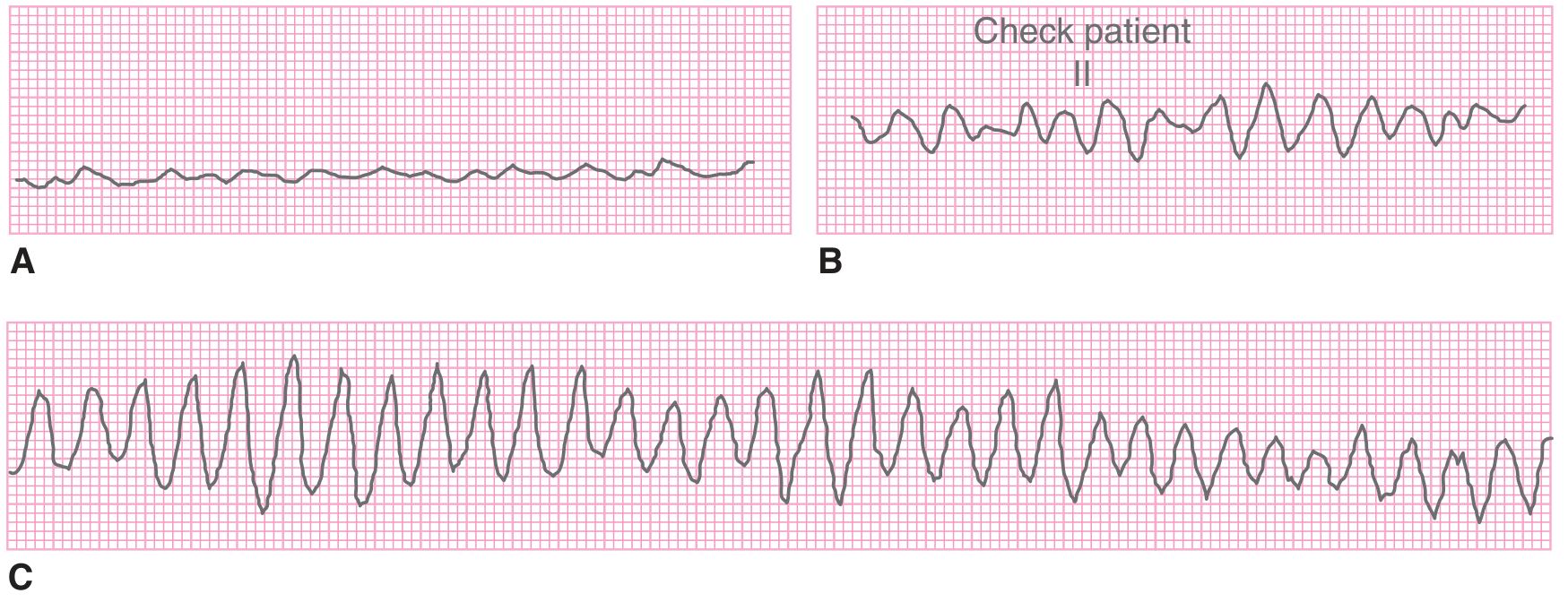
VF ECG Waveform: Three Subtypes (by Amplitude)
From Tintinalli's Emergency Medicine, VF waveforms are classified by amplitude:
| Type | Description | Clinical Significance |
|---|---|---|
| Coarse VF | High-amplitude (>0.5 mV) irregular waves | Early VF; more likely to respond to defibrillation |
| Intermediate VF | Medium-amplitude waves | Intermediate duration |
| Fine VF | Low-amplitude (<0.2 mV) irregular undulations | Late VF; may mimic asystole; poor prognosis |
Voltage Decay Over Time
From Guyton & Hall:
- At onset: ~0.5 mV amplitude
- After 20-30 seconds: 0.2-0.3 mV
- After several minutes: as low as 0.1 mV or less (fine VF, may appear nearly flat)
This is why early defibrillation is critical - coarse VF responds much better than fine VF to electrical shock.
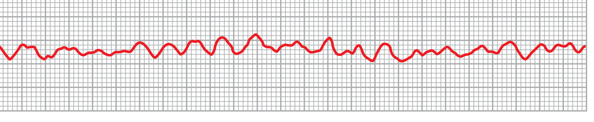
ECG Image - Classic VF (Lead II from Guyton & Hall):
Summary: What You See vs. What You Don't
You DON'T see:
- P waves
- QRS complexes
- T waves
- Any organized rhythm
- Isoelectric baseline
You DO see:
- Irregular, chaotic, high-frequency undulations
- Constantly changing amplitude, morphology, and cycle length
- No two "waves" look alike
Physiological Basis (Guyton & Hall)
VF results from "circus movement" - reentrant electrical impulses continuously re-exciting ventricular muscle. Triggered by:
- Ischemia (most common - severe CAD, acute MI)
- Electrical shock
- Prolonged pathway (dilated hearts)
- Decreased conduction velocity
- Shortened refractory period
The ventricles never fully contract or relax - they remain in a state of partial, uncoordinated contraction. No blood is pumped. Unconsciousness occurs within 4-5 seconds; irreversible brain damage begins within minutes without CPR/defibrillation.
Clinical Context (Tintinalli's)
- Primary VF: Sudden onset without preceding hemodynamic deterioration (typically ischemic)
- Secondary VF: Follows prolonged left ventricular failure or circulatory shock
- Other triggers: Cardiac catheterization, electrocution, commotio cordis (blunt chest trauma)
Treatment: Immediate defibrillation (biphasic 200 J, escalating to 360 J if needed) + CPR + epinephrine/amiodarone per ACLS protocol. This is the #1 shockable rhythm.
Sources: Guyton and Hall Textbook of Medical Physiology; Tintinalli's Emergency Medicine, 9th Ed.; Braunwald's Heart Disease
This is a shared conversation. Sign in to Orris to start your own chat.