
consolidation vs infiltrate va ggo on chest x ray
consolidation vs ground glass opacity chest X-ray CT comparison
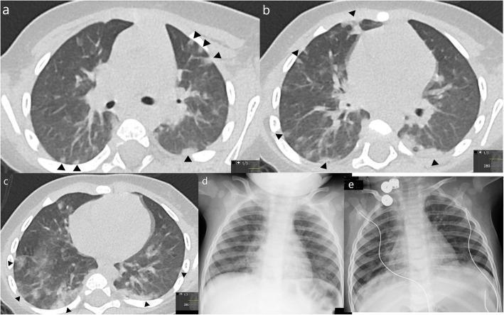
This composite figure presents diagnostic imaging findings in a 10-month-old infant with confirmed COVID-19. Panels (a-c) are axial non-contrast chest CT slices demonstrating patchy consolidations and ground-glass opacities (GGO) with a characteristic subpleural and peripheral distribution throughout the upper and lower lobes. Black arrowheads highlight the 'halo sign,' where central areas of consolidation are surrounded by peripheral rings of ground-glass opacity. Panels (d) and (e) show a longitudinal comparison of chest X-rays. Image (d), taken one day prior to the CT, shows subtle, diffuse bilateral pulmonary opacities. Image (e), performed four days after the initial X-ray, illustrates a partial resolution of these bilateral opacities, indicating clinical improvement. This series highlights the classic pediatric radiological presentation of SARS-CoV-2 infection, emphasizing peripheral lung involvement and the evolution of findings over a short clinical course.
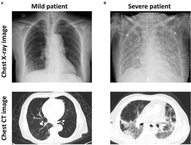
This diagnostic comparison image demonstrates the radiological findings of COVID-19 across two different levels of severity using chest X-ray and CT imaging. Section (A) represents a 'Mild patient,' showing localized patchy shadows and peripheral ground-glass opacities (GGO) primarily in the lower lung fields on X-ray, with corresponding focal GGO visible on the axial CT slice. In contrast, section (B) displays a 'Severe patient' characterized by extensive, bilateral, and confluent pulmonary opacities. The severe X-ray reveals a significant loss of lung lucency and obscuration of the cardiophrenic angles, while the axial CT shows widespread, dense consolidation and diffuse GGO throughout both lung parenchyma. These images highlight the progressive pulmonary damage associated with severe SARS-CoV-2 infection, specifically illustrating how increased opacity density and distribution correlate with clinical severity in viral pneumonia. The material is suitable for medical education in pulmonology and radiology to distinguish disease staging via imaging modalities.
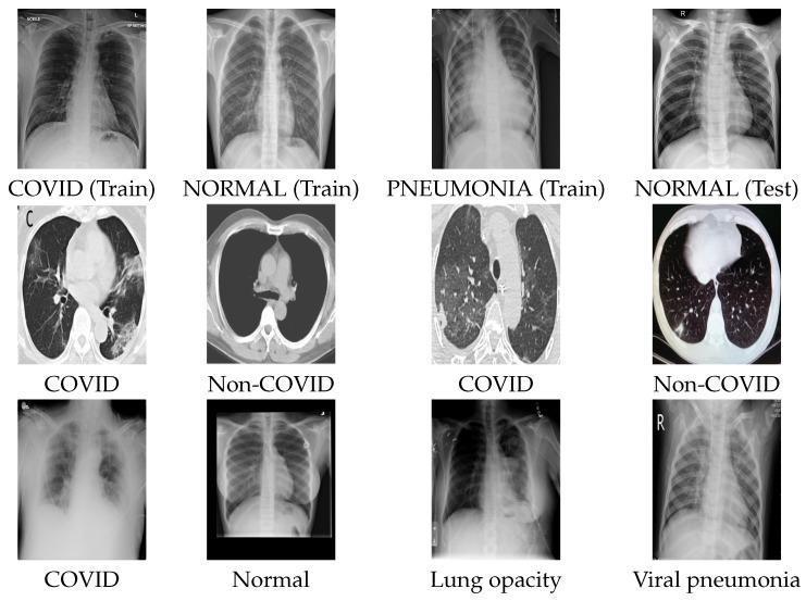
This composite educational graphic presents a comparison of diagnostic imaging for COVID-19 and other respiratory conditions across three distinct datasets. The first and third rows feature Chest X-ray (CXR) images. Row one displays 'COVID (Train)' showing peripheral ground-glass opacities, 'NORMAL (Train)' with clear lung fields, 'PNEUMONIA (Train)' with dense bilateral consolidation, and 'NORMAL (Test)'. Row three contrasts 'COVID' presenting with diffuse, hazy pulmonary infiltrates against 'Normal', 'Lung opacity' (localized dense consolidation), and 'Viral pneumonia' (interstitial markings). The second row utilizes Axial Computed Tomography (CT) scans to contrast 'COVID' pathology with 'Non-COVID' controls. The COVID-positive CT images demonstrate characteristic multifocal ground-glass opacities (GGO) and areas of consolidation within the lung parenchyma, while the Non-COVID images show clear parenchyma and normal thoracic anatomy. This visual aid is designed for medical training in radiological differentiation between COVID-19, typical viral pneumonia, and healthy thoracic structures, highlighting key diagnostic features such as opacity distribution, texture, and lung field clarity.
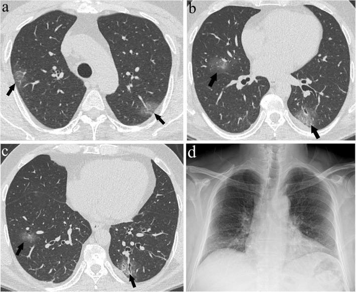
This composite medical image presents a comparison between high-resolution axial CT scans and a posteroanterior chest X-ray (CXR) from a 56-year-old male with COVID-19 pneumonia. Panels (a), (b), and (c) display axial CT slices at the levels of the upper, middle, and lower lung zones, respectively. These images reveal multifocal, bilateral ground-glass opacities (GGOs) indicated by black arrows. The GGOs appear as hazy, increased attenuation of the lung parenchyma without obscuring the underlying vascular structures, characteristic of grade 1 lesions. The distribution is notably peripheral and involves all three lung zones, yielding a total CT Score (CTS) of 6. In contrast, panel (d) shows the corresponding CXR, which appears largely unremarkable with clear lung fields and no discernible signs of pneumonia or hazy opacities. This comparison highlights the superior sensitivity of CT over CXR in detecting early or mild manifestations of viral pneumonia, such as small areas of ground-glass opacity that do not significantly alter the overall radiographic density on conventional X-ray.
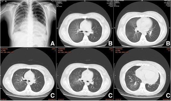
This composite figure presents serial diagnostic imaging of the chest, including a posteroanterior chest X-ray (Panel A) and multiple axial computed tomography (CT) lung window slices (Panels B and C) spanning approximately five months. The primary finding across all modalities is a persistent, localized area of ground-glass opacity (GGO) involving the right lung lobes. In Panel A, the chest radiograph shows increased hazy density in the right middle and lower lung zones without a discrete mass or consolidation. The subsequent axial CT images in Panel B (obtained 10 days later) and Panel C (obtained 5 months later) confirm diffuse, geographic ground-glass opacities within the right lung parenchyma. Notably, the underlying bronchovascular structures remain visible through the hazy density, which is characteristic of GGO rather than dense consolidation. The longitudinal nature of these images demonstrates the chronic persistence of these right-sided pulmonary exudative changes. These findings are clinically relevant for evaluating chronic interstitial processes, localized inflammatory conditions, or rare disorders such as chylous reflux syndrome affecting the pulmonary system.
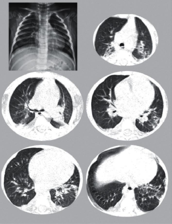
This composite diagnostic image features a pediatric chest X-ray in the top left and five axial CT scans of the chest from the same patient. The chest X-ray demonstrates diffuse, subtle increased density within the lung fields and a prominent cardiac silhouette. The axial CT images, presented in lung window settings, reveal bilateral ground-glass opacities (GGO) with a predominantly peripheral and posterior distribution. Several areas show more dense consolidative changes, and some lesions exhibit the 'halo sign,' characterized by a nodular opacity surrounded by a ring of ground-glass attenuation. The heart appears moderately enlarged relative to the thoracic cavity. These findings are consistent with multi-system inflammatory syndrome in children (MIS-C) related to COVID-19, highlighting the spectrum of pulmonary involvement ranging from interstitial inflammation to parenchymal consolidation.
Consolidation vs. Infiltrate vs. Ground-Glass Opacity (GGO) on Chest Imaging
1. Consolidation
- Increased opacity that obscures the underlying pulmonary vessels and bronchial walls
- Margins of blood vessels are completely lost within the opacity
- Air bronchogram - bronchi remain air-filled and visible as dark branching channels cutting through the white opacity (a hallmark sign)
- Well-defined outer borders (especially when lobar), or ill-defined at the advancing edge
- Silhouettes adjacent structures: obliterates the diaphragm, cardiac border, or intrapulmonary vessels when adjacent
- CXR: Dense white opacity, often lobar or segmental; obscures the hemidiaphragm (silhouette sign)
- CT: Homogeneous increased attenuation with complete vessel obscuration; air bronchograms often more visible
"Air space consolidation, by definition, is seen when alveolar air is replaced by fluid, cells, or other material. On HRCT, consolidation results in an increase in lung opacity associated with obscuration of underlying vessels." - Murray & Nadel's Textbook of Respiratory Medicine
2. Ground-Glass Opacity (GGO)
- Hazy "misty" increase in lung density - like frosted glass
- Vessels and bronchi still visible through the opacity (this is the defining feature)
- "Black bronchus sign": on CT, airway lumen appears darker than adjacent abnormal lung (reversed from normal, where they are similar density)
- Can be focal, multifocal, or diffuse
- CXR: Hazy increased density where vessel margins are obscured (reduced contrast resolution means GGO actually looks more like consolidation on plain film - this is an important pitfall)
- CT/HRCT: Hazy increase in attenuation, but vessels and bronchial walls clearly visible through it
"A ground-glass pattern on HRCT is defined as a generalised increase in opacity that does not obscure pulmonary vessels and bronchial walls. At a microscopic level, the changes responsible for ground-glass opacity include partial filling of the air spaces, thickening of the interstitium, or a combination of the two." - Grainger & Allison's Diagnostic Radiology
"On plain radiography, ground-glass opacification is seen as hazy, increased lung opacity in which the margins of pulmonary vessels are obscured. Because of the greater contrast resolution, ground-glass opacification on CT appears as a hazy increase in lung attenuation but without obscuration of bronchial and vascular markings." - Grainger & Allison's Diagnostic Radiology
3. Infiltrate
- It is vague and non-specific - it does not describe a specific radiological pattern
- Different radiologists and clinicians use it to mean different things (sometimes consolidation, sometimes GGO, sometimes a combination)
- The Fleischner Society and modern radiology guidelines discourage its use in favor of precise descriptors (consolidation, GGO, reticular opacity, nodule, etc.)
- Still widely used clinically, especially in the phrase "new pulmonary infiltrate" to mean "new opacity suggesting pneumonia"
Quick Comparison Table
| Feature | Consolidation | GGO | "Infiltrate" |
|---|---|---|---|
| Vessel visibility | Obscured | Visible | Not specified |
| Density | Dense/white | Hazy/"misty" | Variable |
| Air bronchogram | Common | Absent/uncommon | Not specified |
| Silhouette sign | Present | Absent | Not specified |
| Pathology | Complete airspace filling | Partial filling or interstitial thickening | Nonspecific |
| Best modality | CXR or CT | CT (poor on CXR) | - |
| Specificity as term | Precise | Precise | Vague/imprecise |
Spectrum and Mixed Patterns
- "Crazy paving": GGO + superimposed interlobular septal thickening (classically seen in PCP, alveolar proteinosis)
- "Halo sign": GGO surrounding a nodule/consolidation (classically invasive aspergillosis - hemorrhagic halo)
- "Reverse halo / atoll sign": Central GGO ringed by peripheral consolidation (classically organizing pneumonia)
- "Perilobular consolidation": Consolidation smeared around the lobule - characteristic of organizing pneumonia
Clinical Pearl


Image