


129055.jpg
129053.jpg
What is this disease?
I can see the images clearly. Let me analyze them and look up relevant medical information simultaneously.
analyse_media
analyse_media
Reading File
Now let me search the medical library and find images for a comprehensive answer.
Finding Sources
Searching Images
spider veins telangiectasia varicose veins leg
Reading File
Reading File
I have everything I need to give a thorough answer. Here is the analysis:
Diagnosis: Spider Veins (Telangiectasias) with Reticular Veins
Both images show the classic appearance of telangiectasias (spider veins) and reticular veins on the lower limb - a presentation of superficial chronic venous disease (CVD).
What You Are Seeing
| Finding | Description |
|---|---|
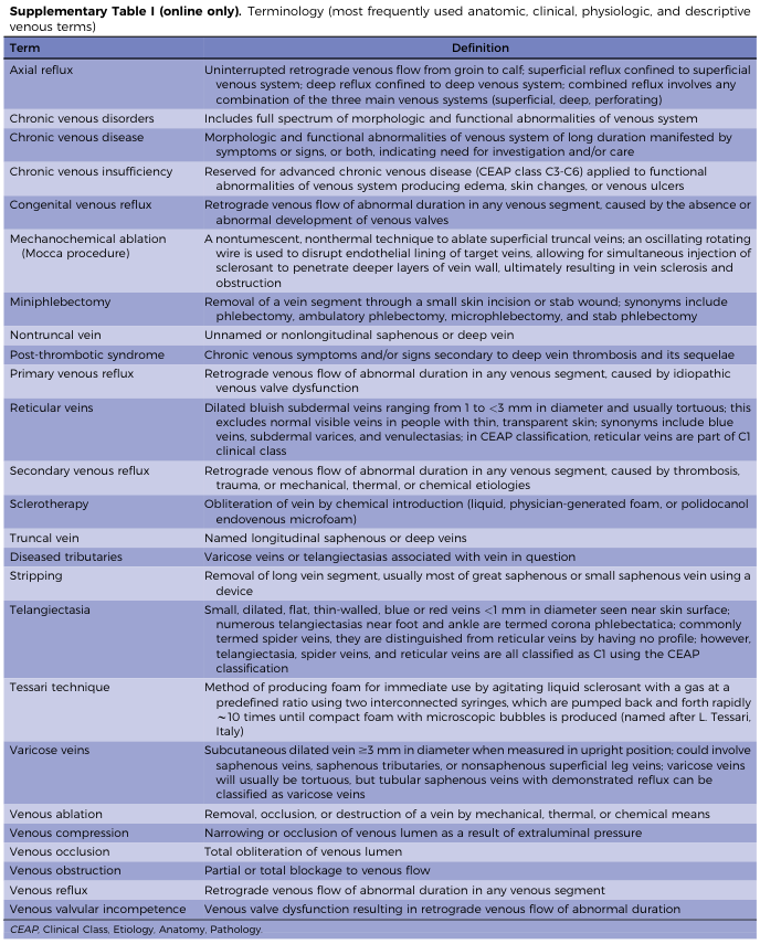
| Telangiectasias / Spider veins | Tiny intradermal venules < 1 mm in diameter, forming web-like red/purple/blue patterns on the surface |
| Reticular veins | Slightly larger, bluish subdermal veins 1–2.9 mm in diameter, tortuous, visible beneath the skin |
| Small red dot (cherry angioma) | A benign capillary collection - harmless, seen on the thigh in image 2 |
As described in Bailey and Love's Short Practice of Surgery (28th Ed): "Telangiectasia (thread veins, spider veins and hyphen webs): represent tiny intradermal venules less than 1 mm in diameter."
Why Does This Happen?
The one-way valves inside the superficial veins weaken, allowing blood to flow backward (reflux) and pool. This raises pressure in the small superficial vessels, causing them to dilate and become visible through the skin. Contributing factors include:
- Genetics / family history (most common cause)
- Prolonged standing or sitting (occupational)
- Hormonal changes - pregnancy, oral contraceptives, menopause
- Aging - valve weakness increases with age
- Obesity - increased abdominal pressure on leg veins
- Female sex - hormones make vein walls more lax
Grading - CEAP Classification
This presentation corresponds to CEAP Class C1 (telangiectasias/reticular veins), the mildest end of the spectrum of Chronic Venous Disease. The spectrum goes:
- C1 - Spider/reticular veins (as shown here)
- C2 - Varicose veins (> 3 mm, bulging/tortuous)
- C3 - Edema
- C4 - Skin changes (pigmentation, eczema, lipodermatosclerosis)
- C5-C6 - Healed/active venous ulcers
Symptoms to Watch For
Spider veins are often purely cosmetic, but can cause:
- Aching, heaviness, or fatigue in the legs - especially after prolonged standing
- Mild burning or itching around the veins
- Ankle swelling toward the end of the day
Red Flags - See a Doctor Urgently If:
- Sudden one-sided leg swelling (possible deep vein thrombosis / DVT)
- Warmth, redness, or tenderness over a vein (possible thrombophlebitis)
- Brown/dark skin discoloration around the ankle
- Non-healing sores or ulcers
Treatment Options
(Per Pfenninger & Fowler's Procedures for Primary Care, 3rd Ed.)
| Option | Details |
|---|---|
| Conservative | Graduated compression stockings (15–30 mmHg), leg elevation, regular walking/exercise |
| Sclerotherapy | Injection of a sclerosant solution (most effective for spider/reticular veins, ~70–90% success) |
| Laser/IPL therapy | Surface laser for tiny superficial vessels, no needles needed |
| Radiofrequency ablation | For larger feeding reticular veins |
Note: This is a visual clinical assessment only and does not replace an in-person evaluation. A vascular specialist or dermatologist can perform a venous duplex ultrasound to check for any underlying valve incompetence before deciding on treatment.
This is a shared conversation. Sign in to Orris to start your own chat.