
Describe c.o.p.d for medical exam in detail for full mark que.
"chronic obstructive pulmonary disease"[MeSH Terms] AND management
COPD emphysema chronic bronchitis pathology diagram
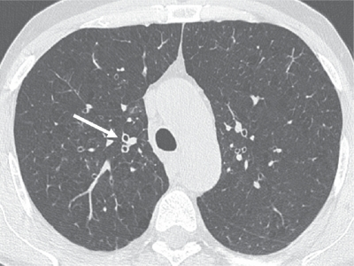
**Imaging Modality:** High-resolution thin-section computed tomography (HRCT) of the chest. **Anatomical Region:** Axial section at the level of the upper lobes and the trachea. **Observed Pathology:** Chronic Obstructive Pulmonary Disease (COPD) with associated chronic bronchitis and early-stage emphysema. **Characteristic Visual Features:** * **Bronchial Wall Thickening:** There is visible thickening of the segmental and subsegmental bronchial walls (indicated by the white arrow). These appear as prominent "ring shadows" when visualized in cross-section, reflecting airway inflammation and remodeling. * **Parenchymal Changes:** Small, scattered areas of low attenuation (lucency) without visible walls are distributed in a centrilobular pattern throughout the lung parenchyma, consistent with early centrilobular emphysema. * **Vascular Markings:** Peripheral pulmonary vasculature remains present but is set against a background of mildly decreased lung attenuation. **Key Diagnostic Features:** The combination of thickened bronchial walls and centrilobular lucencies in a patient with a smoking history is highly characteristic of the airway-predominant and parenchymal-destructive phenotypes of COPD. These findings differentiate the condition from pure bronchiectasis or asthma, which typically lack the centrilobular destructive component.
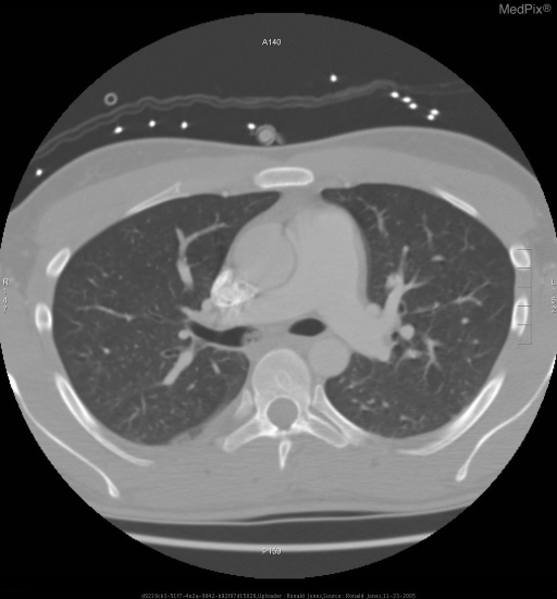
**Imaging Modality:** Axial Computed Tomography (CT) scan of the chest. **Anatomical Region:** Thorax at the level of the pulmonary artery bifurcation and carina. **Observed Pathology:** The lungs exhibit diffuse, bilateral hyperlucency and a paucity of peripheral vascular markings, characteristic of pulmonary emphysema. There is evidence of bronchial wall thickening, suggesting chronic bronchitis, consistent with Chronic Obstructive Pulmonary Disease (COPD). **Characteristic Visual Features:** - **Lung Parenchyma:** Increased radiolucency with attenuation of the pulmonary vasculature. - **Mediastinum:** Prominent main pulmonary artery trunk, which may indicate pulmonary hypertension secondary to chronic lung disease. A dense, calcified focus is noted within the right hilar region, likely representing a calcified lymph node or vascular calcification. - **Thoracic Cage:** Mild increase in the anteroposterior diameter of the chest. - **Pleura:** A small, dependent pleural effusion or pleural thickening is visible in the posterior right hemithorax. **Diagnostic Context:** These radiologic findings are highly suggestive of chronic obstructive airway disease with significant emphysematous changes and associated airway inflammation.
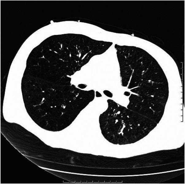
This diagnostic image is an axial chest computed tomography (CT) scan displayed in a lung window, showcasing the thoracic cavity at the level of the main bronchi. The primary findings are consistent with chronic obstructive pulmonary disease (COPD), specifically emphysema and chronic bronchitis. Both lung fields exhibit hyperlucency and attenuated vascular markings, indicative of parenchymal destruction. Notably, there are multiple large, thin-walled, air-filled spaces known as bullae scattered throughout both lungs. The bronchial structures show evidence of wall thickening and subtle dilation, suggesting associated chronic bronchitis and potential bronchiectasis. Areas of increased opacity representing fibrous foci are visible, reflecting underlying chronic inflammatory or scarring processes. The scan demonstrates the classic structural remodeling associated with long-term obstructive airway disease, including the presence of bullous emphysema. This visual material is intended for medical education regarding the radiological manifestations of end-stage chronic respiratory conditions and the identification of bullous disease on CT imaging.
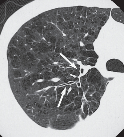
**Imaging Modality:** High-resolution computed tomography (HRCT), axial section. **Anatomical Region:** Right lung parenchyma. **Observed Pathology:** Advanced pulmonary emphysema with comorbid small airway disease. **Characteristic Visual Features:** * **Emphysema:** The lung field exhibits diffuse, multi-focal areas of low attenuation (lucency) representing alveolar destruction. Findings are consistent with centrilobular and paraseptal emphysema, appearing as multiple small, thin-walled cystic spaces and bullae. * **Bronchial Wall Thickening:** There is conspicuous thickening of the bronchial walls (indicated by white arrows). The increased wall-to-lumen ratio suggests chronic inflammatory changes or remodeling. * **Luminal Narrowing:** Associated with the wall thickening, several bronchioles demonstrate a narrowed caliber (stenosis), contributing to a "signet ring" appearance in some cross-sections but with a reduced internal diameter. * **Vascular Attenuation:** Peripheral pulmonary vasculature appears attenuated and displaced by the hyperinflated, emphysematous lung parenchyma. **Diagnostic Significance:** This combination of parenchymal hyperlucency and bronchial wall thickening is characteristic of chronic obstructive pulmonary disease (COPD) with a significant chronic bronchitis component. The image illustrates the coexistence of large-space destruction and airway remodeling.
centriacinar panacinar emphysema histology comparison

This diagnostic image consists of four high-resolution computed tomography (HRCT) axial sections of the lung parenchyma, illustrating the primary radiological patterns of emphysema in Chronic Obstructive Pulmonary Disease (COPD). Images A and B demonstrate centriacinar emphysema, characterized by small, focal areas of low attenuation (lucent airspaces) located in the center of the secondary pulmonary lobules, typically surrounding the central bronchovascular bundle. Image C displays panacinar emphysema, showing a more uniform and diffuse destruction of the entire acinus, resulting in generalized lung hyperlucency and attenuation of the pulmonary vasculature. Image D illustrates paraseptal emphysema (distal acinar emphysema), where focal lucent airspaces are distributed preferentially along the subpleural surfaces and interlobular septa, often demarcated by thin walls. Key educational concepts include the distinction between lobular distributions and the resulting architectural distortion and vascular attenuation associated with progressive alveolar wall destruction.
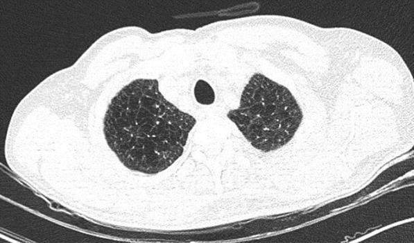
This diagnostic image is an axial non-contrast high-resolution computed tomography (HRCT) scan of the thorax at the level of the upper lobes. The image demonstrates bilateral, diffuse, and homogeneous areas of abnormally decreased lung attenuation (hyperlucency). These findings are characteristic of panacinar (panlobular) emphysema, where there is a uniform destruction of the pulmonary acinus. Key features include a simplified lung architecture with a notable paucity of pulmonary vascular markings throughout the affected parenchyma. Unlike centriacinar emphysema, which often shows focal areas of low attenuation, this presentation shows a generalized darkening of the lung fields without well-defined walls or cystic structures. No acute infiltrates, ground-glass opacities, or consolidations are present, which in a clinical context (such as COVID-19 screening) helps differentiate chronic obstructive pulmonary disease (COPD) from acute infectious processes. The image serves as a classic educational example of end-stage alveolar destruction and hyperinflation seen in severe emphysema.
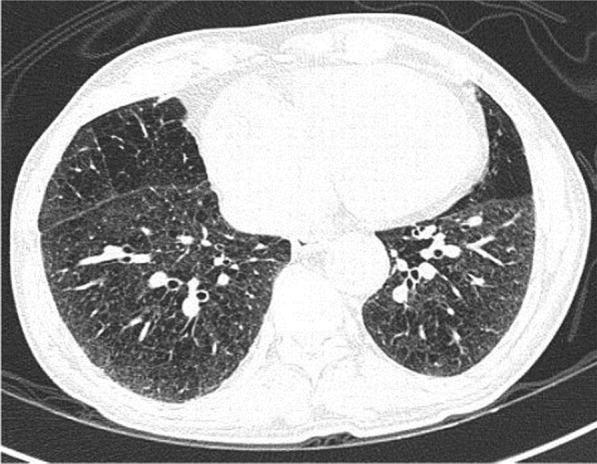
This diagnostic image is an axial non-contrast chest CT scan at the level of the lower lobes, demonstrating characteristic features of panacinar emphysema. The lung parenchyma exhibits a diffuse, generalized decrease in attenuation (hypodensity) bilaterally, resulting in a significantly darker appearance than normal lung tissue. Structurally, there is extensive destruction of the alveolar walls, leading to the formation of numerous small, round, and irregularly shaped airspaces that give the parenchyma a coarse, 'swiss cheese' or porous texture. Unlike centriacinar emphysema which typically affects the upper lobes, these changes are distributed uniformly across the visible lung segments without regional predilection. Key landmarks include the central mediastinum with a visible heart and descending aorta, and the bilateral major fissures. There is a notable absence of peripheral ground-glass opacities or consolidations, distinguishing this chronic obstructive pulmonary disease (COPD) manifestation from acute infectious processes like COVID-19 bronchopneumonia.
Chronic Obstructive Pulmonary Disease (COPD) - Comprehensive Medical Exam Answer
1. DEFINITION
- Chronic Bronchitis (clinical definition): Productive cough persisting for at least 3 consecutive months in at least 2 consecutive years, in the absence of other explanations.
- Emphysema (pathological definition): Permanent, abnormal enlargement of airspaces distal to the terminal bronchioles due to destruction of their walls, without obvious fibrosis.
2. EPIDEMIOLOGY
- Third most common cause of death in the USA
- Accounts for >$40 billion/year in direct and indirect healthcare costs
- Affects approximately 15-30% of habitual smokers (though radiographic studies now suggest significant progressive changes even in smokers with normal spirometry)
- More common in males, though incidence in females is rising
3. ETIOLOGY / RISK FACTORS
| Risk Factor | Notes |
|---|---|
| Cigarette smoking | By far the most important (responsible for ~85-90% of cases) |
| α1-antitrypsin (AAT) deficiency | Causes panacinar emphysema; autosomal codominant |
| Occupational dust/fumes | Coal, silica, cadmium, grain dust |
| Air pollution | Biomass fuel smoke (especially in developing countries) |
| Airway hyperresponsiveness | Pre-existing asthma increases risk |
| Genetic factors | Beyond AAT deficiency |
| Recurrent respiratory infections | In childhood impair lung development |
4. PATHOGENESIS
A. Protease-Antiprotease Imbalance (Emphysema)
- Cigarette smoke triggers recruitment of neutrophils and macrophages into the lung
- These cells release elastase, MMP-2, MMP-9, cathepsins - proteases that destroy elastin in alveolar walls
- Normally, α1-antitrypsin (AAT) neutralizes neutrophil elastase
- In AAT deficiency OR when smoke overwhelms AAT, unbalanced proteolysis destroys alveolar walls
- Result: loss of elastic recoil, air trapping, hyperinflation
B. Oxidative Stress
- Cigarette smoke contains free radicals that overwhelm antioxidant defenses
- Oxidants inactivate AAT further compounding the protease-antiprotease imbalance
C. Mucus Dysfunction (Chronic Bronchitis)
- Smoke causes surface epithelial mucous metaplasia and submucosal gland hyperplasia
- MUC5AC increases 10-fold and MUC5B increases 3-fold in severe COPD
- Acrolein (in cigarette smoke) is a potent inducer of MUC5AC production
- Cigarette smoke also reduces CFTR function and increases ENaC function → decreases airway surface liquid → impaired mucociliary clearance
- Results in persistent airway infections (especially Haemophilus influenzae)
D. Inflammation
- COPD features neutrophilic inflammation (unlike asthma's eosinophilic inflammation)
- Key mediators: IL-8, TNF-α, LTB4
- Leads to small airway fibrosis and remodeling
5. MORPHOLOGY / PATHOLOGY
A. Emphysema - Types
| Type | Location | Cause | Features |
|---|---|---|---|
| Centriacinar (Centrilobular) | Central/proximal acinus; respiratory bronchioles | Cigarette smoking | Most common; upper lobe predominant |
| Panacinar (Panlobular) | Entire acinus uniformly | AAT deficiency | Lower lobe predominant; more severe destruction |
| Paraseptal (Distal Acinar) | Distal acinus; near pleura/septa | Unknown; associated with bullae | Can cause spontaneous pneumothorax |
| Irregular (Paracicatricial) | Irregular; near fibrosis | Scarring | Adjacent to scars |
B. Emphysema Gross and Microscopic Features
- Gross: Voluminous, pale, hyperinflated lungs; loss of normal fine texture
- Microscopy: Enlarged airspaces, destruction of alveolar walls, loss of elastic tissue; no significant fibrosis
- DLCO is low (reduced alveolar surface area)
- Static lung compliance (CstL) is increased (reduced elastic recoil)
C. Chronic Bronchitis Pathology
- Mucous gland hyperplasia in trachea and large airways (Reid index >0.4: ratio of gland thickness to bronchial wall thickness)
- Goblet cell metaplasia in small airways (normally absent)
- Chronic bronchiolitis: Small airway inflammation and fibrosis - the main cause of airflow obstruction
- Airway wall smooth muscle hypertrophy
- Bronchitic patients tend to develop hypoxemia and hypercapnia ("Blue Bloater")
6. CLINICAL FEATURES
Classic Presentation: "Pink Puffer" vs "Blue Bloater"
| Feature | Pink Puffer (Emphysema) | Blue Bloater (Chronic Bronchitis) |
|---|---|---|
| Build | Thin, cachectic | Overweight, cyanotic |
| Cough | Minimal | Productive, chronic |
| Sputum | Minimal | Copious, purulent |
| Dyspnea | Severe | Moderate |
| PaO2 | Near normal (at rest) | Low (hypoxemia) |
| PaCO2 | Low/normal | High (hypercapnia) |
| Cyanosis | Absent | Present |
| Cor pulmonale | Late | Early |
| Polycythemia | Absent | Present |
| Pursed lip breathing | Present | Absent |
| DLCO | Reduced | Normal/near-normal |
Typical Symptoms
- Dyspnea: Progressive, initially on exertion; the cardinal symptom
- Chronic productive cough: "Morning cough" is characteristic
- Wheeze: Especially on exertion or during exacerbations
- Exercise intolerance: Due to dynamic hyperinflation and ventilation-perfusion mismatch
Physical Signs
- Barrel chest (increased A-P diameter due to hyperinflation)
- Hyper-resonance on percussion
- Reduced breath sounds, prolonged expiratory phase
- Wheeze and coarse crackles
- Pursed-lip breathing (maintains PEEP, prevents airway collapse)
- Use of accessory muscles of respiration
- Signs of cor pulmonale: Raised JVP, peripheral edema, loud P2, right ventricular heave
7. INVESTIGATIONS
A. Spirometry (Definitive for Diagnosis)
- FEV1/FVC ratio < 0.70 post-bronchodilator confirms airflow obstruction
- FEV1 is reduced; FVC is normal or mildly reduced
- Obstruction is NOT fully reversible (distinguishes from asthma)
- TLC, RV, FRC are increased (air trapping and hyperinflation)
GOLD Severity Classification (Based on Post-BD FEV1 % predicted)
| GOLD Grade | FEV1 % predicted | Severity |
|---|---|---|
| GOLD 1 | ≥80% | Mild |
| GOLD 2 | 50-79% | Moderate |
| GOLD 3 | 30-49% | Severe |
| GOLD 4 | <30% | Very severe |
B. Arterial Blood Gas (ABG)
- Type 1 respiratory failure (hypoxemia alone) in mild-moderate disease
- Type 2 respiratory failure (hypoxemia + hypercapnia) in severe disease
- Chronic hypercapnic patients: metabolic compensation (raised HCO3, near-normal pH)
- In the Costanzo physiology case example: PaO2 = 60 mmHg, PaCO2 = 30 mmHg (hyperventilating to compensate), elevated A-a gradient confirms V/Q mismatch
C. Chest X-Ray
- Hyperinflated lungs (flattened diaphragms, increased A-P diameter)
- Increased retrosternal airspace
- Barrel-shaped chest
- Bullae (especially in emphysema)
- Peripheral vascular marking attenuation
D. HRCT Chest
- Gold standard for characterizing emphysema type and extent
- Centrilobular emphysema: scattered focal lucencies around bronchovascular bundles (upper lobe)
- Panacinar emphysema: diffuse homogeneous hyperlucency, paucity of vasculature
- Bronchial wall thickening, air trapping, bullae
E. Other Tests
- DLCO: Reduced in emphysema, near-normal in pure chronic bronchitis
- ECG: Right axis deviation, P pulmonale (right atrial enlargement), RBBB in cor pulmonale
- FBC: Polycythemia (secondary to chronic hypoxemia); eosinophilia may suggest asthma-COPD overlap
- Sputum culture: During exacerbations (H. influenzae, S. pneumoniae, M. catarrhalis)
- Alpha-1 antitrypsin level: In patients <45 years, non-smokers, or lower-lobe predominant emphysema
8. COMPLICATIONS
- Acute exacerbations (most common) - often triggered by viral/bacterial infection
- Respiratory failure (Type 1 and Type 2)
- Cor pulmonale (right heart failure due to pulmonary hypertension)
- Polycythemia (secondary, due to hypoxemia)
- Pneumothorax (rupture of bullae)
- Lung cancer (3-4x increased risk, especially squamous cell and small cell)
- Pulmonary hypertension (hypoxic vasoconstriction - reversed by O2 therapy)
- Malnutrition and muscle wasting (systemic inflammation)
- Depression and anxiety
- Osteoporosis (steroid use, smoking, inactivity)
9. MANAGEMENT
A. Stepwise Pharmacological Treatment (GOLD Guidelines)
Bronchodilators (Mainstay of Treatment)
- SABA (Short-Acting Beta-2 Agonist): Salbutamol (albuterol) - for acute symptom relief
- SAMA (Short-Acting Muscarinic Antagonist): Ipratropium bromide
- Combination SABA + SAMA is more effective than either alone
- LABA (Long-Acting Beta-2 Agonist): Salmeterol, formoterol, indacaterol
- LAMA (Long-Acting Muscarinic Antagonist): Tiotropium, glycopyrronium, umeclidinium
- LAMA + LABA combination is preferred for moderate-severe COPD
Inhaled Corticosteroids (ICS)
- Less central than in asthma; lower efficacy in COPD
- Associated with increased risk of bacterial pneumonia
- Recommended ONLY for:
- Severe airflow obstruction (FEV1 <50%)
- History of frequent exacerbations
- High blood eosinophil count (eosinophils ≥300 cells/μL suggest benefit)
- Concurrent asthma (Asthma-COPD Overlap, ACO)
- Triple therapy (LABA + LAMA + ICS) for frequent exacerbators with high eosinophils
Other Pharmacological Agents
- Roflumilast (PDE-4 inhibitor): Reduces exacerbation frequency; approved for severe COPD with chronic bronchitis phenotype
- Azithromycin (long-term): Reduces exacerbation frequency; risk of hearing loss, resistance
- N-acetylcysteine (mucolytic): Limited evidence
- Theophylline: Historically used but recent large RCT failed to show benefit on exacerbation frequency; narrow therapeutic window; now rarely used
B. Non-Pharmacological Treatment
- Smoking cessation: Single most important intervention; slows FEV1 decline
- Pulmonary rehabilitation: Improves exercise capacity, dyspnea, quality of life
- Long-term oxygen therapy (LTOT): For PaO2 ≤55 mmHg (or ≤60 mmHg with cor pulmonale/polycythemia); minimum 15 hours/day; reduces mortality in hypoxemic patients
- Vaccinations: Annual influenza vaccine; pneumococcal vaccine
- Nutritional support
- Non-invasive ventilation (NIV/BiPAP): For stable hypercapnic patients
C. Surgical/Interventional Options
- Lung volume reduction surgery (LVRS): Removes most diseased lung tissue; improves mechanics; suitable for upper-lobe emphysema with poor exercise capacity
- Bronchoscopic lung volume reduction: Endobronchial valves or coils
- Bullectomy: For giant bullae compressing normal lung
- Lung transplantation: For end-stage COPD (GOLD 4)
10. ACUTE EXACERBATIONS OF COPD (AECOPD)
Definition
Causes (Triggers)
- Respiratory tract infection (most common): 50% bacterial (H. influenzae, S. pneumoniae, M. catarrhalis), 50% viral (Rhinovirus most common)
- Air pollution, cold weather
- Pulmonary embolism (important to exclude)
- Discontinuation of medications
Management of AECOPD
- Controlled oxygen therapy: Target SpO2 88-92% (avoid excessive O2 - risk of hypercapnic drive suppression). Use Venturi mask for precise delivery.
- Short-acting bronchodilators: Nebulised salbutamol + ipratropium
- Systemic corticosteroids: Prednisolone 30-40 mg/day for 5 days (reduces recovery time and treatment failure)
- Antibiotics: Amoxicillin/doxycycline/co-amoxiclav if signs of infection (purulent sputum, increased dyspnea, increased sputum volume - Anthonisen criteria); β-lactams commonly used
- Non-invasive ventilation (NIV/BiPAP): Indicated if pH <7.35 with PaCO2 >6.0 kPa despite controlled O2 and bronchodilators
- Invasive mechanical ventilation: If NIV fails or patient cannot protect airway
- DVT prophylaxis: Heparin
- Treat precipitants: Chest physiotherapy, treat pneumonia, exclude PE
11. DIFFERENTIATING COPD FROM ASTHMA
| Feature | COPD | Asthma |
|---|---|---|
| Age of onset | >40 years | Any age, often childhood |
| Smoking history | Usually present | Not required |
| Airflow obstruction | Partially/not reversible | Fully reversible |
| Inflammation | Neutrophilic | Eosinophilic |
| ICS response | Poor (unless high eos) | Excellent |
| Progression | Relentlessly progressive | Variable, episodic |
| Diurnal variation | Minimal | Marked (worse at night/morning) |
| Triggers | Infections, exercise | Allergens, exercise, cold |
| DLCO | Reduced (emphysema) | Normal |
| Atopy/IgE | Absent | Often present |
12. PROGNOSIS
- BODE Index: Body mass index, Obstruction (FEV1), Dyspnea (MRC scale), Exercise capacity (6-min walk test) - predicts mortality better than FEV1 alone
- Median survival of ~3 years for GOLD 4 without intervention
- LTOT improves survival in hypoxemic patients
- Smoking cessation is the only intervention that slows FEV1 decline significantly
KEY EXAM POINTS TO REMEMBER
- COPD = obstructive (FEV1/FVC <0.70), NOT FULLY reversible - this distinguishes it from asthma
- Centriacinar emphysema = smoking (upper lobes); Panacinar = AAT deficiency (lower lobes)
- Reid index >0.4 = chronic bronchitis (mucous gland hypertrophy)
- AAT deficiency - test all young COPD patients, non-smokers, or lower-lobe disease
- "Blue bloater" (bronchitis) = hypoxemia + hypercapnia + cor pulmonale early; "Pink puffer" (emphysema) = near-normal gases, pursed lips
- LTOT indication: PaO2 ≤55 mmHg (or ≤60 with polycythemia/cor pulmonale), minimum 15 h/day
- NIV/BiPAP indication in AECOPD: pH <7.35 with hypercapnia despite medical therapy
- Target SpO2 in COPD exacerbation: 88-92% (NOT 100% - risk of hypercapnia)
- Roflumilast - the only PDE-4 inhibitor approved for COPD; use with chronic bronchitis phenotype
- ICS use guided by blood eosinophil count; associated with increased pneumonia risk in COPD
Give me pathophysiology in schematic presentation
A detailed medical schematic/flowchart diagram showing the pathophysiology of COPD (Chronic Obstructive Pulmonary Disease). The diagram should have two main parallel pathways starting from cigarette smoke and noxious particles at the top: LEFT PATHWAY - Emphysema: Cigarette Smoke → Neutrophil/Macrophage Recruitment → Release of Elastases, MMPs, Cathepsins → Protease-Antiprotease Imbalance (AAT overwhelmed/deficient) → Destruction of Alveolar Walls & Elastic Tissue → Enlargement of Airspaces (Emphysema) → Loss of Elastic Recoil → Air Trapping & Hyperinflation → Static & Dynamic Hyperinflation → Barrel Chest, Increased TLC/RV → V/Q Mismatch → Hypoxemia RIGHT PATHWAY - Chronic Bronchitis: Cigarette Smoke → Airway Epithelial Inflammation → Mucous Gland Hyperplasia + Goblet Cell Metaplasia → Excess Mucus Production (MUC5AC ↑10x, MUC5B ↑3x) → Ciliary Dysfunction + Impaired Mucociliary Clearance → Mucus Plugging of Small Airways → Persistent Bacterial Infection (H. influenzae) → Chronic Inflammation → Airway Wall Fibrosis → Airflow Obstruction → Hypoxemia + Hypercapnia BOTTOM CONVERGENCE - Both pathways converge to: AIRFLOW OBSTRUCTION (FEV1/FVC <0.70, Not Fully Reversible) → then branches to: 1. Pulmonary Hypertension (Hypoxic Vasoconstriction) → Cor Pulmonale 2. Respiratory Failure (Type 1 & Type 2) 3. Polycythemia (Chronic Hypoxemia → EPO ↑ → RBC ↑) Include oxidative stress as a cross-linking mechanism between both pathways. Use color coding: blue for emphysema pathway, orange for chronic bronchitis pathway, red for complications. Clean white background, medical textbook style, clear boxes with arrows, professional typography.
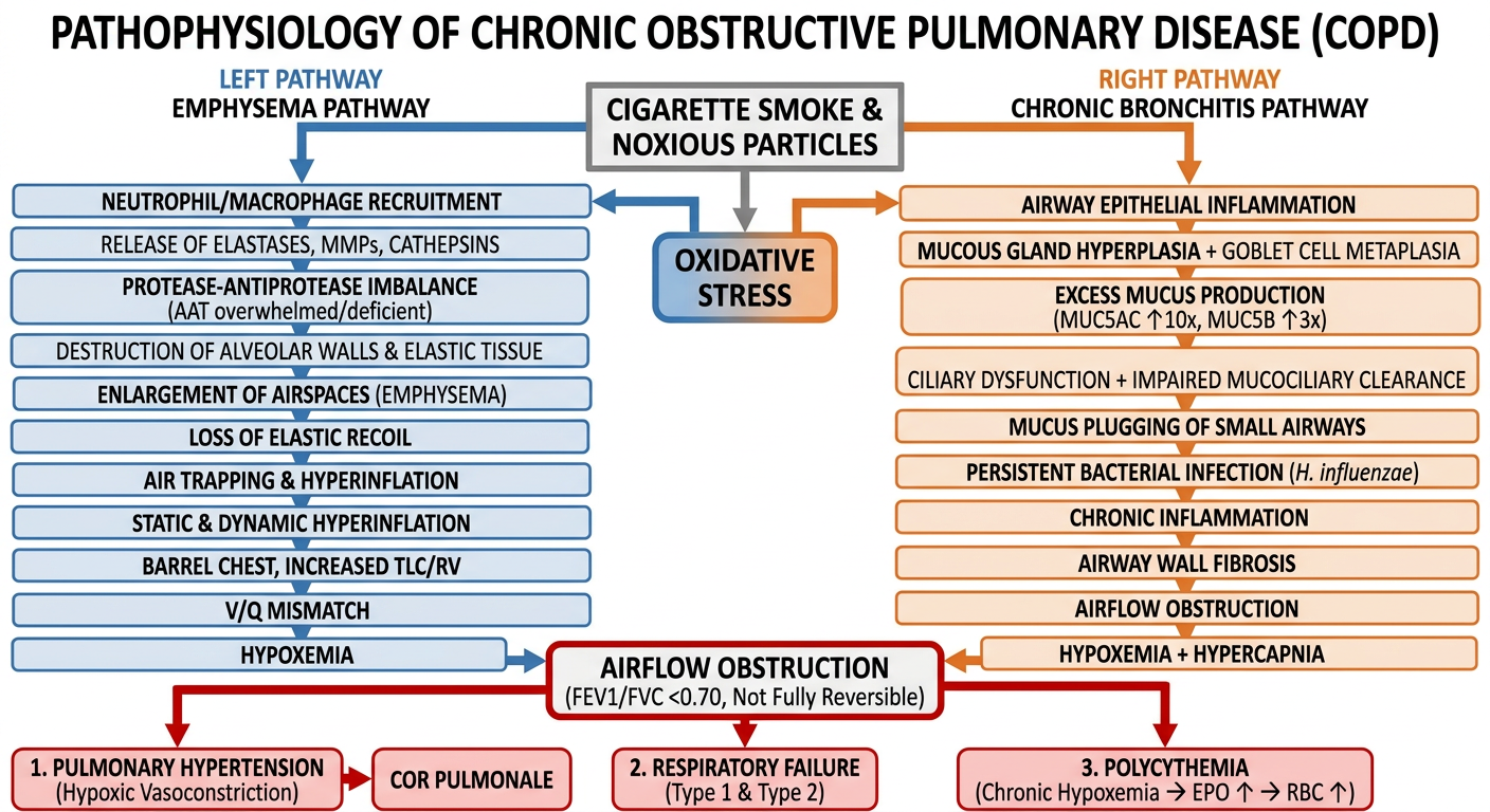
COPD Pathophysiology - Schematic Flowchart
            CIGARETTE SMOKE + NOXIOUS PARTICLES (Primary Trigger)
                         +  OXIDATIVE STRESS
                              |
            ┌─────────────────┴──────────────────┐
            ▼                                    ▼
   ══ EMPHYSEMA PATHWAY ══            ══ CHRONIC BRONCHITIS PATHWAY ══
   (Blue / Parenchymal)               (Orange / Airway)

Neutrophil + Macrophage             Airway Epithelial Inflammation
    Recruitment                              │
        │                                    ▼
        ▼                       Mucous Gland Hyperplasia
 Release of Elastases,          + Goblet Cell Metaplasia
 MMPs, Cathepsins                            │
        │                                    ▼
        ▼                        Excess Mucus (MUC5AC ↑10×
α1-Antitrypsin Overwhelmed           MUC5B ↑3×)
 (or Deficient in AAT-D)             + Ciliary Dysfunction
        │                                    │
        ▼                                    ▼
Alveolar Wall Destruction          Impaired Mucociliary
  (Loss of Elastin)                   Clearance
        │                                    │
        ▼                                    ▼
 Enlarged Airspaces              Mucus Plugging of Small
  (Emphysema)                         Airways
        │                                    │
        ▼                                    ▼
 Loss of Elastic Recoil          Persistent Infection
   ↑ Lung Compliance             (H. influenzae, etc.)
        │                                    │
        ▼                                    ▼
  Air Trapping &                  Chronic Airway
  Hyperinflation                  Inflammation
  ↑ TLC, ↑ RV, ↑ FRC                       │
        │                                    ▼
        ▼                          Airway Wall Fibrosis
 Dynamic Hyperinflation             + Bronchoconstriction
  (Exercise)                                │
        │                                    ▼
        ▼                         Small Airway Narrowing
  ↑ Work of Breathing             (Airflow Obstruction)
        │                                    │
        └────────────────┬───────────────────┘
                         ▼
         ══════════════════════════════════
          AIRFLOW OBSTRUCTION
          FEV1/FVC < 0.70 (Post-BD)
          NOT Fully Reversible
          V/Q Mismatch
         ══════════════════════════════════
                         │
         ┌───────────────┼───────────────┐
         ▼               ▼               ▼
   HYPOXEMIA        HYPERCAPNIA      AIR TRAPPING
  (PaO2 ↓)         (PaCO2 ↑)        Barrel Chest
         │               │
         ▼               ▼
 Hypoxic Pulm.    Respiratory
 Vasoconstriction  Acidosis
         │
         ▼
 Pulmonary HTN
         │
         ▼
   Cor Pulmonale          ┌─── Polycythemia
 (RVH → RV Failure)       │    (Hypoxemia → EPO↑
         │                │     → RBC↑)
         ▼                │
  Peripheral Edema, ◄─────┘
  Raised JVP, ↑P2
Key Molecular Cross-Links
| Mechanism | Role |
|---|---|
| Oxidative stress | Inactivates AAT, amplifies both emphysema and bronchitis |
| NF-κB activation | Drives inflammatory cytokine release (IL-8, TNF-α, LTB4) |
| Protease-antiprotease imbalance | Core of emphysematous destruction |
| Mucus hyperconcentration | Adhesion to airway surfaces, plugging small airways |
| Hypoxic vasoconstriction | Bridges airway disease to pulmonary hypertension |