
Details pf aspiration pneumonia
"aspiration pneumonia"[MeSH Terms] AND management
aspiration pneumonia chest X-ray radiograph
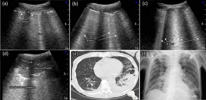
A multi-modal comparison of thoracic imaging findings in aspiration pneumonia, featuring point-of-care ultrasound (POCUS), computed tomography (CT), and chest X-ray (CXR). Panels (a)-(d) demonstrate a spectrum of lung ultrasound findings: (a) shows a single vertical B-line artifact originating from the pleural line; (b) displays multiple (≥3) B-lines; (c) shows confluent (≥5) B-lines creating a 'white lung' appearance; and (d) identifies sub-pleural consolidation (a hypoechoic area) and adjacent pleural effusion. Panel (e) is an axial CT scan of the chest showing significant consolidation and air bronchograms in the left lung, typical of aspiration pneumonia. Panel (f) is a posterior-anterior chest radiograph from the same patient, illustrating a loss of the silhouette of the left diaphragmatic arch behind the heart, indicating left lower lobe pathology. The collection serves as an educational comparison of diagnostic modalities for identifying pulmonary edema, interstitial syndrome, and consolidation in a clinical setting.
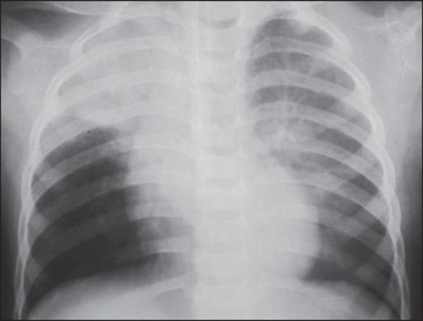
This diagnostic image is an anteroposterior (AP) chest X-ray of an 18-month-old pediatric patient. The radiograph demonstrates focal areas of increased density, specifically consolidative opacities located in the right upper lobe and the upper segment of the left lower lobe. These radiographic findings are highly characteristic of aspiration pneumonia or atelectasis occurring while the patient is in the supine (dorsal decubitus) position. The mediastinal shadow, cardiac silhouette, and diaphragmatic contours appear within normal limits for the patient's age. The skeletal structures, including the ribs and clavicles, show no fractures or deformities. The localized nature of the opacities in dependent lung segments provides significant clinical evidence for aspiration following a vomiting event, especially in the context of recovering from anesthesia. This visual serves as an educational example of typical radiographic patterns in pediatric aspiration-related pulmonary complications.
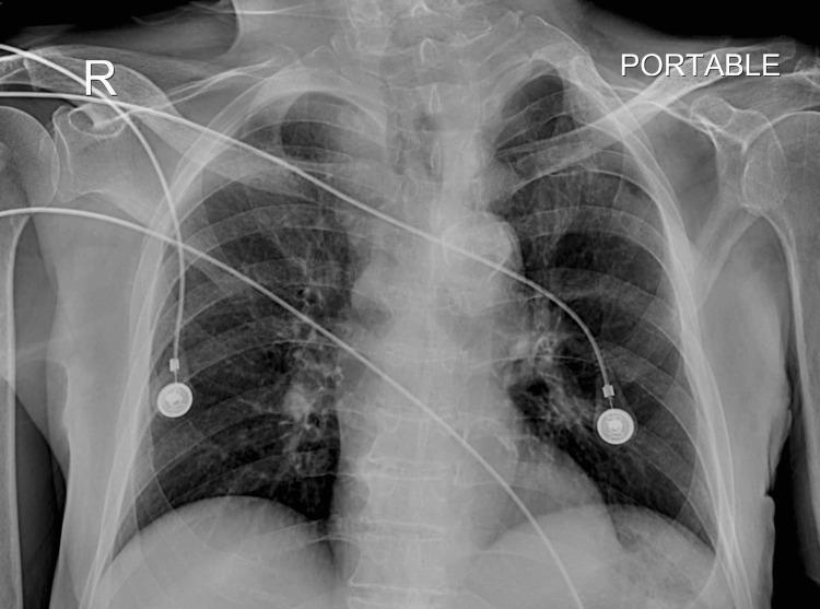
Anterior-posterior (AP) portable chest radiograph. The image depicts the thoracic cage, including the ribs, clavicles, scapulae, and thoracic spine. A right-side marker ('R') and 'PORTABLE' label are visible. The central mediastinal silhouette, including the heart and great vessels, appears within normal limits for this projection. The trachea is centrally located and patent, visible as a radiolucent column extending toward the carina. The lung parenchyma shows bilateral pulmonary vascular markings without clear evidence of consolidation or large pleural effusions. Several external medical monitoring devices are present: multiple radiopaque ECG leads and two circular cutaneous electrode pads are positioned over the chest wall. The radiograph is used clinically to confirm tracheal patency and assess for complications like aspiration pneumonia in patients with chronic dysphagia and instrumentation. The educational focus is on the interpretation of a standard follow-up bedside chest X-ray and identifying normal airway anatomy and medical monitoring hardware.
Aspiration Pneumonia
Definition
- Tintinalli's Emergency Medicine, p. 488: "Aspiration alone does not cause pneumonia" - the aspirated material must be infectious, large enough in volume, or sufficiently noxious to overwhelm pulmonary defences.
Epidemiology
- Approximately 50% of healthy adults aspirate small amounts during sleep; it is the pathological aspirate that matters.
- Leading cause of death in nursing home patients and the leading cause of transfer from nursing home to hospital. It is the second most common infection in nursing homes after UTI.
- Accounts for roughly 5-10% of community-acquired pneumonias.
- 2025 meta-analysis (PMID: 41062168) shows that aspiration pneumonia carries higher mortality and longer hospital stays than standard community-acquired pneumonia.
Pathophysiology
| Mechanism | Driver |
|---|---|
| Chemical | Gastric acid (pH <2.5), activated charcoal, contrast agents - cause direct mucosal inflammation and chemical pneumonitis |
| Bacterial | Oral flora, GI organisms - inoculate the lower airways and cause parenchymal infection |
- Goldman-Cecil Medicine, p. 1002: "Oftentimes the pneumonia is not infectious but rather a chemical response to gastric acid or gastric contents."
- Robbins Pathology (10th ed.), p. 478: "The resultant pneumonia is partly chemical due to the irritating effects of gastric acid and partly bacterial (from the oral flora). Typically more than one organism is recovered on culture."
Risk Factors
Conditions impairing protective airway reflexes:
- Alcohol use disorder, drug overdose, opioid use
- Seizures, stroke, coma, general anaesthesia
- Dementia, altered mental status
- Neuromuscular diseases (polymyositis-dermatomyositis, myasthenia gravis, ALS)
- Scleroderma (impaired esophageal peristalsis)
Anatomic / GI factors:
- Esophageal obstruction, stricture, or dysmotility
- Severe GERD, hiatal hernia
- Nasogastric tube, PEG tube, prolonged supine position
Oral hygiene factors (Tintinalli):
- 4 or more decayed teeth, gingivitis, periodontitis, dental plaque
- Medications reducing saliva (diuretics, anticholinergics, antipsychotics, levodopa)
Age-related:
- Decreased cough reflex threshold in elderly
- Decreased mucociliary function, decreased immune response
Microbiology
Community-acquired aspiration pneumonia:
- Anaerobes (classically): Bacteroides, Fusobacterium, Peptostreptococcus - common in periodontal disease, foul-smelling sputum
- Aerobes (increasingly recognised): Streptococcus pneumoniae, viridans streptococci, Haemophilus influenzae
Hospital-acquired / healthcare-associated aspiration:
- Gram-negative enteric bacilli (49% in one large study): Klebsiella pneumoniae, E. coli, Pseudomonas aeruginosa, Enterobacter, Serratia, Proteus
- Staphylococcus aureus (12%), including MRSA in risk groups
- Anaerobes (16%)
Important caveat (Fishman's Pulmonary, p. 1031): Specific anaerobic coverage is NOT routinely needed for all aspiration pneumonia. Anaerobes are of low individual virulence; they cause disease synergistically. Reserve anaerobic coverage for: (1) periodontal disease, (2) putrid/foul-smelling sputum, (3) necrotizing pneumonia or lung abscess on imaging.
Aspiration Syndromes (Full Spectrum)
| Syndrome | Mechanism | Key Feature |
|---|---|---|
| Acute airway obstruction | Mechanical blockage | Immediate, life-threatening |
| Aspiration pneumonitis | Chemical injury | Resolves in 24-48 h, no antibiotics |
| Aspiration pneumonia | Bacterial infection | Progressive, requires antibiotics |
| Lung abscess | Necrotizing infection | Thick-walled cavity, foul sputum |
| Exogenous lipoid pneumonia | Oil aspiration | Chronic, bilateral infiltrates |
| Diffuse aspiration bronchiolitis | Recurrent small-volume aspiration | Elderly, tree-in-bud pattern |
| Chronic interstitial fibrosis | Chronic chemical injury | Long-term, irreversible |
Clinical Features
Onset and presentation:
- Acute presentation: fever, productive cough (sometimes foul-smelling), dyspnea, tachypnea, pleuritic chest pain, leukocytosis
- Subacute in nursing home patients: decreased appetite, weakness, altered sensorium - vital signs may initially be normal or show only low-grade fever
- Hypoxemia develops over hours to days
- Physical examination: coarse rhonchi, rales, and wheezes in affected dependent lung zones
Complications:
- Lung abscess - most common serious complication; foul-tasting/smelling sputum is a hallmark
- Empyema - pus in the pleural space, requiring drainage
- ARDS - in severe cases with large volume aspiration
- Respiratory failure
Diagnosis
Imaging - Key Concept: Dependent Segments
| Patient position during aspiration | Affected segment |
|---|---|
| Supine | Superior segments of both lower lobes + posterior segments of upper lobes |
| Upright/semi-recumbent | Basal segments of lower lobes |
| Right-sided predominance | Due to the more vertical angle of the right mainstem bronchus |
Imaging findings:
- CXR: bronchopneumonic patchy infiltrates in dependent zones; thick-walled abscess with air-fluid level confirms lung abscess
- CT: more sensitive; shows ground-glass opacities, consolidation, cavitation, air bronchograms, tree-in-bud pattern
- Lung ultrasound: B-lines, sub-pleural consolidation, adjacent effusion
Laboratory:
- CBC: leukocytosis with left shift
- Blood cultures (before antibiotics, but do not delay treatment)
- Sputum Gram stain and culture (if obtainable)
- Pulse oximetry; ABG if hypoxemic or severely ill
- No gold-standard diagnostic test - diagnosis is clinical inference in a patient with aspiration risk factors + compatible infiltrate in a dependent segment
Treatment
Step 1 - Stabilisation
- Assess airway, breathing, circulation
- Supplemental oxygen; consider NIV or intubation if respiratory failure
- Pulse oximetry; selective ABG
- Bronchodilators for aspiration-induced bronchospasm
Step 2 - Antibiotics
| Setting | Antibiotic Choice |
|---|---|
| Community-acquired, otherwise healthy | Narrow-spectrum: amoxicillin-clavulanate or ceftriaxone ± metronidazole |
| Community-acquired, standard | Clindamycin, OR ampicillin-sulbactam, OR moxifloxacin, OR carbapenem |
| Healthcare-associated / hospital-acquired | Third-generation cephalosporin OR fluoroquinolone OR piperacillin-tazobactam OR carbapenem |
| MRSA risk factors | Add vancomycin or linezolid |
| Lung abscess / necrotising / periodontal disease | Ensure anaerobic coverage (clindamycin or metronidazole + beta-lactam) |
- Treatment duration: typically 7-14 days; prolonged (3-6 weeks) for lung abscess, empyema, or necrotizing pneumonia
- Empyema requires pleural drainage in addition to antibiotics
Step 3 - Dysphagia Management
- All elderly patients with pneumonia, recent stroke, or degenerative neurologic disease should have a formal swallow evaluation by a speech-language pathologist (SLP)
- Formulate an individualised feeding/swallowing strategy
- Bedside cough/gag reflex assessment alone is unreliable for screening aspiration risk
Step 4 - Prevention
- Improve oral hygiene (major modifiable risk factor)
- Head-of-bed elevation (>30-45 degrees) for bed-bound and tube-fed patients
- Behavioural modification for GERD/scleroderma (fundoplication in refractory cases)
- Reduce/rotate medications that impair swallowing or reduce salivary flow
- Chlorhexidine oral decontamination in ventilated patients to reduce VAP risk
- Minimise unnecessary prolonged supine positioning
Pathology
- Macroscopic: Patchy bronchopneumonia in dependent segments; necrotizing pneumonia in severe cases; thick-walled abscesses with foul-smelling contents in established lung abscess
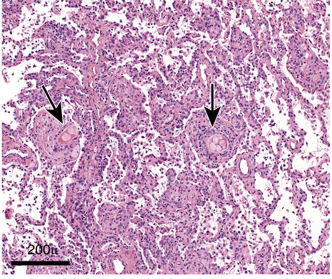
- Microscopic: Exudative alveolar infiltrate with neutrophils; areas of necrosis; foreign body giant cell granulomas with multinucleated cells in microaspiration (poorly formed, non-necrotizing)
- Robbins Pathology: "This type of pneumonia is often necrotizing, pursues a fulminant clinical course, and is a frequent cause of death."
Prognosis
- High mortality compared to CAP - particularly in elderly nursing home patients
- Predictors of poor outcome: advanced age, healthcare setting, MRSA, gram-negative organisms, lung abscess, respiratory failure
- 2025 meta-analysis (PMID: 41062168): aspiration pneumonia is associated with significantly higher hospital mortality and longer LOS vs. CAP
Key Distinctions Summary
| Feature | Aspiration Pneumonitis | Aspiration Pneumonia |
|---|---|---|
| Aspirate | Sterile gastric acid | Oropharyngeal bacteria |
| Onset | Rapid (within hours) | Gradual (days) |
| pH of aspirate | <2.5 | Variable |
| Fever | Low-grade | High, progressive |
| Resolution | 24-48 h without antibiotics | Requires antibiotics |
| Infiltrate | Bilateral, basal | Dependent segment, often unilateral early |
| Antibiotics | Not routinely needed | Essential |
Pathophysiology of aspiration pneumonia
"aspiration pneumonia"[MeSH Terms] AND pathophysiology
aspiration pneumonia histopathology lung alveoli neutrophil inflammation
Pathophysiology of Aspiration Pneumonia
Overview
- Fishman's Pulmonary Diseases & Disorders, p. 1195: "The nature of the pneumonia that develops depends on the inoculum and the host response."
Part 1 - Failure of Normal Lung Defences
| Defence Mechanism | How it protects |
|---|---|
| Glottic closure (cricopharyngeal-laryngeal muscles) | Seals the airway during swallowing |
| Cough reflex | Expels material that breaches the glottis |
| Mucociliary escalator | Moves particles/bacteria trapped in mucus up and out |
| Alveolar macrophages | First-line phagocytosis of aspirated bacteria |
| Surfactant proteins (SP-A, SP-D) | Opsonise bacteria; modulate inflammatory response |
| Secretory IgA | Neutralises pathogens in airway secretions |
- Goldman-Cecil Medicine, p. 973: "Any disturbance of these protective mechanisms can result in aspiration injury to the lungs. An altered level of consciousness can impair normal swallowing and suppress the cough reflex."
Part 2 - The Swallowing Mechanism and Its Failure (Dysphagia)
- Oropharyngeal phase: Chewing → tongue propels bolus → larynx elevates and moves anteriorly → epiglottis deflects → glottis closes → bolus passes the UES
- Esophageal phase: Peristalsis + relaxation of lower esophageal sphincter → food enters stomach
- Acute stroke: 40-70%
- Parkinson's disease: 52-82%
- Alzheimer's disease: 84%
- Elderly institutionalised patients: >60%
Part 3 - The Cough Reflex and Substance P
Part 4 - Chemical Arm (Aspiration Pneumonitis - Mendelson's Syndrome)
Triggers:
- Gastric fluid with pH < 2.5
- Large volume aspirate
- Particulate food matter (even if pH > 2.5, particulate matter dramatically amplifies the inflammatory response)
- Tube feeds and blood do NOT cause chemical pneumonitis (higher pH)
Two-Phase Injury Model (Fishman's, p. 1196):
- Acid causes a direct caustic burn of airway epithelium and alveolar cells
- Epithelial and type I pneumocyte injury → breakdown of the alveolar-capillary barrier
- Capillary leak → noncardiogenic pulmonary oedema (increased permeability oedema)
- Injury is analogous to a chemical burn - Goldman-Cecil: "Acid rapidly injures airway epithelial and alveolar cells; within hours, cells become dysfunctional and capillary leak occurs, resulting in profound noncardiogenic pulmonary edema."
- Type II pneumocyte dysfunction → surfactant depletion → alveolar collapse and increased surface tension
- Damaged epithelium releases pro-inflammatory cytokines: TNF-α, IL-8, IL-1β
- Neutrophil recruitment begins at 3-4 h and peaks at 4-6 h
- Neutrophils produce:
- Reactive oxygen species (ROS): via NADPH oxidase (primary source) and xanthine oxidase
- ROS activate NF-κB → amplifies TNF-α and IL-8 production
- Proteases (serine proteases, elastase, MMP) → degrade alveolar matrix
- Neutrophil Extracellular Traps (NETs): decondensed DNA-histone-granule protein complexes that cause collateral lung damage
- The intensity of alveolar neutrophil infiltration correlates directly with the severity of acute lung injury
Progression to ARDS:
Chemical burn → capillary leak → alveolar flooding → surfactant loss → atelectasis → V/Q mismatch → hypoxaemia → neutrophil influx → NF-κB/cytokine storm → diffuse alveolar damage → ARDS
Part 5 - Bacterial Arm (Aspiration Pneumonia Proper)
Step 1 - Oropharyngeal Colonisation
- Normal oral flora: viridans streptococci, anaerobes
- In healthy adults the lower airways are sterile because small microaspirations are cleared by mucociliary action + macrophages
- Pathogenic colonisation occurs with: poor oral hygiene, dental plaque, periodontal disease, hospitalisation, PPI/H2 blocker use (raises gastric pH → gram-negative gastric colonisation → retrograde pharyngeal colonisation), enteral feeding, advanced age
Step 2 - Inoculation of the Lower Airways
- Small inocula are cleared by alveolar macrophages
- Large inocula overwhelm macrophage capacity → bacterial replication begins
Step 3 - Innate Immune Response
- Pattern recognition receptors (TLRs, NLRs) on alveolar macrophages recognise bacterial PAMPs (LPS, peptidoglycan, flagellin)
- NF-κB activation → TNF-α, IL-1β, IL-6, IL-8 release
- IL-8 is the major chemokine attracting neutrophils from the pulmonary capillaries
- Massive neutrophil influx into alveoli → exudative alveolar filling
- Neutrophils and macrophages release proteases, ROS → tissue damage
- Complement activation → opsonisation of bacteria and MAC-mediated lysis
Step 4 - Bacterial Synergy (Anaerobic-Aerobic Co-Infection)
- Why anaerobes alone are of low virulence
- Why polymicrobial infection is the rule in aspiration pneumonia
- Why anaerobic-specific coverage is only needed in specific subsets (periodontitis, foul sputum, necrotising pneumonia)
Step 5 - Tissue Injury and Necrotisation
- Severe cases progress to necrotising pneumonia: liquefactive necrosis of lung parenchyma
- Cavities form → lung abscess (thick-walled, air-fluid level on imaging)
- Spread to pleural space → empyema
- Bacteraemic spread → sepsis
Part 6 - Role of Gastric pH and Colonisation
| Gastric condition | Effect |
|---|---|
| Normal acidic stomach (pH <2) | Sterile; aspirate causes chemical pneumonitis only |
| Antacids / H2 blockers / PPIs | Raised gastric pH → gram-negative bacterial gastric overgrowth → aspirate is now both chemical AND bacterial |
| Enteral feeding / gastroparesis | Same as above |
| Small bowel obstruction | Bile reflux + bacterial overgrowth → severe mixed injury |
- Fishman's: "Colonization of the gastric contents by potentially pathogenic organisms may occur when the gastric pH is increased by the use of antacids, H2 receptor blockers, or proton pump inhibitors... In these circumstances, the pulmonary inflammatory response is likely to result from both bacterial infection and the inflammatory response of the gastric particulate matter."
Part 7 - Segment Distribution (Gravitational Pathophysiology)
| Patient's position during aspiration | Dependent segments affected |
|---|---|
| Supine | Posterior segments of upper lobes + superior segments of lower lobes |
| Upright/semi-recumbent | Basal segments of lower lobes |
| Right-sided predominance | Right mainstem bronchus is more vertical and direct continuation of trachea |
Part 8 - Histopathology
| Finding | Significance |
|---|---|
| Neutrophilic alveolar exudate | Active bacterial infection / chemical injury |
| Type II pneumocyte hyperplasia | Reparative response after type I cell loss |
| Alveolar macrophage accumulation | Phagocytic response to aspirated material |
| Organising pneumonia pattern | Sub-acute/healing phase |
| Foreign body giant cell reaction | Response to food particles, lipid, or other foreign material |
| Necrotising changes | Severe infection; progression to abscess |
| Hyaline membranes | Diffuse alveolar damage / ARDS pattern in fulminant cases |
Integrated Pathophysiological Sequence
PREDISPOSING CONDITION
(stroke, dementia, intoxication, sedation, dysphagia)
↓
FAILURE OF AIRWAY PROTECTIVE REFLEXES
(impaired swallowing, reduced cough threshold, reduced substance P)
↓
ASPIRATION of oropharyngeal / gastric contents
↓
┌──────────────────────┬───────────────────────┐
CHEMICAL ARM BACTERIAL ARM MECHANICAL
(gastric acid pH <2.5) (colonised oral flora) (large bolus)
↓ ↓ ↓
Direct burn of Inoculation of Airway
alveolar epithelium lower airways obstruction
↓ ↓
Capillary leak TLR/NF-κB activation
Oedema ↓
Surfactant loss Cytokines: TNF-α, IL-8
↓ ↓
Neutrophil influx ←──── Neutrophil recruitment
(3-6 h peak) from capillaries
↓ ↓
NADPH oxidase ROS Proteases / Phagocytosis
NETs, proteases ↓
↓ ─────────────────
ALVEOLAR DAMAGE → Polymicrobial synergy
↓ ↓
V/Q mismatch Bacterial replication
Hypoxaemia Tissue invasion
↓ ↓
┌── ARDS (severe) Necrotising pneumonia
│ ↓
└──────────────────► Lung abscess / Empyema / Sepsis
Key Molecular Players
| Mediator | Role |
|---|---|
| TNF-α | Master pro-inflammatory cytokine; recruits neutrophils, activates endothelium |
| IL-8 (CXCL8) | Primary neutrophil chemoattractant into alveoli |
| NF-κB | Central transcription factor amplifying the inflammatory cascade |
| NADPH oxidase | Major ROS source in activated neutrophils |
| Neutrophil Extracellular Traps (NETs) | DNA-histone-granule protein complexes; antimicrobial but also cause collateral lung damage |
| Substance P | Mediates cough reflex; depleted in elderly patients with aspiration pneumonia |
| Surfactant SP-A/SP-D | Opsonise bacteria; modulate macrophage responses; depleted in injury |
| Complement (C3b, MAC) | Opsonisation and bacterial lysis |
Chext xray pattern of aspiration pneumonia
aspiration pneumonia chest X-ray consolidation dependent segments
aspiration pneumonia CT scan tree in bud consolidation lower lobe
Chest X-Ray Pattern of Aspiration Pneumonia
The Fundamental Principle: Gravity-Driven Distribution
- Harrison's Principles of Internal Medicine (2025): "Chest x-rays show consolidation in dependent pulmonary segments: in the basilar segments of the lower lobes if the patient has aspirated while upright and in either the posterior segment of the upper lobe (usually on the right side, given that the right mainstem bronchus has a more vertical orientation) or the superior segment of the lower lobe if the patient has aspirated while supine."
Segment Distribution by Patient Position
| Patient position at time of aspiration | Affected segments |
|---|---|
| Supine (most common - bedbound, comatose, post-op) | Superior (apical) segments of both lower lobes + Posterior segments of upper lobes |
| Upright / semi-recumbent | Basal segments of the lower lobes |
| Right-sided predominance in all positions | Right mainstem bronchus is wider, shorter, and more vertically oriented (more direct continuation of trachea) |
Why Right-Sided?
Classic CXR Patterns - From Early to Late
1. Patchy Bronchopneumonic Consolidation (Most Common)
- Heterogeneous, patchy, ill-defined opacities in dependent segments
- Does NOT conform to a single lobe (unlike lobar pneumonia from pneumococcus)
- Bronchopneumonic distribution - involves the bronchi and surrounding parenchyma
- Can be unilateral (early/milder) or bilateral (larger volume aspiration)
- Air bronchograms may be present within consolidation
2. Small Centrilobular Nodules
- Small nodules (3-5 mm) scattered around airways in dependent zones
- Represent inflammation centred on small bronchioles (bronchiolitis component)
- Best seen on CT; may not be visible on plain CXR
3. Volume Loss / Atelectasis
- Consolidation may be accompanied by ipsilateral volume loss
- Trachea and mediastinum shift toward the affected side
- Right lower lobe is the single most commonly affected lobe
Radiographic Image: Aspiration Pneumonia (Murray & Nadel's, eFigure 46.4)
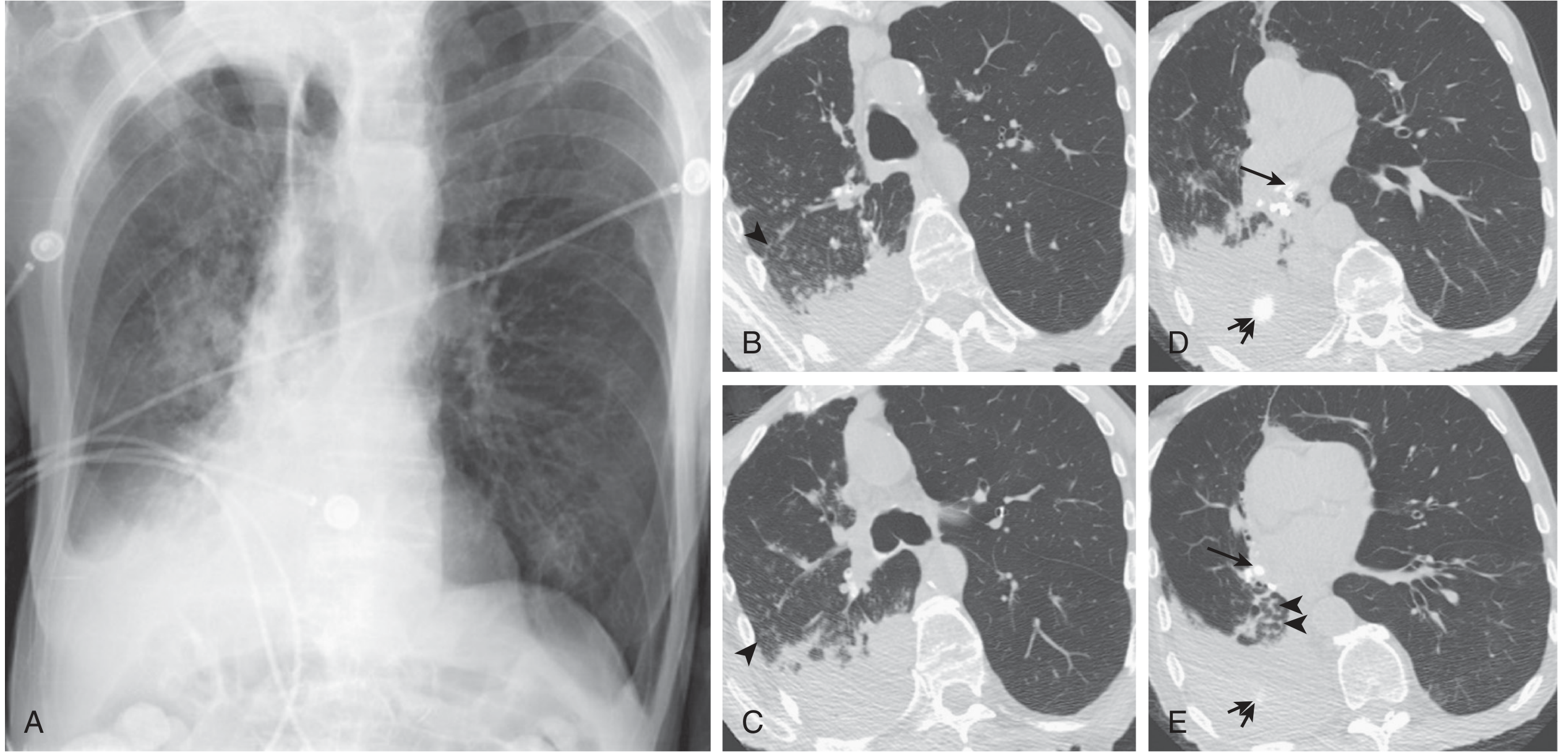
CT Scan Findings (More Sensitive Than CXR)
| CT Finding | Significance |
|---|---|
| Patchy consolidation in dependent segments | Hallmark finding |
| Tree-in-bud opacities | Mucus/exudate in small airways + bronchioles; recurrent aspiration |
| Ground-glass opacity (GGO) | Early inflammatory change; partial airspace filling |
| Centrilobular nodules | Bronchiolitis component |
| Air bronchograms | Airspace consolidation with patent large airways |
| Bilateral distribution | Large-volume or chemical aspiration |
| Crazy paving pattern | Lipoid pneumonia (oil/mineral aspiration) - GGO + interlobular septal thickening |
| Bronchiectasis | Chronic/recurrent aspiration |
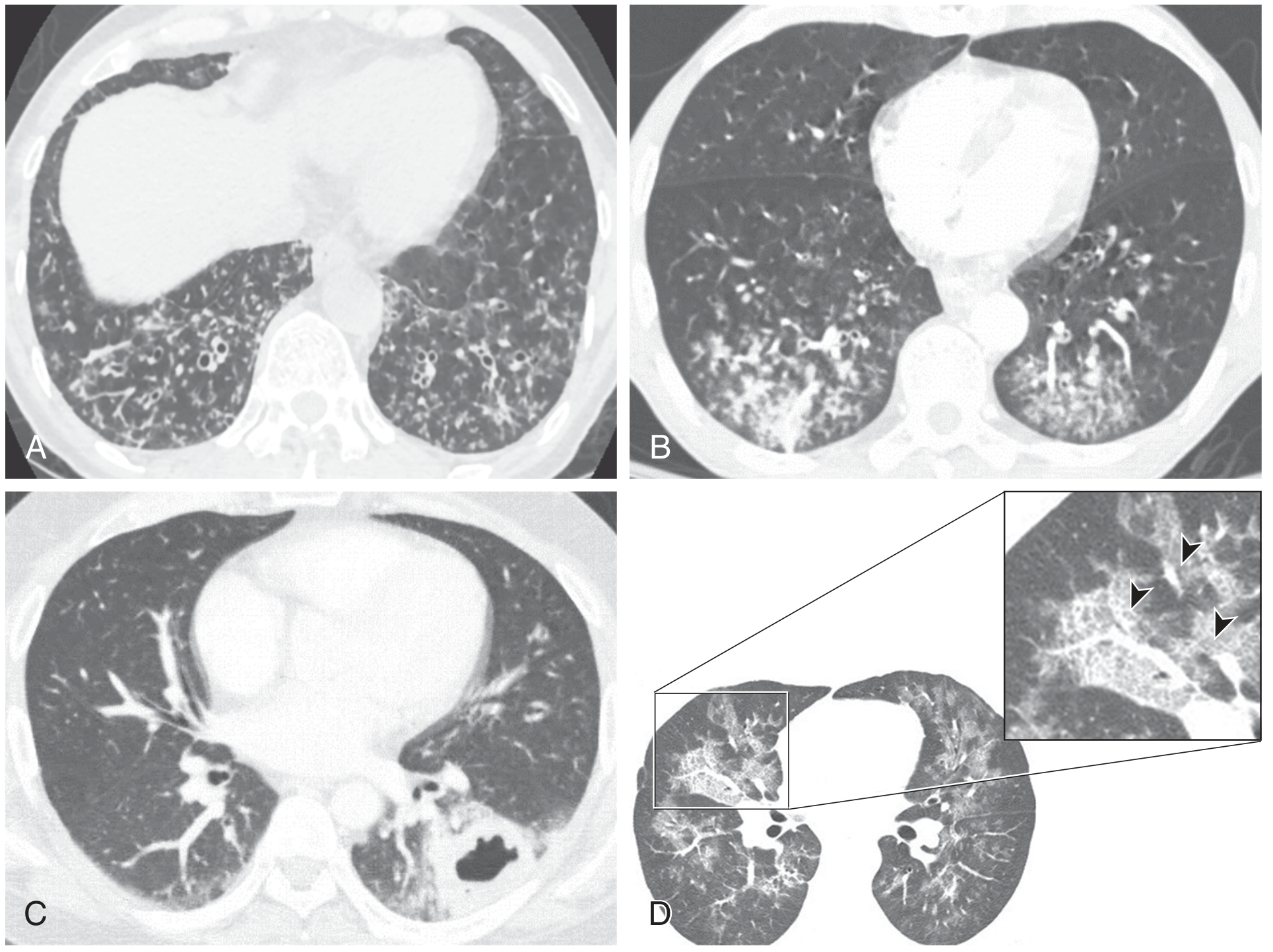
Complications on Imaging
Lung Abscess
- Thick-walled cavity with irregular internal lining
- Air-fluid level (pathognomonic of cavitation with necrotic debris)
- Surrounding infiltrate/consolidation
- Located in the same dependent aspiration-prone segments
- Round area of low attenuation (fluid-density content) surrounded by consolidation
- Air-fluid level within the cavity
- Ragged/shaggy inner wall (distinguishes abscess from malignancy)
- Does NOT compress surrounding lung (unlike empyema, which is oblong + compresses lung)
- Reactive lymph node enlargement may be seen
| Feature | Lung Abscess | Empyema | Malignant Cavity |
|---|---|---|---|
| Shape | Round | Oblong/lenticular | Variable |
| Wall | Thick, ragged/shaggy inner wall | Smooth inner wall | Nodular, wall >16 mm |
| Effect on lung | Necrosis (no compression) | Compresses adjacent lung | Variable |
| Air-fluid level | Yes | Sometimes | Rare |
| Location | Parenchymal | Pleural space | Any |
Other Complications Visible on Imaging
| Complication | CXR Finding |
|---|---|
| Pleural effusion | Blunting of costophrenic angle; layering on decubitus views |
| Empyema | Loculated pleural opacity; "split pleura" sign on CT |
| ARDS | Bilateral diffuse airspace opacification ("whiteout") |
| Pneumothorax | (rare) from necrotising pneumonia or abscess rupture |
Multi-Modal Imaging: POCUS + CT + CXR
The Silhouette Sign
| Silhouette obliterated | Lobe affected |
|---|---|
| Right heart border | Right middle lobe |
| Left heart border | Lingula |
| Right hemidiaphragm | Right lower lobe |
| Left hemidiaphragm | Left lower lobe |
Aspiration Pneumonitis (Mendelson's) vs. Aspiration Pneumonia - Radiographic Differences
| Feature | Aspiration Pneumonitis | Aspiration Pneumonia |
|---|---|---|
| Onset of CXR changes | Within 1-2 hours | 24-48+ hours after aspiration |
| Distribution | Bilateral, diffuse - can progress to "whiteout" | Dependent, unilateral or bilateral |
| Pattern | Bilateral alveolar opacities; can resemble pulmonary oedema | Patchy bronchopneumonic consolidation |
| Evolution | Clears in 24-72 h (if no superinfection) | Persists, may progress; cavitation possible |
| Complications | ARDS | Lung abscess, empyema |
Diffuse Aspiration Bronchiolitis (Chronic Recurrent Aspiration)
- Widespread centrilobular nodules
- Diffuse tree-in-bud opacities - bilateral, predominantly lower lobe
- May show mild bronchiectasis
- Fishman's: "Diffuse aspiration bronchiolitis presents with radiographic evidence of widespread centrilobular and tree-in-bud opacities"
Summary: Key Radiographic Points
- Right lower lobe - single most commonly affected lobe on CXR
- Posterior segments of upper lobes + superior segments of lower lobes - dominant in supine patients
- Basal lower lobes - dominant in upright/semi-recumbent patients
- Pattern: patchy bronchopneumonia (not lobar consolidation)
- Bilateral and right-sided predominance in larger-volume aspirations
- Lung abscess = thick-walled cavity with air-fluid level in a dependent segment - strongly suggests aspiration aetiology
- Tree-in-bud on CT = small airway involvement; seen in both acute and chronic aspiration
- No pathognomonic pattern - Grainger & Allison: "The pattern is very variable, dependent on the quantity and quality of aspirated material"
Anaerobic pneumonia the same as aspiration
"anaerobic pneumonia"[MeSH Terms] AND aspiration
"aspiration pneumonia" AND anaerobic AND microbiology
Are Anaerobic Pneumonia and Aspiration Pneumonia the Same?
Understanding the Relationship
ASPIRATION PNEUMONIA (broad clinical syndrome)
│
├── Chemical arm (aspiration pneumonitis / Mendelson's)
│ → No infection; sterile gastric acid injury
│
└── Bacterial arm
├── Aerobic gram-negative bacilli (most common in hospital)
├── Aerobic gram-positives (S. pneumoniae, S. aureus, viridans strep)
└── ANAEROBIC organisms ← "Anaerobic pneumonia" lives here
→ Fusobacterium nucleatum
→ Bacteroides / Prevotella species
→ Peptostreptococcus
→ (often polymicrobial - mixed anaerobic + aerobic)
- Murray & Nadel's: "Anaerobic infections are the second most common cause of severe aspiration pneumonia, after aerobic gram-negative bacilli, and are often polymicrobial infections."
Historical Context
Three Distinct Aspiration-Associated Anaerobic Syndromes
| Syndrome | Timing | Character | Dominant feature |
|---|---|---|---|
| Chemical pneumonitis | Within hours | Sterile initially; may be complicated by anaerobes | Acute hypoxaemia, dry cough, ARDS risk |
| Aspiration pneumonia | Days | Mixed aerobic-anaerobic infection | Fever, purulent sputum, bronchopneumonic infiltrate |
| Anaerobic pleuropneumonia | 1-2+ weeks | Predominantly anaerobic; necrotising | Putrid sputum, cavitation, empyema, weight loss |
Key Differences: Anaerobic Pneumonia vs. Aspiration Pneumonia (General)
| Feature | Anaerobic Pneumonia (Classic) | Aspiration Pneumonia (Broad) |
|---|---|---|
| Causative organisms | Strictly/predominantly anaerobes | Mixed - aerobes often dominant (especially hospital-acquired) |
| Onset | Subacute / indolent - days to weeks | Can be acute (hours-days) or subacute |
| Sputum | Putrid, foul-smelling (pathognomonic of anaerobic) | Purulent but NOT necessarily foul |
| Course | Slow, constitutional symptoms | Variable; can be rapid |
| Weight loss / malaise | Prominent (weeks of illness) | Less prominent early on |
| Necrotising tendency | High - lung abscess, empyema are hallmarks | Present in severe cases; less universal |
| Dental disease | Strongly associated | Associated but not always present |
| Setting | Often community, alcoholism, seizure | Both community and hospital (especially elderly, nursing home) |
| Lung abscess | Common (anaerobes in 93% of lung abscesses) | A complication, not the rule |
| Response to penicillin | Classically good response | Depends on organisms; hospital-acquired may need broader coverage |
The Organisms: What Makes Anaerobic Pneumonia Distinctive
| Organism | Notes |
|---|---|
| Fusobacterium nucleatum | Most virulent; can cause disease alone; associated with Lemierre syndrome |
| Prevotella melaninogenica (formerly Bacteroides melaninogenicus) | Very common; responsible for characteristic foul odour |
| Peptostreptococcus spp. | Gram-positive anaerobic cocci; common in mixed infections |
| Bacteroides spp. (usually NOT B. fragilis) | Component of polymicrobial infections |
| Porphyromonas spp. | Periodontal pathogen |
- Fishman's: "In lung abscesses, anaerobes are recoverable from up to 93% of patients."
- Harrison's: "A putrid lung abscess refers to cases with foul-smelling breath, sputum, or empyema; these manifestations are essentially diagnostic of an anaerobic lung abscess."
The Synergy Principle
- Mixed cultures are the rule, not the exception
- Broad-spectrum coverage targeting the polymicrobial mix is needed
The Putrid Sputum Clue
Putrid (foul-smelling) sputum = anaerobic infection until proven otherwise
Why Anaerobic Coverage Is NOT Routine for All Aspiration Pneumonia
- Most patients with aspiration pneumonia have risk factors for aerobic gram-negative colonisation
- Anaerobes are of low individual virulence
- Community-acquired aspiration in otherwise healthy patients does NOT routinely require specific anti-anaerobic therapy
- Anaerobic coverage is specifically indicated when:
- Periodontal disease is present
- Putrid/foul-smelling sputum is observed
- Necrotising pneumonia on imaging
- Lung abscess or empyema is present
What Conditions Favour Anaerobic Over Aerobic Aspiration Pneumonia?
| Favours anaerobic dominance | Favours aerobic dominance |
|---|---|
| Community-acquired | Hospital-acquired / healthcare-associated |
| Poor dental hygiene / periodontitis | Good oral hygiene |
| Alcoholism, seizures (prolonged unconsciousness) | Recent antibiotics / PPIs (select resistant aerobes) |
| Slow, subacute onset | Acute, rapid onset |
| Foul-smelling sputum | No putrid smell |
| Lung abscess / empyema | Straightforward consolidation |
| No recent hospitalisation | Nursing home / ICU patient |
Spectrum Summary: From Aspiration to Anaerobic Pleuropneumonia
ACUTE (hours) CHRONIC (weeks)
│ │
Chemical Aspiration Mixed aerobic- Anaerobic
pneumonitis → pneumonia → anaerobic → pleuropneumonia
(Mendelson) (acute bacterial) pneumonia + lung abscess
+ empyema
Sterile Aerobes + Equals parts Predominantly
gastric acid anaerobes aerobes / anaerobes
(both) anaerobes Putrid sputum
Weeks of symptoms
Recent Evidence
- The microbiological profile of aspiration pneumonia has shifted over recent decades
- Aerobic gram-negative bacteria dominate in healthcare-associated cases
- Modern 16S rRNA molecular techniques detect anaerobes in a higher proportion of CAP than classical culture methods suggested - meaning anaerobes may play a larger background role than previously appreciated
Bottom Line
| Concept | Verdict |
|---|---|
| Are they the same? | No |
| Is aspiration a prerequisite for anaerobic pneumonia? | Yes - virtually always |
| Is all aspiration pneumonia anaerobic? | No - aerobes often dominate, especially in hospital settings |
| When does anaerobic pneumonia specifically arise? | When large inocula of gingival bacteria (periodontitis, poor hygiene) are aspirated, especially with prolonged unconsciousness |
| What clinically distinguishes it? | Putrid sputum, indolent course, necrotising tendency, lung abscess/empyema |
| Does all aspiration pneumonia need anti-anaerobic antibiotics? | No - only when specific features (putrid sputum, abscess, periodontitis, empyema) are present |