
Significance of pulse pressure
Pulse pressure is the difference between systolic and diastolic blood pressure (e.g., 120/80 mmHg gives a pulse pressure of 40 mmHg). Normal range is 40-60 mmHg. A wide pulse pressure (>60 mmHg) suggests increased stroke volume or decreased aortic compliance, seen in aortic regurgitation, atherosclerosis, and thyrotoxicosis. A narrow pulse pressure (<25 mmHg) indicates reduced stroke volume, seen in cardiac tamponade, severe aortic stenosis, or heart failure.
Searching sources...
Reading File
Finding Sources
Searching PubMed
"pulse pressure" cardiovascular risk significance
Reading File
Reading File
Reading File
Finding Sources
Reading File
Reading File
Reading File
Reading File
Now I have comprehensive material from multiple authoritative textbooks. Let me compile the full answer.
Loading Image
Here is a complete, textbook-grounded answer on the significance of pulse pressure.
Significance of Pulse Pressure
Definition
Pulse pressure (PP) is the difference between systolic blood pressure (SBP) and diastolic blood pressure (DBP).
PP = SBP - DBP
Example: If BP = 120/80 mmHg, then PP = 40 mmHg.
- Normal range: 30-40 mmHg (commonly stated up to 60 mmHg).
Physiological Determinants
Two major factors determine pulse pressure (Guyton & Hall, Medical Physiology):
- Stroke volume output - the greater the stroke volume, the more blood that must be accommodated in the arterial tree per beat, raising pulse pressure.
- Compliance (distensibility) of the arterial tree - the less compliant (stiffer) the arteries, the greater the pressure rise for the same stroke volume.
This is summarized as:
Pulse Pressure ≈ Stroke Volume / Arterial Compliance
Pulse pressure also serves as a surrogate for stroke volume: assuming arterial compliance is constant, the magnitude of PP directly reflects the volume ejected per beat. It contributes to mean arterial pressure (MAP) via the formula:
MAP = Diastolic Pressure + 1/3 Pulse Pressure
- Costanzo Physiology 7th Ed., p. 135; Guyton and Hall Medical Physiology, p. 189
Wide (High) Pulse Pressure (>60 mmHg)
A widened PP occurs when stroke volume increases OR arterial compliance falls.
| Condition | Mechanism |
|---|---|
| Aortic regurgitation | Blood flows back into LV in diastole → diastolic pressure falls to near zero; no dicrotic notch. PP can reach 80-100 mmHg |
| Patent ductus arteriosus | ≥50% of aortic blood shunts back into pulmonary circulation → very low diastolic pressure |
| Arteriosclerosis / aging | Stiff, noncompliant arteries → systolic pressure rises steeply for same stroke volume; PP may double |
| Aortic aneurysm | Loss of aortic wall elasticity |
| Anemia | Reduced blood viscosity + compensatory high CO |
| Hyperthyroidism | High cardiac output state |
| Exercise | Increased stroke volume |
| Arteriovenous fistula | Run-off in diastole lowers DBP |
| Increased intracranial pressure | Cushing reflex - wide PP with bradycardia |
| Fever / sepsis (hyperdynamic) | Vasodilation + high CO |
- Roberts and Hedges' Clinical Procedures in Emergency Medicine, p. 43
Narrow (Low) Pulse Pressure (<25 mmHg)
A narrow PP signals reduced stroke volume or very high peripheral resistance.
| Condition | Mechanism |
|---|---|
| Cardiac tamponade | Pericardial fluid compresses heart → severely reduced stroke volume. Classic finding in tamponade |
| Aortic stenosis | Reduced outflow → low SBP; PP falls significantly |
| Severe heart failure | Poor LV contractility → low stroke volume |
| Hypovolemia / hemorrhagic shock | Reduced venous return → reduced stroke volume. An early, sensitive indicator of reduced central blood volume even before conventional vital signs change |
| Increased peripheral resistance | As in early septic shock or severe vasoconstriction |
Evidence suggests that a reduction in pulse pressure is an early indicator of reduced central blood volume even when traditional vital signs (SBP, SpO2) remain stable - making it a sensitive bedside hemodynamic marker.
- Roberts and Hedges' Clinical Procedures in Emergency Medicine, p. 43
Pulse Pressure Variation (PPV) in Critical Care
In mechanically ventilated patients, the respiratory variation in pulse pressure (PPV) is used to predict fluid responsiveness:
-
PP is directly proportional to stroke volume; mechanical ventilation cyclically alters RV preload.
-
A PPV >13% predicts fluid responsiveness (i.e., the patient is on the ascending limb of the Frank-Starling curve and will respond to a fluid bolus with increased cardiac output).
-
This is more reliable than static measures like CVP or PCWP.
-
Roberts and Hedges' Clinical Procedures in Emergency Medicine, p. 348-352
Pulse Pressure as a Cardiovascular Risk Marker
From Fuster and Hurst's The Heart (15th Ed.) and Goldman-Cecil Medicine:
- Systolic BP rises steadily with age; diastolic BP peaks around age 50 and then falls - resulting in a progressively widening PP in older adults.
- PP is a measure of aortic stiffness (vascular stiffness).
- In the Framingham Heart Study, widened PP (reflecting lower DBP at any SBP ≥120 mmHg) was associated with significantly increased coronary heart disease risk in adults aged 50-79 years, and PP was not inferior to either SBP or DBP alone for predicting CHD in this age group.
- The REGARDS study showed PP was independently associated with incident coronary heart disease even after adjustment for SBP.
- Factors associated with lower DBP and wide PP: older age, female sex, and diabetes.
- In chronic kidney disease (CKD), vascular calcification causes reduced compliance → widened PP → increased cardiac afterload and LVH.
Abnormal Pulse Pressure Contours (Summary Diagram)
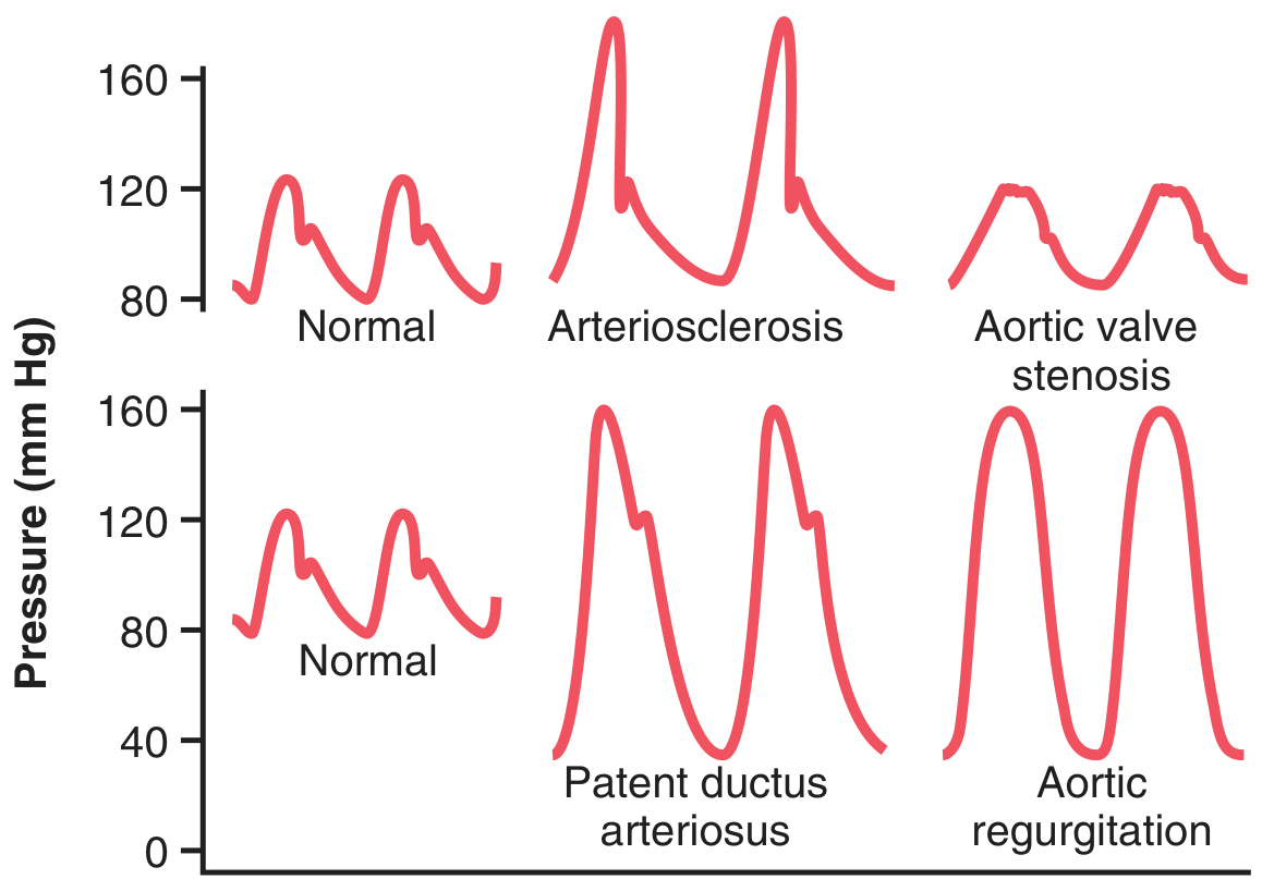
Figure: Aortic pressure pulse contours comparing normal with arteriosclerosis (doubled PP), aortic stenosis (narrow PP), patent ductus arteriosus (very wide PP, low diastole), and aortic regurgitation (near-zero diastolic pressure, absent dicrotic notch). - Guyton and Hall Textbook of Medical Physiology, p. 189
Summary Table
| Feature | Wide PP (>60 mmHg) | Narrow PP (<25 mmHg) |
|---|---|---|
| Stroke volume | High | Low |
| Arterial compliance | Low (stiff) | Normal/not the issue |
| Key causes | AR, PDA, arteriosclerosis, thyrotoxicosis, anemia, AV fistula, ICP | Tamponade, AS, HF, hypovolemia, shock |
| Clinical implication | Vascular stiffness, high CV risk | Reduced preload/output - urgent cause |
Recent evidence note: A 2024 meta-analysis (PMID 39039284) explored outcome-driven thresholds for pulse pressure amplification, confirming that PP metrics carry prognostic weight beyond simply SBP or DBP alone, particularly in older hypertensive populations.
Central blood. Volume means blood flow to brain
No — "central blood volume" does not mean blood flow to the brain. It refers to the volume of blood within the thoracic (chest) compartment — specifically the heart, pulmonary circulation, and great vessels.
Searching sources...
Finding Sources
Reading File
Clarification: What "Central Blood Volume" Actually Means
Central blood volume (CBV) refers to the volume of blood within the heart chambers and pulmonary circulation (the thoracic compartment). It does NOT mean blood flow to the brain.
Specifically, it includes:
- Right atrium and right ventricle
- Pulmonary arteries, pulmonary capillaries, pulmonary veins
- Left atrium and left ventricle
This represents approximately ~360 mL (heart) + ~440 mL (pulmonary) = ~800 mL, or roughly 16% of total blood volume in a 70 kg person.
- Medical Physiology (Boron & Boulpaep), p. 654
Why It Matters
Central blood volume functions as the filling reservoir for the left heart. Think of it as the "upstream tank" that determines how much preload the left ventricle receives.
| Central Blood Volume | Effect |
|---|---|
| Reduced (hypovolemia, hemorrhage, venodilation) | Less preload → lower stroke volume → lower pulse pressure and BP |
| Increased (fluid overload, heart failure, venoconstriction) | More preload → increases stroke volume (up to a point) |
In left-sided heart failure, the careful regulation of central blood volume breaks down, leading to pulmonary congestion and edema.
- Medical Physiology (Boron & Boulpaep), p. 654
The Confusion - "Central" Does Not Mean "Brain"
The word "central" here means central circulation (thorax), as opposed to "peripheral" circulation (limbs, splanchnic vessels, skin). The brain is supplied by the cerebral circulation, which is a separate concept governed by cerebral autoregulation.
| Term | Meaning |
|---|---|
| Central blood volume | Blood in heart + pulmonary vessels (thoracic reservoir) |
| Cerebral blood flow (CBF) | Blood flowing to the brain (~750 mL/min, ~15% of CO) |
| Cerebral perfusion pressure | MAP minus intracranial pressure |
Clinical Context (from the previous discussion on Pulse Pressure)
In the Roberts & Hedges emergency medicine text, the phrase you saw - "reduction in central blood volume" - meant that the patient's thoracic filling volume was dropping (due to bleeding or fluid loss), which causes a narrowed pulse pressure even before SBP visibly falls. That is why a falling pulse pressure is an early warning sign: it reflects reduced stroke volume from the heart's depleted filling reservoir, not reduced brain blood flow.
- Roberts and Hedges' Clinical Procedures in Emergency Medicine, p. 43
This is a shared conversation. Sign in to Orris to start your own chat.