
Bristol stool score7 + mild reddish stool with mild tensmus
bloody diarrhea tenesmus acute colitis management
dysentery bloody stool colitis differential diagnosis
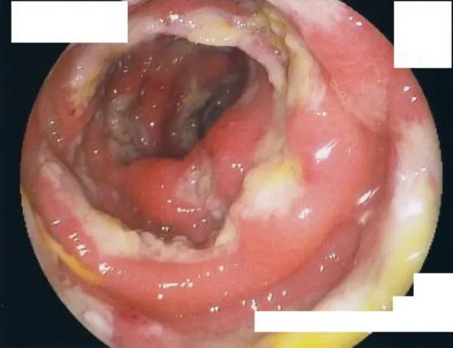
Endoscopic colonoscopic image of the right colon (cecum and ascending colon) from an immunocompromised patient with amebic dysentery. Modality: colonoscopy with white-light endoscopy; gold standard diagnosis remains colonic biopsy. Macroscopic mucosal changes are characterized by multiple punctate ulcers measuring 2–10 mm, with adherent yellowish exudate and surrounding erythema. In advanced areas, ulcers may coalesce to create large necrotic cavitations with relative sparing of normal intervening mucosa. The mucosa appears inflamed, edematous, friable, and susceptible to superficial bleeding on instrumentation. This distribution and morphology—predominantly in the right colon—are classic for amebic colitis, though overlap with other infectious or inflammatory conditions occurs. Endoscopic assessment guides biopsy targeting and therapeutic planning; histopathology or molecular testing corroborates Entamoeba histolytica infection. Diagnostic significance is high in immunocompromised individuals because amebic disease can progress to fulminant colitis and require surgical resection. Differential diagnoses include bacterial colitis (Salmonella, Shigella, Campylobacter), cytomegalovirus colitis, invasive fungal infections, and Crohn’s disease. Clinically, patients may present with abdominal pain, fever, diarrhea with blood, and weight loss. This image serves educational purposes, illustrating characteristic colonic ulcers with exudate and necrosis, supporting differential diagnosis and biopsy site selection, and highlighting the severe endoscopic phenotype that can precipitate colectomy.
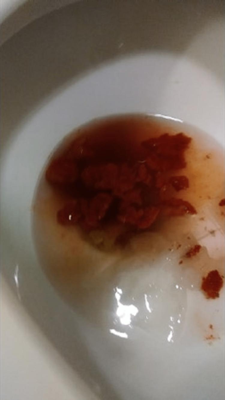
This clinical photograph shows a stool sample within a white porcelain toilet bowl, exhibiting a distinct reddish-orange discoloration. The stool appears as fragmented, semi-solid clumps with a highly saturated, uniform red-orange pigment. The surrounding water displays a gradient of the same reddish-orange tint, suggesting the leaching of pigments from the fecal matter into the liquid. Clinically, this presentation is significant for the differential diagnosis of hematochezia (bloody stool) versus food-induced pseudohematochezia. The artificial, overly saturated hue and fragmented consistency, combined with a patient history of ingesting specific dyed snacks (such as Flamin' Hot Cheetos), point toward a dietary etiology rather than an acute gastrointestinal bleed. This image serves as an educational tool for gastroenterology and primary care in identifying non-pathological causes of stool discoloration that mimic lower gastrointestinal hemorrhage.
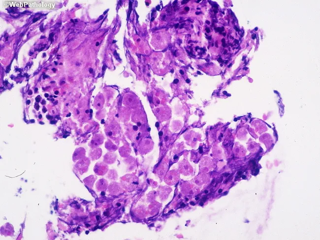
This is a bright-field light microscopy image of a colonic mucosal biopsy stained with Hematoxylin and Eosin (H&E), at high magnification (approximately 400x total). The histology shows inflamed colonic mucosa with an inflammatory exudate in the lamina propria and within the ulcerated mucosal surface. Within this exudate, round to oval trophozoites with granular cytoplasm and a small, centrally placed karyosome are evident, consistent with Entamoeba histolytica. These trophozoites may contain ingested erythrocytes, a characteristic diagnostic clue for invasive amebiasis. Alveolated borders (pseudopodia) are not readily visible in all fields but may be inferred by amoeboid cytoplasm. The surrounding tissue demonstrates acute and chronic inflammatory infiltrates, with neutrophils, macrophages, and occasional eosinophils; focal mucosal necrosis and ulceration are possible. The image highlights tissue invasion by protozoa within a colitis framework, supporting a diagnosis of amebic colitis rather than noninvasive Entamoeba species. Clinically, this histopathologic pattern correlates with dysentery, abdominal pain, and potential systemic spread if untreated. Diagnostic significance lies in identifying Entamoeba histolytica trophozoites in tissue, guiding antiparasitic therapy (metronidazole or tinidazole) followed by luminal eradication therapy. This image is relevant for educational pathology teaching, differential diagnosis of colitis, and research on invasive enteric protozoa. This educational image aids clinical correlation.
Bristol Stool Type 7 + Reddish (Blood-tinged) Stool + Mild Tenesmus
What This Triad Means
| Feature | Significance |
|---|---|
| BSS Type 7 (entirely liquid, watery) | Severe secretory or inflammatory diarrhea |
| Reddish / blood-tinged stool | Lower GI bleeding - points to colonic source (hematochezia) |
| Tenesmus (urge to defecate with little passage) | Rectal/colonic inflammation, particularly proctitis or distal colitis |
Inflammatory vs. Noninflammatory Diarrhea
| Feature | Inflammatory (this case) | Noninflammatory |
|---|---|---|
| Stool character | Bloody, small-volume | Large-volume, watery |
| Site | Colon | Small intestine |
| Fecal leukocytes | Frequently present | Small numbers only |
| Key symptoms | Tenesmus, fever, cramps | Nausea, vomiting, generalized cramps |
Differential Diagnosis
1. Shigella (Bacillary Dysentery) - Most likely
- The classic cause of dysentery: watery diarrhea progressing to bloody stools + tenesmus + fever
- Only 50-100 organisms needed to cause infection (most efficient enteric pathogen)
- S. flexneri and S. dysenteriae cause more severe bloody dysentery; S. sonnei (75% of US cases) causes mostly watery diarrhea
- Rosen's Emergency Medicine: "Patients with dysentery have grossly bloody diarrhea, tenesmus, and constitutional symptoms such as fever, nausea, vomiting, headache, or myalgias"
2. Entamoeba histolytica (Amebic Dysentery)
- Suspect if: travel history, poor sanitation exposure, immunocompromised
- Can present identically to bacterial dysentery
- Tenesmus is a recognized feature - Harrison's: "Tenesmus...may be a feature of cases with proctitis, as in shigellosis or amebiasis"
3. Campylobacter jejuni
- Second most common cause of bloody diarrhea (after Shigella)
- Profuse watery diarrhea + fever initially, may become bloody; associated with poultry/raw milk
- Campylobacter can occasionally cause frank dysentery
4. Salmonella spp.
- Inflammatory diarrhea, may be bloody; longer incubation (>16 h), associated with eggs/poultry/dairy
5. Enterohemorrhagic E. coli (EHEC / O157:H7)
- Grossly bloody diarrhea, often WITHOUT fever (important distinguishing feature)
- Associated with undercooked ground beef; can progress to HUS
- No tenesmus typically - but can occur
6. Clostridioides difficile Colitis
- Consider if recent antibiotic use or hospitalization
- Can cause pseudomembranous colitis with bloody stool
7. Ulcerative Colitis / IBD Flare
- Proctitis pattern: "frequent painful bowel movements with blood, pus, and mucus; tenesmus often prominent" (Sleisenger & Fordtran)
- Consider if history of persistent/intermittent episodes without infection risk factors
8. Ischemic Colitis
- Typically in older patients with vascular risk factors; sudden onset bloody diarrhea, left-sided pain
Red Flags Requiring Urgent Evaluation
- Fever > 38.5°C / systemic toxicity
- Signs of dehydration (orthostatic hypotension, tachycardia)
- Severe abdominal pain
- Immunocompromised state
- Age extremes (very young or elderly)
- Symptoms lasting > 3-5 days without improvement
- Suspected EHEC (risk of HUS - avoid antibiotics in this case)
Workup
- Stool culture + stool PCR multiplex panel (covers Shigella, Salmonella, Campylobacter, EHEC, C. diff, etc.) - preferred; PCR sensitivity 93-97%
- Fecal leukocytes or fecal calprotectin - confirms inflammatory pattern
- Stool microscopy for ova and parasites (if amebiasis suspected)
- Stool for C. diff toxin (if antibiotic history)
- CBC, CMP, CRP - assess severity, dehydration, systemic response
- Sigmoidoscopy/colonoscopy if IBD suspected or not resolving
Diagnostic evaluations, whenever possible, should precede empiric antibiotic therapy. (Harrison's Principles of Internal Medicine, 22e)
Management
| Priority | Action |
|---|---|
| Rehydration | Oral rehydration therapy (ORS) first-line; IV fluids if severely dehydrated |
| Antibiotics (empiric) | Ciprofloxacin 500 mg BID x 3-5 days for suspected bacterial dysentery; adjust based on sensitivities |
| Amebic dysentery | Metronidazole 500-750 mg TID x 7-10 days, followed by luminal agent (paromomycin) |
| C. diff | Oral vancomycin or fidaxomicin; stop precipitating antibiotic |
| Avoid antimotility agents alone | Loperamide alone (without antibiotics) is contraindicated in dysentery - prolongs fever/bacteremia; safe only when co-administered with appropriate antibiotics |
| EHEC | Antibiotics are controversial / potentially harmful - supportive care; monitor for HUS |
Pathognomonic Images
Bottom Line
- Rosen's Emergency Medicine, Concepts and Clinical Practice
- Harrison's Principles of Internal Medicine, 22e
- Sleisenger and Fordtran's Gastrointestinal and Liver Disease
- Symptom to Diagnosis: An Evidence-Based Guide, 4th Edition
Without fever but mucus and foul smell is present 7-8 times per day since 3 days and pain in abdomen and hip region during defecation and pain in bilateral calf muscle continuous no signs of dehydration appetite is good
Refined Clinical Analysis: Updated Symptom Profile
- BSS Type 7 (watery) + blood + mucus + foul smell
- Afebrile (no fever)
- 7-8 stools/day x 3 days (subacute onset)
- Abdominal pain + pain in hip/perineal region during defecation
- Bilateral calf muscle pain (continuous)
- No dehydration, appetite preserved
Most Likely Diagnosis: Intestinal Amebiasis (Amebic Colitis)
Key Differentiating Table (Amebic vs. Bacterial Dysentery)
| Feature | Amebic Colitis | Bacterial Dysentery |
|---|---|---|
| Fever >38°C | Minority | Majority |
| Onset | Gradual (days-weeks) | Abrupt |
| Duration | >7 days | 2-7 days |
| Fecal occult blood | 100% | 40% |
| Abdominal pain | 12-80% | ~50% |
| Weight loss | Common | Unusual |
| Mucus in stool | Present | Present |
Why the Other Features Fit
| Symptom | Explanation |
|---|---|
| Foul-smelling stool | Tissue necrosis and proteolytic activity of E. histolytica trophozoites; also seen in Giardia ("rotten egg/sulfur" smell) |
| Mucus in stool | Mucosal invasion and inflammation of colon; classic in amebic/inflammatory colitis |
| Hip/perineal pain during defecation | Rectal and pelvic floor inflammation - referred pain from sigmoid colon + rectal involvement; tenesmus with sacral/perineal radiation is characteristic of colonic/rectal pathology |
| Bilateral calf muscle pain (continuous) | This is a significant finding - discussed separately below |
| No dehydration, appetite preserved | Consistent with non-toxigenic, non-secretory colitis; amebic colitis does not produce enterotoxins causing massive fluid loss like cholera or ETEC |
The Bilateral Calf Muscle Pain - Important Differentials
1. Reactive Arthritis / Enteric Reactive Arthropathy
- Occurs 1-4 weeks after enteric infections (Shigella, Salmonella, Campylobacter, Yersinia)
- Can cause asymmetric oligoarthritis, enthesitis, and muscle pain / myalgia in lower limbs
- Though classically post-infectious, can appear during the infection
2. Electrolyte Imbalance (Hypokalemia / Hypomagnesemia)
- Even without clinical dehydration, significant potassium and magnesium losses occur with frequent diarrhea (7-8x/day)
- Hypokalemia causes muscle cramps, weakness, and myalgia - especially prominent in calf muscles
- This is the most immediately likely cause of the calf pain in this context
3. Campylobacter Infection with Myalgia
- Campylobacter jejuni characteristically causes prominent myalgia as a prodromal and accompanying symptom
- Calf muscle pain is well-recognized in Campylobacter enteritis
- However, Campylobacter usually causes fever
4. Parasitic Causes (Less likely but possible)
- Trichinella can cause myositis with calf involvement + GI symptoms - but usually follows ingestion of undercooked pork and causes fever
- Giardia causes foul-smelling, greasy stool but no blood
5. Unrelated Vascular/MSK Cause
- Deep vein thrombosis (DVT) - bilateral is less typical
- Peripheral vascular disease
- Must be examined clinically
Revised Differential Diagnosis (Most to Least Likely)
| Rank | Diagnosis | Key Supporting Features |
|---|---|---|
| 1 | Amebic colitis (E. histolytica) | Afebrile, gradual onset, blood + mucus + foul stool, no dehydration |
| 2 | Campylobacter enteritis | Myalgia/calf pain prominent, bloody mucoid stool - but usually febrile |
| 3 | C. difficile colitis | Mucus, foul-smelling stool, abdominal pain - ask about recent antibiotic use |
| 4 | Ulcerative colitis (IBD) | Chronic relapsing course, tenesmus, bloody mucoid stool, no fever common in mild-moderate disease |
| 5 | Yersinia enterocolitica | Abdominal pain, diarrhea, can mimic appendicitis, hip pain may reflect mesenteric adenitis |
Investigations to Order (Priority)
Immediate
- Stool microscopy x3 (fresh warm stool) - look for E. histolytica trophozoites with ingested RBCs (pathognomonic)
- Stool antigen test (ELISA) for E. histolytica - sensitivity ~85-95%, specificity ~93-99%; preferred over microscopy
- Stool culture - for Shigella, Salmonella, Campylobacter, Yersinia
- Multiplex stool PCR panel - detects most bacterial and parasitic causes simultaneously
Blood Work
- Serum electrolytes (K+, Mg2+, Na+) - check for hypokalemia/hypomagnesemia explaining calf pain
- CBC with differential - eosinophilia suggests parasitic cause
- CRP, ESR - assess inflammatory burden
- Serum anti-amebic antibodies (IgG) - useful if >7 days of illness; supports E. histolytica diagnosis
- Liver function tests - rule out early amebic liver abscess (right lobe pain, elevated ALP)
If Initial Tests Negative
- Colonoscopy with biopsy - flask-shaped amebic ulcers are pathognomonic; also diagnoses IBD
Treatment
If Amebic Colitis Confirmed (or strongly suspected)
- Metronidazole 500-750 mg three times daily x 7-10 days (oral or IV if severe)
- Alternative: Tinidazole 2g once daily x 3 days (better tolerated, higher cure rate)
- Paromomycin 25-35 mg/kg/day in 3 divided doses x 7 days
- Alternative: Diloxanide furoate 500 mg TID x 10 days
Tissue amebicide alone is insufficient - luminal agent must follow to prevent recurrence and ongoing transmission.
Supportive Care
- Oral rehydration - even without dehydration signs, maintain fluid intake given 7-8 stools/day
- Potassium supplementation if hypokalemia found - will also resolve calf pain
- Avoid antimotility agents (loperamide) - contraindicated in invasive/inflammatory diarrhea
Do NOT use
- Steroids (risk of fulminant amebic colitis if misdiagnosed as IBD without first ruling out amebiasis)
Critical Caution
If IBD (ulcerative colitis) is being considered, always rule out amebiasis first before starting steroids - glucocorticoids can precipitate fulminant, potentially fatal amebic colitis.
Summary
- Sleisenger and Fordtran's Gastrointestinal and Liver Disease
- Rosen's Emergency Medicine, Concepts and Clinical Practice
- Harrison's Principles of Internal Medicine, 22e
Aalegra
Drug name
Aalegra drug medication composition uses
Aalegra (Fexofenadine)
Drug Class
Composition
- Fexofenadine hydrochloride
- Available strengths: 30 mg, 60 mg, 120 mg, 180 mg
Mechanism of Action
Indications
| Indication | Dose | Duration |
|---|---|---|
| Seasonal allergic rhinitis (hay fever) - sneezing, runny/itchy nose, watery eyes | 120 mg once daily | As needed / seasonal |
| Perennial allergic rhinitis | 120 mg once daily | Continuous |
| Chronic idiopathic urticaria (hives) | 180 mg once daily | Until resolution |
| Children 6-11 years (allergic rhinitis/urticaria) | 30 mg twice daily | - |
| Children 2-11 years (urticaria) | 30 mg twice daily | - |
Pharmacokinetics
| Parameter | Value |
|---|---|
| Onset of action | ~1-3 hours |
| Peak effect | ~2-3 hours |
| Half-life | 11-15 hours |
| Dosing frequency | Once daily |
| Protein binding | ~60-70% (albumin, α1-acid glycoprotein) |
| Metabolism | Minimal hepatic metabolism (~5%) - excreted largely unchanged |
| Excretion | Feces (80%), urine (11%) |
Dosing
- Adults & children ≥12 years: 120 mg once daily (rhinitis) or 180 mg once daily (urticaria)
- Take with water on an empty stomach (or at least not with fruit juice)
- Tablets should be swallowed whole - do NOT crush or chew
Side Effects
- Headache
- Nausea, dyspepsia
- Dizziness (mild)
- Fatigue
- Drowsiness (rare - significantly less than 1st generation)
- Back pain, myalgia
Important Drug Interactions
| Interacting Drug | Effect |
|---|---|
| Erythromycin / Ketoconazole | Increase fexofenadine plasma levels (increased side effects) |
| Antacids (Al/Mg-containing) | Reduce fexofenadine absorption - separate by 2 hours |
| Fruit juices (orange, apple, grapefruit) | Reduce bioavailability up to 36% - take with water only |
| Rifampicin | Decreases fexofenadine levels |
Contraindications / Cautions
- Hypersensitivity to fexofenadine
- Renal impairment - dose reduce (120 mg starts as 60 mg once daily); drug is renally excreted
- Pregnancy - Category C (use only if benefit outweighs risk)
- Breastfeeding - not recommended (excreted in breast milk)
- Elderly - use with caution (renal clearance may be reduced)
Key Advantages Over 1st-Generation Antihistamines
- Non-sedating - safe to use while driving or operating machinery
- Once-daily dosing - improves compliance
- No anticholinergic effects - no dry mouth, urinary retention, constipation
- No cardiac toxicity (unlike terfenadine)
- Safe in patients with BPH, glaucoma (unlike older antihistamines)
Context Note (Relevance to Current Case)
- Dermatology 2-Volume Set, 5e
- Drugs.com - Allegra (fexofenadine)
- Cleveland Clinic - Fexofenadine
Repeated episode of diarrhea after cake and pastry consumption
Repeated Diarrhea After Cake and Pastry - Clinical Analysis
| Ingredient | Condition it Triggers |
|---|---|
| Milk / cream / butter | Lactose intolerance |
| Wheat flour | Celiac disease / Gluten sensitivity |
| High fructose corn syrup / sugar | Fructose malabsorption |
| Sorbitol / mannitol / xylitol (sugar-free varieties) | Sugar alcohol malabsorption |
| High fat content | Fat-induced gastrocolic reflex / exocrine pancreatic insufficiency |
| Eggs, nuts, wheat | IgE-mediated food allergy |
Differential Diagnosis (Most to Least Likely)
1. Lactose Intolerance (Most Common)
- Lactase enzyme deficiency → undigested lactose remains in intestinal lumen
- Acts as osmotic load drawing fluid into the bowel
- Colonic bacteria ferment lactose → gas (H2, CO2, methane), short-chain fatty acids → bloating, cramping, watery diarrhea
"Lactose is present in many dairy products but is also a hidden component of a vast number of processed foods." - Harrison's Principles of Internal Medicine, 22e
- Lactose exclusion diet (rapid and effective)
- Hydrogen breath test (>20 ppm rise above baseline = positive)
- Lactose tolerance test (serum glucose rise <20 mg/dL = positive)
- Lactose-free diet; lactase enzyme supplements (OTC) before dairy ingestion
- Calcium + Vitamin D supplementation if dairy eliminated
2. Celiac Disease (Gluten Enteropathy) - High Priority
- Immune-mediated T-cell response to gliadin (gluten component) in genetically susceptible individuals (HLA-DQ2/DQ8)
- Villous atrophy + crypt hyperplasia + intraepithelial lymphocytosis in duodenum/proximal jejunum
- Loss of absorptive surface → malabsorption of fat, carbohydrates, vitamins, iron, calcium
- Diarrhea, bloating, fatigue
- Iron deficiency anemia (most common presentation in adults)
- Folate / B12 deficiency
- Weight loss, steatorrhea (fatty, pale, floating stools)
- Dermatitis herpetiformis (pruritic blistering rash) in ~10%
"In adults, celiac disease manifests most commonly between 30 and 60 years of age with anemia (due to iron deficiency), diarrhea, bloating, and fatigue. Many cases escape clinical attention for extended periods because of atypical presentations." - Robbins & Kumar Basic Pathology
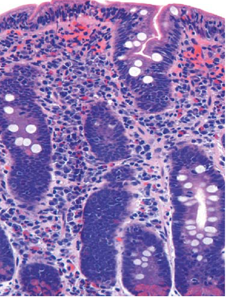
- Serology (first-line): IgA anti-tissue transglutaminase (anti-tTG IgA) - most sensitive; anti-endomysial antibody (EMA-IgA) - most specific; deamidated gliadin peptide (DGP) antibodies
- Duodenal biopsy (gold standard) - villous atrophy, crypt hyperplasia, increased intraepithelial lymphocytes
- Check total IgA level first (IgA deficiency causes false negatives)
- Strict lifelong gluten-free diet (wheat, barley, rye avoided)
- Monitor with repeat serology for dietary compliance
- Supplement iron, folate, B12, calcium, Vitamin D
3. Fructose / Sugar Alcohol Malabsorption
- Fructose absorbed by limited-capacity facilitated diffusion → easily overwhelmed by processed foods
- Sorbitol/mannitol/xylitol are poorly absorbed → osmotic diarrhea + fermentation in colon
- Sorbitol also worsens fructose malabsorption when both are present together
"Sugar alcohol malabsorption also may cause diarrhea: sorbitol, mannitol, and xylitol are poorly absorbed nonnutritive sweeteners...and may cause diarrhea when ingested in excess." - Sleisenger & Fordtran's GI and Liver Disease
4. Non-Celiac Gluten Sensitivity (NCGS)
- Diarrhea, bloating, fatigue with gluten ingestion
- Celiac serology negative, biopsy normal or near-normal
- No autoimmune mechanism confirmed
- May be partly due to fructan (poorly absorbable carbohydrate in wheat) rather than gluten itself
- Diagnosis: exclusion of celiac disease + food allergy, then gluten-free diet trial
5. IgE-Mediated Food Allergy
- True food allergy to wheat, eggs, milk, nuts (common cake ingredients)
- Symptoms within minutes to 2 hours: diarrhea + urticaria + angioedema + flushing
- More common in children; 1-2% of adults
- Diagnosis: Skin prick test, serum specific IgE (RAST), oral food challenge
- Treatment: Strict allergen avoidance; epinephrine auto-injector if anaphylaxis risk
6. Exocrine Pancreatic Insufficiency (EPI)
- High-fat content of cakes/pastries overwhelms reduced pancreatic lipase
- Steatorrhea (greasy, pale, foul-smelling, floating stools), abdominal cramps after fatty meals
- Associated with chronic pancreatitis, cystic fibrosis, previous pancreatic surgery
- Diagnosis: Fecal elastase-1 (low <100 µg/g), Sudan stain for fecal fat, fecal fat quantification
- Treatment: Pancreatic enzyme replacement therapy (PERT) with meals
Investigation Pathway
Repeated diarrhea after cake/pastry
          |
          ├── Check for anemia, iron deficiency → ? Celiac
          |
          ├── Bloating + gas predominantly → ? Lactose or Fructose
          |
          ├── Hives/urticaria also present → ? Food allergy
          |
          ├── Oily/pale/floating stools → ? EPI or Celiac
          |
          └── Sugar-free products specifically → ? Sugar alcohols
| Test | What It Diagnoses |
|---|---|
| Anti-tTG IgA + total serum IgA | Celiac disease screening |
| Hydrogen breath test (lactose) | Lactose intolerance |
| Hydrogen breath test (fructose) | Fructose malabsorption |
| Duodenal biopsy | Celiac (gold standard) |
| Fecal elastase-1 | Exocrine pancreatic insufficiency |
| Specific IgE / skin prick test | IgE food allergy |
| CBC, iron studies, folate, B12 | Nutritional deficiencies from malabsorption |
| Food diary + elimination diet | Practical first step for any food intolerance |
Practical First Step
- First eliminate lactose (easiest, most common)
- If symptoms persist, eliminate gluten
- Simultaneously keep a food and symptom diary
- Send celiac serology before starting gluten-free diet (diet normalizes antibodies and biopsy)
Key rule: Always do celiac blood tests first before going gluten-free - otherwise results become falsely negative.
- Harrison's Principles of Internal Medicine, 22e
- Robbins & Kumar Basic Pathology
- Sleisenger and Fordtran's Gastrointestinal and Liver Disease
- Yamada's Textbook of Gastroenterology, 7e
Medicine what to choose after consuming pastry and cake causing Diarrhea
Medicines for Post-Cake/Pastry Diarrhea
STEP 1 - Immediate Symptomatic Relief (Any Cause)
A. Oral Rehydration Solution (ORS) - FIRST PRIORITY
- Replace fluid and electrolytes lost with diarrhea
- Even without obvious dehydration, ongoing stool losses deplete sodium, potassium, bicarbonate
- WHO ORS formula or commercial sachets (Electral, Pedialyte)
- Drink after every loose stool
B. Loperamide (Imodium) - Symptomatic Anti-diarrheal
- 4 mg initially, then 2 mg after each loose stool
- Maximum 16 mg/day
- Stop if no improvement within 48 hours
- Active bloody diarrhea / dysentery (inflammatory colitis)
- Suspected infectious colitis (Shigella, C. diff)
- In IBD: risk of toxic megacolon
- Avoid exceeding dose - FDA black box warning: overdose can cause cardiac arrhythmias (torsades de pointes)
STEP 2 - Cause-Specific Medicines
If Cause = Lactose Intolerance
| Medicine | How to Use |
|---|---|
| Lactase enzyme supplement (Lactaid, Dairy Ease) | Take with the first bite of lactose-containing food; available OTC |
| Calcium + Vitamin D supplement | If dairy is eliminated from diet |
| Loperamide | For breakthrough symptoms |
Best approach: Take lactase enzyme before eating cake/pastry with dairy. Prevents diarrhea entirely if lactose is the trigger.
If Cause = Celiac Disease / Gluten Sensitivity
| Medicine / Action | Notes |
|---|---|
| Strict gluten-free diet (primary treatment) | No specific drug; diet IS the medicine |
| Iron + Folate + B12 supplements | For deficiencies from malabsorption |
| Calcium + Vitamin D | Bone density protection |
| Loperamide | Short-term symptom control only |
No pill can substitute for a gluten-free diet in celiac disease. If gluten was accidentally consumed, loperamide + ORS provides symptomatic relief while the gut recovers.
If Cause = Fructose / Sugar Alcohol Malabsorption
| Medicine | Notes |
|---|---|
| Simethicone (Gas-X, Wind-Eze) | 40-125 mg after meals; reduces gas/bloating from bacterial fermentation of unabsorbed sugars |
| Loperamide | For diarrhea episodes |
| Low-FODMAP diet | Avoids fructose, sorbitol, mannitol, fructans |
If Cause = Fat Malabsorption / Exocrine Pancreatic Insufficiency (EPI)
| Medicine | Dose | Notes |
|---|---|---|
| Pancreatic enzyme replacement therapy (PERT) - Pancreatin / Creon | 25,000-40,000 units lipase with each meal | Taken at the start of the meal; replaces missing pancreatic enzymes |
| Fat-soluble vitamin supplements (A, D, E, K) | Daily | Lost due to fat malabsorption |
If Cause = IgE Food Allergy (rare, with hives/urticaria)
| Medicine | When to Use |
|---|---|
| Antihistamine (cetirizine, fexofenadine/Aalegra 120 mg) | Mild allergic reaction with diarrhea + itching/hives |
| Prednisolone 20-40 mg | Moderate allergic reaction |
| Epinephrine auto-injector (EpiPen) | Anaphylaxis - emergency use only |
Quick Reference Summary Table
| Trigger | Best Preventive Medicine | Best Rescue Medicine |
|---|---|---|
| Lactose (milk/cream/butter in cake) | Lactase enzyme before eating | Loperamide + ORS |
| Gluten (wheat flour) | Gluten-free diet (no drug) | Loperamide + ORS |
| Fructose/Sorbitol | Low-FODMAP diet | Simethicone + Loperamide |
| High fat | Eat smaller portions | Loperamide; if EPI: Pancreatin |
| Food allergy | Strict avoidance | Antihistamine; Epi if severe |
| Any acute episode | - | ORS always first |
Key Rules
- ORS before any anti-diarrheal - replace fluids first
- Lactase supplement taken with food is the most practical preventive strategy if lactose intolerance is suspected
- Never take loperamide if stool is bloody - this points to the dysentery picture discussed earlier, where it is contraindicated
- Identify and eliminate the trigger - medicines only treat symptoms, not the root cause
- Celiac disease requires serology testing before going gluten-free - don't self-treat with gluten-free diet until blood tests are done
Blood in stool after repeated episodes of loose stool