
Mechanism of innte and acquired immunity according to apurba sastry , give me an easy to retain conscise accurate answer
Note: Apurba Sastry's Essentials of Medical Microbiology is not in the textbook library, but the content below is drawn from authoritative sources (Cellular and Molecular Immunology, Jawetz, Robbins) and closely mirrors Sastry's approach - same concepts, same flow.
Innate vs. Acquired (Adaptive) Immunity
Quick Memory Hook
Innate = BORN with it, FAST, NON-SPECIFIC Acquired = LEARNED, SLOW first time, SPECIFIC + REMEMBERED
1. INNATE IMMUNITY (Non-specific / Natural)
Definition
Key Features (remember: GRIM)
| Feature | Detail |
|---|---|
| Germline-encoded | Receptors are fixed in DNA - no gene rearrangement needed |
| Recognizes PAMPs | Pathogen-Associated Molecular Patterns (e.g., LPS of gram-negatives, lipoteichoic acid of gram-positives, dsRNA of viruses, unmethylated CpG DNA of bacteria) |
| Immediate | Response in minutes-hours (0-96 hours) |
| Memory: NONE | Same response every time; no enhancement on re-exposure |
Components (3 layers)
- Skin (keratin layer, tight junctions)
- Mucus + cilia (mucociliary escalator)
- Antimicrobial peptides: Defensins (alpha and beta) and Cathelicidins - punch holes in microbial membranes
- Low pH (stomach acid, skin), lysozyme (tears, saliva), normal flora
- Neutrophils - first to arrive at infection; phagocytose and kill via ROS (oxidative burst), NETs
- Macrophages - tissue-resident sentinels; phagocytosis + cytokine production (TNF-alpha, IL-1, IL-6)
- Dendritic Cells (DCs) - bridges innate to adaptive; present antigens to T cells
- NK Cells - kill virus-infected cells and tumor cells (recognize "missing self" - cells lacking MHC-I)
- Mast Cells - release histamine; important in allergy and defense at barriers
- Eosinophils/Basophils - anti-parasitic functions
- Complement system (C1-C9): lysis, opsonization, chemotaxis (the 3 Ls - Lysis, Label/opsonize, Lure/chemotaxis)
- Acute phase proteins: C-reactive protein (CRP), Mannose-binding lectin (MBL), Serum amyloid A
- Cytokines: Type I interferons (IFN-alpha, IFN-beta) - antiviral defense
Pattern Recognition Receptors (PRRs)
- TLRs (Toll-Like Receptors): recognize extracellular/endosomal PAMPs (e.g., TLR4 recognizes LPS)
- NLRs (NOD-Like Receptors): cytosolic; form inflammasomes (activate IL-1beta and IL-18)
- RLRs (RIG-Like Receptors): detect cytosolic viral RNA
- Also recognizes DAMPs (Damage-Associated Molecular Patterns) from dead/injured host cells (e.g., HMGB1, uric acid crystals, ATP)
2. ACQUIRED IMMUNITY (Adaptive / Specific)
Definition
Key Features (remember: SMED)
| Feature | Detail |
|---|---|
| Specificity | Each lymphocyte has receptors for ONE antigen (clonal distribution) |
| Memory | Long-lived memory cells - basis of vaccination |
| Expansion | Clonal proliferation of antigen-selected lymphocytes |
| Diversity | ~10^7-10^9 distinct antigens can be recognized |
Two Branches
A. Humoral Immunity (B cells - antibody-mediated)
- B lymphocytes mature in bone marrow; activated by antigen (+/- T cell help)
- Differentiate into plasma cells → secrete antibodies (Ig)
- Antibody functions: Neutralization, Opsonization, Complement activation, ADCC
- Immunoglobulin classes: IgM (first responder), IgG (most abundant, crosses placenta), IgA (mucosal), IgE (allergy/parasites), IgD (B cell surface receptor)
B. Cell-Mediated Immunity (T cells)
- T lymphocytes mature in thymus; recognize antigen only when presented by MHC molecules on APCs
- CD4+ T helper cells (recognize MHC Class II):
- Th1 → activate macrophages (IFN-gamma) → kill intracellular bacteria/viruses
- Th2 → help B cells make antibodies; IL-4, IL-5, IL-13
- Th17 → mucosal/neutrophilic defense (IL-17)
- T-reg → suppress immune responses (IL-10, TGF-beta)
- CD8+ Cytotoxic T cells (CTL, recognize MHC Class I) → kill virus-infected cells and tumor cells via perforin/granzymes
3. Comparison Table (Exam Favorite)
| Feature | Innate | Acquired |
|---|---|---|
| Present at birth | Yes | No (develops after antigen contact) |
| Specificity | Broad (classes of microbes ~1000 PAMPs) | Narrow (individual antigens >10^7) |
| Receptors | Germline-encoded, invariant (TLR, NLR) | Somatic recombination (TCR, BCR) - diverse |
| Memory | None | Yes (memory B and T cells) |
| Speed | Minutes to hours | Days to weeks (primary); hours (secondary) |
| Key cells | Neutrophils, Macrophages, NK cells, DCs | B cells, T cells |
| Key molecules | Complement, defensins, interferons | Antibodies (Ig), cytokines |
| Self-nonself | Yes (recognizes healthy "self" signals) | Yes (clonal deletion of autoreactive cells) |
4. How They Connect (Critical Integration Point)
Microbe enters
      ↓
INNATE responds immediately → inflammation, complement, NK cells
      ↓
Dendritic cells pick up antigen → migrate to lymph nodes
      ↓
Present antigen to T cells (MHC-peptide + costimulation)
      ↓
ADAPTIVE response begins → T and B cell activation
      ↓
Antibodies + CTLs clear infection
      ↓
Memory cells remain → faster response on re-exposure
Memory Mnemonics
- Innate cells: "NMD NK" - Neutrophils, Macrophages, Dendritic cells, NK cells, Mast cells
- PRRs: "TEN" - TLRs, (N)LRs, RLRs
- Ig classes: "GAMED" - IgG, IgA, IgM, IgE, IgD
- T helper subtypes: "1-2-17-Reg" - Th1 (intracellular), Th2 (antibody/allergy), Th17 (extracellular bacteria/fungi), Treg (suppress)
- Antibody functions: "NOCA" - Neutralization, Opsonization, Complement activation, ADCC
Innate and acquired immunity mechanism
Mechanisms of Innate & Acquired Immunity
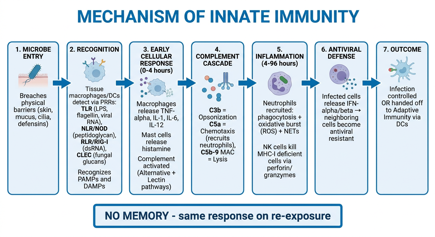
INNATE IMMUNITY - Mechanism
Step-by-step:
- Skin, mucus, cilia, low pH, lysozyme physically block microbe entry
- Antimicrobial peptides (defensins, cathelicidins) punch holes in microbial membranes
- Tissue-resident cells (macrophages, DCs, mast cells) detect microbes via Pattern Recognition Receptors (PRRs):
- TLR4 → recognizes LPS (gram-negative bacteria)
- TLR3/7/8 → recognizes viral RNA
- NOD1/NOD2 (NLRs) → detect bacterial peptidoglycan fragments in cytosol
- RIG-I (RLRs) → detects cytosolic viral dsRNA
- Recognizes PAMPs (microbial structures) and DAMPs (from damaged host cells)
- Macrophages → phagocytose + destroy via ROS, lysosomal enzymes; release TNF-α, IL-1, IL-6, IL-12
- Neutrophils → recruited by chemokines; phagocytosis + oxidative burst + NETs
- NK cells → kill cells lacking MHC-I (virus-infected/tumor cells) via perforin/granzymes
- Complement → activated by 3 pathways (Classical/Lectin/Alternative) → Opsonization (C3b) + Chemotaxis (C5a) + Lysis (MAC = C5b-9)
- Interferons (IFN-α, IFN-β) → released by infected cells → make neighboring cells antiviral resistant
- Cytokines (IL-1, TNF, IL-6) → fever, acute phase proteins (CRP, fibrinogen), vasodilation, leukocyte recruitment
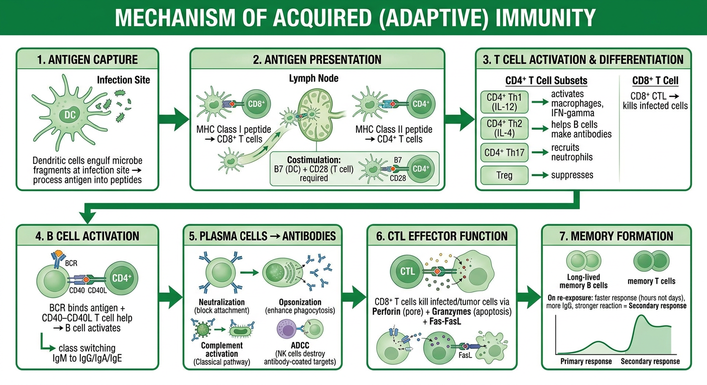
ACQUIRED IMMUNITY - Mechanism
Step-by-step:
- Dendritic cells engulf microbe fragments at infection site
- Migrate to lymph nodes, present peptide-MHC to naïve T cells
- MHC Class I → presents to CD8+ T cells (endogenous/viral antigens)
- MHC Class II → presents to CD4+ T cells (exogenous antigens)
- Costimulatory signals (B7 on DC + CD28 on T cell) required for full activation
- CD4+ T cells differentiate based on cytokine milieu:
| Subset | Cytokine driving it | Function |
|---|---|---|
| Th1 | IL-12, IFN-γ | Activate macrophages → kill intracellular pathogens |
| Th2 | IL-4 | Help B cells → antibodies; defense vs parasites |
| Th17 | IL-6 + TGF-β | Recruit neutrophils; defense vs extracellular bacteria/fungi |
| Treg | TGF-β | Suppress immune responses; prevent autoimmunity |
- CD8+ CTLs → kill infected cells via perforin (pore formation) + granzymes (trigger apoptosis) + Fas-FasL pathway
- B cell recognizes antigen via BCR → gets T cell help (CD40-CD40L interaction + cytokines)
- Differentiates into plasma cells → secrete antibodies
- Class switching (IgM → IgG/IgA/IgE) driven by cytokines
- Neutralization - antibodies block pathogen attachment to host cells
- Opsonization - IgG coats microbe → Fc receptor on phagocytes enhances phagocytosis
- Complement activation - IgM/IgG activate classical pathway → MAC lysis
- ADCC - NK cells/eosinophils destroy antibody-coated targets
- Long-lived memory B and T cells persist
- On re-exposure: faster (hours vs days), stronger, more IgG than IgM
The Bridge Between Both
Innate (immediate, 0-96h)
    ↓  DCs activated by TLR signals
    ↓  Migrate to lymph node
Adaptive (days-weeks, primary)
    ↓  T + B cell activation
    ↓  Effector phase (antibodies + CTLs)
Memory (lifelong)
    ↓  Rapid secondary response
Innate immunity provides the danger signal that licenses DCs to activate T cells. Without innate activation, adaptive responses are weak or absent - this is why adjuvants in vaccines mimic innate signals (e.g., alum activates NLRP3 inflammasome).
- Innate = recognize PAMPs/DAMPs via fixed PRRs → inflammation + complement + interferon → fast but non-specific
- Acquired = recognize specific antigens via TCR/BCR → clonal expansion → specific killing + antibodies + memory
Separte mechanism flowchartfor both
A clean medical education flowchart titled "Mechanism of Innate Immunity" with the following sequential steps connected by arrows: 1. MICROBE ENTRY - Breaches physical barriers (skin, mucus, cilia, defensins) 2. RECOGNITION - Tissue macrophages/DCs detect via PRRs: TLR (LPS, flagellin, viral RNA), NLR/NOD (peptidoglycan), RLR/RIG-I (dsRNA), CLEC (fungal glucans). Recognizes PAMPs and DAMPs 3. EARLY CELLULAR RESPONSE (0-4 hours) - Macrophages release TNF-alpha, IL-1, IL-6, IL-12. Mast cells release histamine. Complement activated (Alternative + Lectin pathways) 4. COMPLEMENT CASCADE - C3b = Opsonization, C5a = Chemotaxis (recruits neutrophils), C5b-9 MAC = Lysis 5. INFLAMMATION (4-96 hours) - Neutrophils recruited: phagocytosis + oxidative burst (ROS) + NETs. NK cells kill MHC-I deficient cells via perforin/granzymes 6. ANTIVIRAL DEFENSE - Infected cells release IFN-alpha/beta → neighboring cells become antiviral resistant 7. OUTCOME - Infection controlled OR handed off to Adaptive Immunity via DCs Box at bottom: NO MEMORY - same response on re-exposure Use blue color scheme, clean white background, bold arrows, medical textbook style
A clean medical education flowchart titled "Mechanism of Acquired (Adaptive) Immunity" with the following sequential steps connected by arrows: 1. ANTIGEN CAPTURE - Dendritic cells engulf microbe fragments at infection site → process antigen into peptides 2. ANTIGEN PRESENTATION - DCs migrate to lymph node. MHC Class I peptide → CD8+ T cells. MHC Class II peptide → CD4+ T cells. Costimulation: B7 (DC) + CD28 (T cell) required 3. T CELL ACTIVATION & DIFFERENTIATION - CD4+ Th1 (IL-12) → activates macrophages, IFN-gamma. CD4+ Th2 (IL-4) → helps B cells make antibodies. CD4+ Th17 → recruits neutrophils. Treg → suppresses. CD8+ CTL → kills infected cells 4. B CELL ACTIVATION - BCR binds antigen + CD40-CD40L T cell help → B cell activates → class switching IgM to IgG/IgA/IgE 5. PLASMA CELLS → ANTIBODIES - Neutralization (block attachment). Opsonization (enhance phagocytosis). Complement activation (Classical pathway). ADCC (NK cells destroy antibody-coated targets) 6. CTL EFFECTOR FUNCTION - CD8+ T cells kill infected/tumor cells via Perforin (pore) + Granzymes (apoptosis) + Fas-FasL 7. MEMORY FORMATION - Long-lived memory B cells + memory T cells persist. On re-exposure: faster response (hours not days), more IgG, stronger reaction = Secondary response Use green color scheme, clean white background, bold arrows, medical textbook style
Flowchart 1 - Innate Immunity Mechanism
Flowchart 2 - Acquired (Adaptive) Immunity Mechanism
Quick recap of the key difference in flow:
| Innate | Acquired | |
|---|---|---|
| Trigger | PAMPs/DAMPs via PRRs | Antigen presented on MHC by DCs |
| Key cells | Macrophages, Neutrophils, NK cells | T cells, B cells |
| Effectors | Complement, ROS, Interferons | Antibodies, CTLs |
| Ends with | Inflammation controlled | Memory cells formed |
Cell mediated immune response and immune mediated immune response
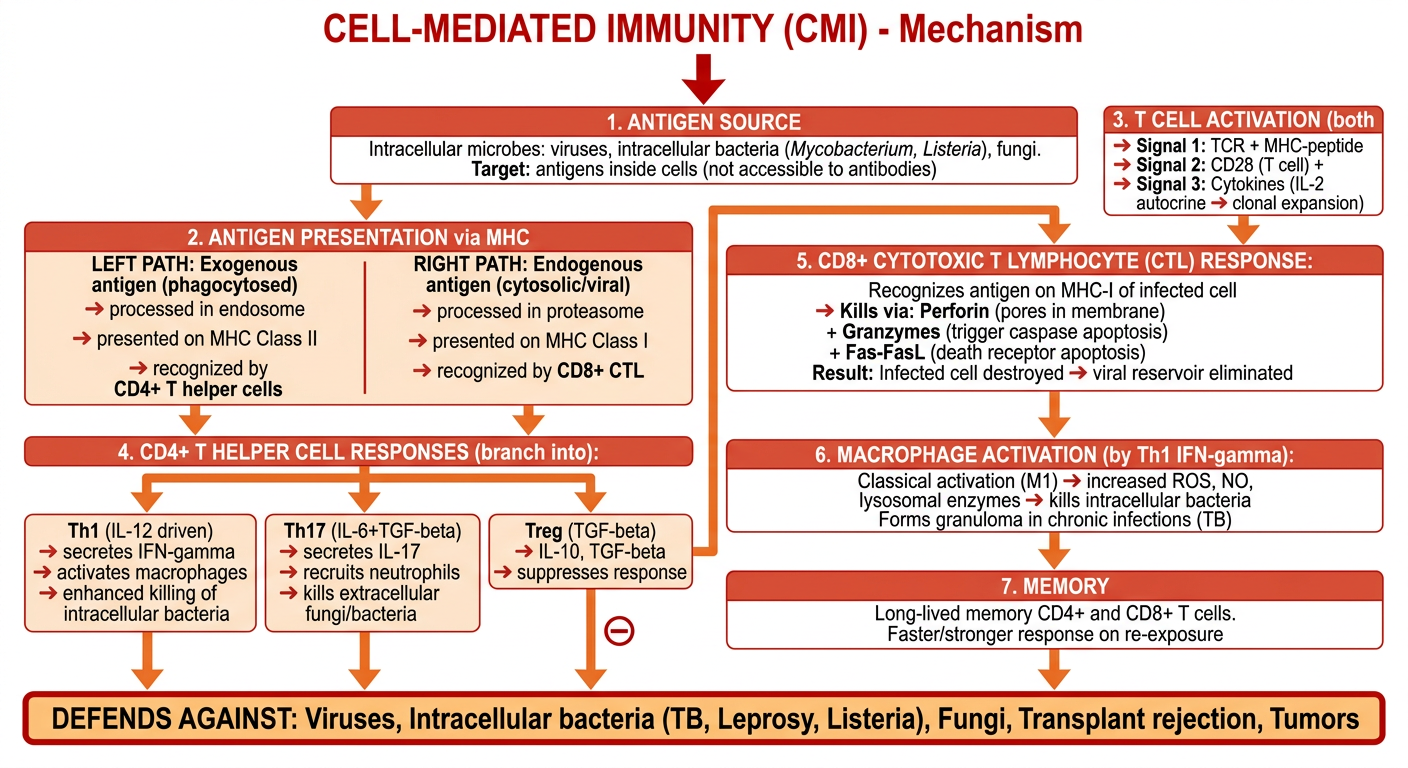
A detailed medical education flowchart titled "CELL-MEDIATED IMMUNITY (CMI) - Mechanism" with a clean vertical flow using arrows, on white background, red and orange color scheme. Flow steps: 1. ANTIGEN SOURCE - Intracellular microbes: viruses, intracellular bacteria (Mycobacterium, Listeria), fungi. Target: antigens inside cells (not accessible to antibodies) 2. ANTIGEN PRESENTATION via MHC - Two parallel paths shown side by side: LEFT PATH: Exogenous antigen (phagocytosed) → processed in endosome → presented on MHC Class II → recognized by CD4+ T helper cells RIGHT PATH: Endogenous antigen (cytosolic/viral) → processed in proteasome → presented on MHC Class I → recognized by CD8+ CTL 3. T CELL ACTIVATION (both paths require) - Signal 1: TCR + MHC-peptide. Signal 2: CD28 (T cell) + B7/CD80/CD86 (APC). Signal 3: Cytokines (IL-2 autocrine → clonal expansion) 4. CD4+ T HELPER CELL RESPONSES (branch into): Th1 (IL-12 driven) → secretes IFN-gamma → activates macrophages → enhanced killing of intracellular bacteria Th17 (IL-6+TGF-beta) → secretes IL-17 → recruits neutrophils → kills extracellular fungi/bacteria Treg (TGF-beta) → IL-10, TGF-beta → suppresses response 5. CD8+ CYTOTOXIC T LYMPHOCYTE (CTL) RESPONSE: Recognizes antigen on MHC-I of infected cell Kills via: Perforin (pores in membrane) + Granzymes (trigger caspase apoptosis) + Fas-FasL (death receptor apoptosis) Result: Infected cell destroyed → viral reservoir eliminated 6. MACROPHAGE ACTIVATION (by Th1 IFN-gamma): Classical activation (M1) → increased ROS, NO, lysosomal enzymes → kills intracellular bacteria Forms granuloma in chronic infections (TB) 7. MEMORY - Long-lived memory CD4+ and CD8+ T cells. Faster/stronger response on re-exposure Box at bottom: DEFENDS AGAINST: Viruses, Intracellular bacteria (TB, Leprosy, Listeria), Fungi, Transplant rejection, Tumors Bold arrows, clear labels, medical textbook quality
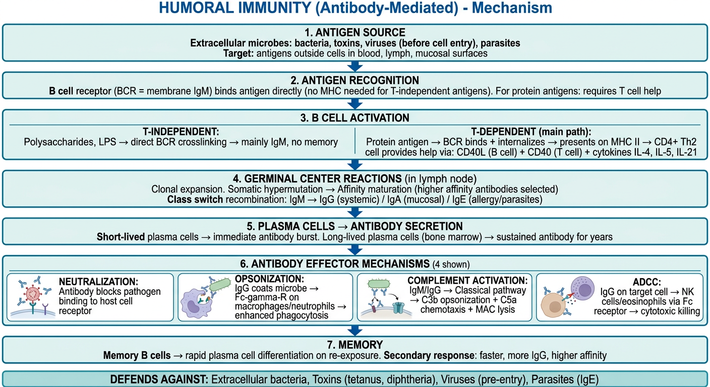
A detailed medical education flowchart titled "HUMORAL IMMUNITY (Antibody-Mediated) - Mechanism" with a clean vertical flow using arrows, on white background, blue and teal color scheme. Flow steps: 1. ANTIGEN SOURCE - Extracellular microbes: bacteria, toxins, viruses (before cell entry), parasites. Target: antigens outside cells in blood, lymph, mucosal surfaces 2. ANTIGEN RECOGNITION - B cell receptor (BCR = membrane IgM) binds antigen directly (no MHC needed for T-independent antigens). For protein antigens: requires T cell help 3. B CELL ACTIVATION - Two types shown: T-INDEPENDENT: Polysaccharides, LPS → direct BCR crosslinking → mainly IgM, no memory T-DEPENDENT (main path): Protein antigen → BCR binds + internalizes → presents on MHC II → CD4+ Th2 cell provides help via: CD40L (B cell) + CD40 (T cell) + cytokines IL-4, IL-5, IL-21 4. GERMINAL CENTER REACTIONS (in lymph node): Clonal expansion Somatic hypermutation → Affinity maturation (higher affinity antibodies selected) Class switch recombination: IgM → IgG (systemic) / IgA (mucosal) / IgE (allergy/parasites) 5. PLASMA CELLS → ANTIBODY SECRETION: Short-lived plasma cells → immediate antibody burst Long-lived plasma cells (bone marrow) → sustained antibody for years 6. ANTIBODY EFFECTOR MECHANISMS (4 shown): NEUTRALIZATION: Antibody blocks pathogen binding to host cell receptor OPSONIZATION: IgG coats microbe → Fc-gamma-R on macrophages/neutrophils → enhanced phagocytosis COMPLEMENT ACTIVATION: IgM/IgG → Classical pathway → C3b opsonization + C5a chemotaxis + MAC lysis ADCC: IgG on target cell → NK cells/eosinophils via Fc receptor → cytotoxic killing 7. MEMORY - Memory B cells → rapid plasma cell differentiation on re-exposure. Secondary response: faster, more IgG, higher affinity Box at bottom: DEFENDS AGAINST: Extracellular bacteria, Toxins (tetanus, diphtheria), Viruses (pre-entry), Parasites (IgE) Bold arrows, clear labels, medical textbook quality
Quick note: you likely meant Cell-Mediated Immunity and Humoral (Antibody-Mediated) Immunity - the two arms of acquired immunity. Here they are:
Flowchart 1 - Cell-Mediated Immunity (CMI)
Flowchart 2 - Humoral (Antibody-Mediated) Immunity
Key Differences at a Glance
| Feature | Cell-Mediated | Humoral |
|---|---|---|
| Mediator | T lymphocytes (CD4+, CD8+) | B lymphocytes → Antibodies |
| Target | Intracellular microbes | Extracellular microbes + toxins |
| Antigen presentation | Via MHC (I or II) required | BCR binds antigen directly |
| Key effector mechanism | Perforin/granzyme killing + macrophage activation | Neutralization, opsonization, complement, ADCC |
| Defends against | Viruses, TB, Leprosy, Listeria, tumors | Extracellular bacteria, toxins, viruses (before entry) |
| Tested by | Mantoux/tuberculin skin test | Serum antibody titres |
| Memory cells | Memory T cells | Memory B cells + long-lived plasma cells |
- CMI = T cells kill what's INSIDE cells (viruses hiding in cells, intracellular bacteria)
- Humoral = Antibodies neutralize what's OUTSIDE cells (bacteria, toxins, free viruses)
Central and peripheral lymphoid organs , thymus , cells of immune system ,and function ,
A comprehensive medical education diagram titled "LYMPHOID ORGANS - Central vs Peripheral" showing two clearly separated columns on white background. LEFT COLUMN (Blue/Purple) - "CENTRAL (PRIMARY) LYMPHOID ORGANS": Box 1: BONE MARROW - Site of hematopoiesis (all blood cells originate here) - B cell maturation (pre-B → immature B → mature naive B) - T cell precursors (prothymocytes) generated here - Provides IL-7 for early lymphocyte development Box 2: THYMUS (below bone marrow with arrow) - Bilobed organ, anterior mediastinum - CORTEX: dense thymocytes, positive selection (learn MHC recognition) - MEDULLA: Hassall corpuscles, negative selection (delete autoreactive T cells) - T cell maturation: CD4-CD8 double negative → double positive → single positive (CD4+ or CD8+) - Involutes after puberty RIGHT COLUMN (Green) - "PERIPHERAL (SECONDARY) LYMPHOID ORGANS": Box 3: LYMPH NODES - Bean-shaped, along lymphatic vessels - Cortex: B cell follicles (germinal centers) - Paracortex: T cell zone - Medulla: plasma cells + macrophages - Filter lymph, trap antigens, activate lymphocytes Box 4: SPLEEN - Filters blood (not lymph) - White pulp: lymphocytes (PALS = T cells around arteriole, follicles = B cells) - Red pulp: destroys old RBCs, reservoir for monocytes - Responds to blood-borne antigens Box 5: MUCOSA-ASSOCIATED LYMPHOID TISSUE (MALT) - Tonsils, Peyer patches (gut), bronchial MALT - First line defense at mucosal surfaces - IgA production Bottom box: KEY RULE - Central organs = where lymphocytes MATURE. Peripheral organs = where lymphocytes RESPOND to antigens. Clean medical textbook style, bold headers, clear arrows showing lymphocyte flow from central to peripheral
Lymphoid Organs + Cells of the Immune System
PART 1 - LYMPHOID ORGANS
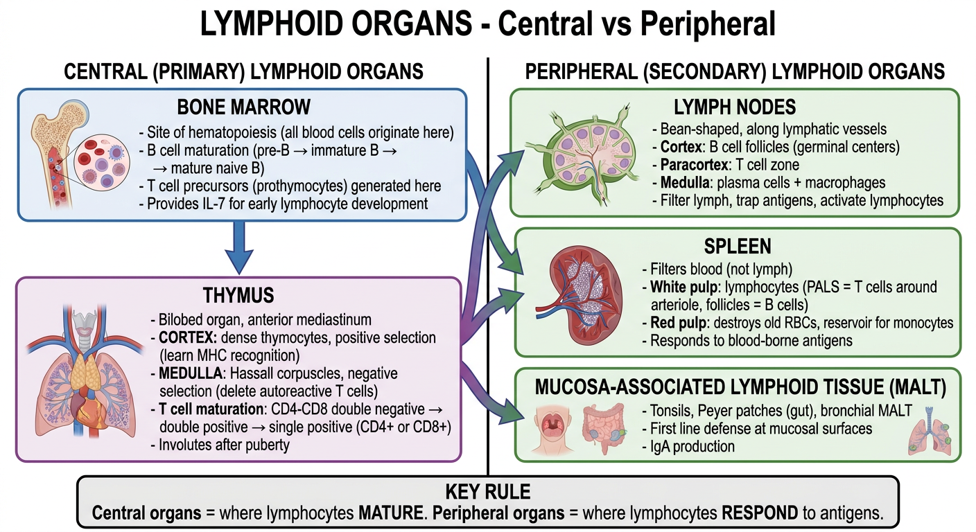
CENTRAL (PRIMARY) LYMPHOID ORGANS
Rule: Where lymphocytes are BORN and MATURE
1. Bone Marrow
| Feature | Detail |
|---|---|
| Location | Flat bones (sternum, ilium, ribs, vertebrae) after puberty |
| Origin | All blood cells from HSC (Hematopoietic Stem Cell) |
| Main function | B cell maturation + generation of T cell precursors |
| B cell stages here | Pro-B → Pre-B → Immature B → Mature naive B (IgM+IgD) |
| T cells | Only precursors made here; they migrate to thymus to mature |
| Selection | Central tolerance: autoreactive B cells deleted here |
2. Thymus
| Feature | Detail |
|---|---|
| Location | Anterior mediastinum (behind sternum) |
| Active period | Birth to puberty; involutes after puberty |
| Function | T cell maturation exclusively |
| CORTEX | Dense thymocytes (immature T cells); cortical epithelial cells produce IL-7 (survival signal); POSITIVE SELECTION occurs here |
| MEDULLA | Sparse T cells; Hassall's corpuscles (whorls of epithelial cells); NEGATIVE SELECTION by medullary thymic epithelial cells (mTECs); DCs and macrophages present |
| Positive selection | T cells that can recognize self-MHC are retained |
| Negative selection | T cells that react too strongly to self-antigens are deleted (clonal deletion) → self-tolerance |
| T cell stages | Double negative (CD4-CD8-) → Double positive (CD4+CD8+) → Single positive (CD4+ or CD8+) → exit to periphery |
| Clinical note | DiGeorge syndrome = thymic aplasia → no T cells |
PERIPHERAL (SECONDARY) LYMPHOID ORGANS
Rule: Where lymphocytes RESPOND to antigens
3. Lymph Nodes
- Bean-shaped organs along lymphatic vessels throughout body
- Cortex - B cell follicles (primary = resting; secondary with germinal center = active response)
- Paracortex - T cell zone; HEVs (High Endothelial Venules) - entry point for naive lymphocytes
- Medulla - plasma cells + macrophages + medullary sinuses
- Function: Filter lymph, trap antigens, site of adaptive immune response initiation
4. Spleen
- Filters blood (not lymph - unlike lymph nodes)
- White pulp - lymphocytes: PALS (Periarteriolar Lymphoid Sheath = T cells around central arteriole) + follicles (B cells)
- Red pulp - destroys senescent RBCs; reservoir for monocytes and platelets
- Marginal zone - interface; macrophages trap blood-borne antigens
- Responds to blood-borne antigens (e.g., encapsulated bacteria)
- Clinical note: Asplenic patients are at high risk for Streptococcus pneumoniae, Haemophilus, Neisseria (encapsulated organisms)
5. MALT (Mucosa-Associated Lymphoid Tissue)
- Tonsils and adenoids (Waldeyer's ring)
- Peyer's patches in small intestine (contain M cells that sample antigens)
- Bronchial MALT (BALT)
- Main output: Secretory IgA for mucosal defense
PART 2 - CELLS OF THE IMMUNE SYSTEM + FUNCTIONS
A. Innate Immune Cells
| Cell | Origin | Key Features | Function |
|---|---|---|---|
| Neutrophils | Bone marrow (myeloid) | Short-lived (1-2 days in tissue); multilobed nucleus; 40-60% of WBC | First responders; phagocytosis + oxidative burst (ROS) + NETs; kill extracellular bacteria/fungi |
| Macrophages | Monocytes (blood) → tissues; some from yolk sac (tissue-resident) | Long-lived (years); M1 (classical) vs M2 (alternative) activation | Phagocytosis; kill intracellular pathogens; cytokine production (TNF, IL-1, IL-6, IL-12); APC function; tissue repair |
| Dendritic Cells (DCs) | Myeloid or lymphoid precursors | Most potent APC; Birbeck granules in Langerhans cells | Capture antigen in tissues → migrate to lymph nodes → present to T cells; bridge innate and adaptive immunity |
| NK Cells | Bone marrow (lymphoid) | No TCR or BCR; CD56+, CD16+; recognize "missing self" | Kill virus-infected cells and tumor cells (MHC-I deficient) via perforin/granzymes; produce IFN-gamma |
| Mast Cells | Mast cell precursors from bone marrow; mature in tissues | Tissue-resident; contain granules with histamine, heparin; IgE receptor (FcεRI) | Allergy/anaphylaxis; defense at epithelial barriers; recruit eosinophils |
| Basophils | Bone marrow | Circulating; <1% of WBC; IgE receptor | Similar to mast cells; allergy; anti-parasitic |
| Eosinophils | Bone marrow | 1-4% of WBC; bilobed nucleus; pink granules (major basic protein) | Kill parasites via MBP, ECP; involved in allergy |
| ILCs (Innate Lymphoid Cells) | Bone marrow (lymphoid) | No antigen receptor; tissue-resident | ILC1 (like Th1), ILC2 (like Th2), ILC3 (like Th17) - early cytokine production |
B. Adaptive Immune Cells
| Cell | Origin/Maturation | Marker | Function |
|---|---|---|---|
| B Lymphocytes | Bone marrow | CD19, CD20, IgM, IgD on surface | Recognize antigen via BCR → differentiate to plasma cells → secrete antibodies; memory B cells |
| Plasma Cells | Differentiated B cells | CD38, CD138 high; Ig low on surface | Antibody-secreting factory; short-lived or long-lived in bone marrow |
| CD4+ T Helper | Thymus; mature in periphery | CD4, CD3, TCR | Orchestrate immune response; help B cells and macrophages; Th1/Th2/Th17/Treg subtypes |
| CD8+ CTL | Thymus; mature in periphery | CD8, CD3, TCR | Kill virus-infected cells and tumors via perforin/granzymes/Fas-FasL |
| Regulatory T cells (Treg) | Thymus or periphery | CD4, CD25, FoxP3 | Suppress immune responses; prevent autoimmunity |
| Memory T cells | Post-activation | CD45RO (memory), CD45RA (naive) | Rapid recall response on re-exposure |
| Memory B cells | Post-activation in germinal center | CD27, class-switched Ig (IgG, IgA, IgE) | Rapid secondary antibody response |
PART 3 - THYMUS IN DETAIL (High-Yield)
Prothymocyte from bone marrow
         ↓
CORTEX - Double Negative (CD4- CD8-)
         ↓  IL-7 → survival
Double Positive (CD4+ CD8+)
         ↓
POSITIVE SELECTION (cortical epithelium + MHC)
"Can this T cell recognize self-MHC?"
YES → survives     NO → dies (neglect ~95%)
         ↓
NEGATIVE SELECTION (mTECs + DCs in medulla)
"Does it react too strongly to self-antigen?"
YES → apoptosis (clonal deletion)
NO → survives → exits as mature single positive T cell
         ↓
CD4+ (recognized MHC II) or CD8+ (recognized MHC I)
         ↓
Enter bloodstream → peripheral lymphoid organs
- Thymosin (α1 and β4) - promotes T cell maturation
- Thymopoietin - differentiation of prothymocytes
- Thymulin - promotes CD4+/CD8+ expression
Quick Memory Table
| Feature | Central Organs | Peripheral Organs |
|---|---|---|
| Examples | Bone marrow, Thymus | Lymph nodes, Spleen, MALT |
| Purpose | Lymphocyte maturation | Antigen response |
| Selection | Yes (central tolerance) | No (effector responses) |
| Filter what? | N/A | Lymph nodes (lymph), Spleen (blood) |
| Antigen needed? | No | Yes |