
Otitis media
Reading File
Now I'll search the medical library and PubMed in parallel for comprehensive coverage of otitis media.
Finding Sources
Searching PubMed
"otitis media"[MeSH Terms] AND management
Good results. Now let me read the most comprehensive sections in parallel - Family Medicine (most detail), Rosen's Emergency Medicine, and the Harriet Lane (pediatric).
Reading File
Reading File
Reading File
The Family Medicine textbook has the most comprehensive content. Let me read more of it to get the full treatment section, plus the Scott-Brown's Otolaryngology content.
Reading File
Reading File
Excellent - I have rich content from the Family Medicine textbook. The Scott-Brown result was about quality of life instruments, not the core clinical content. Let me read more of the Family Medicine textbook to get the OME and complications/recurrent OM sections.
Reading File
I now have comprehensive content to write a thorough, well-structured answer on otitis media. Let me compile it.
Otitis Media
Otitis media (OM) is inflammation of the middle ear, classified into three main types: acute otitis media (AOM), otitis media with effusion (OME), and chronic suppurative otitis media (CSOM). It is the most common infection for which children are seen in a physician's office in the US, with an estimated annual cost of $2.88 billion. By age 7 years, 93% of children have had at least one episode of AOM.
Pathophysiology
The primary mechanism is eustachian tube dysfunction. Abnormal tubal compliance and delayed innervation of the tensor veli palatini muscle lead to tube collapse, impaired drainage, and negative middle ear pressure - allowing nasopharyngeal bacteria to colonize the middle ear space. A viral upper respiratory infection almost always precedes an episode of AOM.
- Textbook of Family Medicine 9e, p. 404
Classification
| Type | Key Feature |
|---|---|
| Acute Otitis Media (AOM) | Acute onset, middle ear effusion + signs of inflammation |
| Otitis Media with Effusion (OME) | "Glue ear" - effusion without acute inflammation; most common cause of hearing loss in children |
| Recurrent AOM | 3+ episodes in 6 months, or 4+ in 12 months |
| Chronic Suppurative OM (CSOM) | Persistent purulent otorrhea through perforated TM or tympanostomy tube |
Common Causative Organisms
The three major bacterial pathogens:
- Streptococcus pneumoniae - 25-40% of cases
- Haemophilus influenzae - 10-30%
- Moraxella catarrhalis - 2-15%
Viral pathogens (RSV, rhinovirus, influenza) also contribute and often precede bacterial superinfection. Resistance of S. pneumoniae to penicillin is an increasing concern.
- Textbook of Family Medicine 9e, p. 404; Rosen's Emergency Medicine, p. 514
Risk Factors
- Male gender
- Child care outside the home (daycare)
- Parental smoking
- Bottle feeding in the supine position
- Genetic and ethnic factors (Inuit, Native American)
- Allergy
- Craniofacial abnormalities (cleft palate)
- Use of a pacifier
- Previous episode of AOM within the preceding 3 months
- Winter season / exposure to URIs
Diagnosis
AOM diagnosis requires all three:
- Acute onset of symptoms (otalgia, ear-tugging in infants, irritability)
- Middle ear effusion (MEE) - confirmed by:
- Visualization of air-fluid levels behind TM
- Bulging drum
- Reduced/absent movement on pneumatic otoscopy
- Flat tympanogram
- Signs of middle ear inflammation:
- TM erythema, bulging, or new-onset otorrhea (not due to otitis externa)
Key distinction: Erythema of the TM without MEE = myringitis/tympanitis (not AOM). Otalgia with a normal, flaccid TM suggests referred otalgia from another source (dental abscess, TMJ, sore throat, etc.).
Bullous myringitis (bullae on TM) may be seen in some cases of AOM - treatment does not differ from standard AOM.
- Rosen's Emergency Medicine, p. 514-518; Textbook of Family Medicine 9e, p. 404
Treatment
AOM - Antibiotic Decision
Most cases in appropriately selected patients can resolve spontaneously. Observation for 2-3 days is an option in some children (not validated in adults).
| Patient Group | Decision |
|---|---|
| Any age, severe symptoms (otalgia + fever >39°C) | Treat immediately |
| < 6 months | Always treat |
| 6 months - 2 years, bilateral AOM | Always treat |
| 6 months - 2 years, unilateral, non-severe | Observation allowed if reliable follow-up |
| > 2 years, non-severe | Observation allowed |
Antibiotic Regimens
| Scenario | Drug of Choice | Dose/Duration |
|---|---|---|
| Low-risk (>6 yr, no recent abx, afebrile, not in daycare) | Amoxicillin | 40-50 mg/kg/day ÷ doses x 5 days |
| High-risk (<2 yr, daycare, recent abx, fever >38°C) | Amoxicillin | 80-90 mg/kg/day ÷ doses x 10 days |
| Treatment failure (symptoms persist >3 days) | Amoxicillin-clavulanate | 80-90 mg/kg/day x 7-10 days |
| OR Cefuroxime axetil | 20-30 mg/kg/day BID x 7-10 days | |
| OR Ceftriaxone IM | 50 mg/kg x 1-3 days | |
| Penicillin allergy | Cefuroxime axetil, Cefpodoxime, or Ceftriaxone | (see above) |
Pain Management
Antibiotics do not reduce pain in the first 24 hours. Always address pain with:
- Acetaminophen or ibuprofen (OTC analgesics)
Special Situations
-
AOM with acute perforation/otorrhea: Treat same as standard AOM; perforations are typically small and self-resolve.
-
Patients with tympanostomy tubes + increased otorrhea: Use ototopical fluoroquinolone drops (e.g., ofloxacin or ciprofloxacin/dexamethasone). Aminoglycoside drops are contraindicated with non-intact TM (ototoxic).
-
Textbook of Family Medicine 9e, p. 405; Rosen's Emergency Medicine, p. 526-532
Otitis Media with Effusion (OME)
- Fluid in the middle ear without signs of acute infection
- Most common cause of acquired hearing loss in children
- Most cases resolve spontaneously within 3 months
- No role for antibiotics, antihistamines, or decongestants in OME
- Indications for tympanostomy tube insertion: bilateral OME with hearing loss >25 dB persisting >3 months, or associated developmental/speech delay
Chronic Suppurative Otitis Media (CSOM)
- Persistent purulent otorrhea through a perforated TM (or tympanostomy tube)
- Pathogens: S. pneumoniae, H. influenzae, S. aureus, Pseudomonas, anaerobes
- Ototopical antibiotics are superior to oral antibiotics (Cochrane evidence)
- Quinolone ototopicals are safe for middle ear use; aminoglycosides carry ototoxicity risk
- Aural toilet with dilute acetic acid solution may help
- Persistent perforation after resolution: consider tympanoplasty
- Refractory cases: tympanomastoidectomy
Recurrent AOM
- Definition: 3+ episodes in 6 months, or 4+ in 12 months
- Tympanostomy tubes (grommets): first-line surgical intervention - reduces frequency of episodes, restores hearing
- Adenoidectomy: considered when adenoiditis acts as a nidus for recurrent infection
- Antibiotic prophylaxis: generally not recommended (risk of resistance)
Complications
Rare since the antibiotic era but potentially serious:
Intratemporal:
- Acute mastoiditis - most common suppurative complication; presents with post-auricular tenderness, erythema, edema, and fever; it is a clinical (not radiologic) diagnosis - CT changes must be correlated with exam
- Facial nerve paralysis - from inflammation along CN VII coursing through the middle ear
- Labyrinthitis / sensorineural hearing loss
- Tympanic membrane perforation, retraction, tympanosclerosis
- Cholesteatoma (keratin-filled sac causing bony erosion)
Intracranial:
- Meningitis
- Epidural abscess
- Brain abscess
- Subdural abscess
- Lateral/sigmoid sinus thrombosis
Red flags for impending complication: increasing pain, vertigo, or facial palsy in a patient with CSOM - require urgent otolaryngology consultation.
- Textbook of Family Medicine 9e, p. 405-406
Prevention
- Pneumococcal conjugate vaccine (PCV13/PCV15) - reduces AOM due to S. pneumoniae
- Influenza vaccine - annual vaccination reduces AOM episodes linked to influenza
- Breastfeeding (protective)
- Avoiding supine bottle-feeding
- Avoiding passive smoke exposure
- Reducing pacifier use after 6 months
Recent Evidence (2024-2026)
- A 2025 Cochrane review (PMID 41307300) confirmed that decongestants and antihistamines have no benefit in treating AOM in children and should not be used.
- A 2026 systematic review (PMID 41764103) examined shorter vs. longer antibiotic courses - evidence supports 5-day courses for low-risk children over 2 years; 10-day courses remain preferred for younger or high-risk children.
- A 2026 meta-analysis (PMID 42359822) found RSV is a significant contributor to AOM in children under 5, suggesting RSV vaccination may reduce AOM burden.
- A 2024 systematic review/meta-analysis (PMID 38666491) confirmed significant quality-of-life improvements after tympanostomy tube surgery in pediatric patients.
Summary: AOM is primarily a disease of young children driven by eustachian tube dysfunction and nasopharyngeal bacterial colonization. Diagnosis requires MEE plus signs of inflammation. First-line treatment is amoxicillin (dose-adjusted by risk), with watchful waiting appropriate in older, low-risk children. Complications are rare but include mastoiditis and intracranial spread. Recurrent disease is managed with tympanostomy tubes.
Adenoids
Finding Sources
Searching PubMed
"adenoids"[MeSH Terms] AND adenoidectomy
Good - I have key sources. Let me read the most relevant sections in parallel - Bailey & Love (enlarged adenoid), Cummings (adenoid contribution), KJ Lee (adenoid facies), and Family Medicine (adenoid hypertrophy).
Reading File
Reading File
Reading File
Reading File
Good content collected. Now let me search for more detailed content on adenoids - anatomy, physiology, adenoidectomy indications - from Cummings and search paragraphs for deeper content.
Finding Sources
Finding Sources
Now let me read the KJ Lee pediatrics section on adenoidectomy indications and fetch the adenoid hypertrophy image from Bailey & Love.
Reading File
Loading Image
Reading File
Excellent - I now have very rich content including the grading table, endoscopic image, and treatment details. Let me read a bit more from Scott-Brown's on medical treatment, and also get the anatomy/Waldeyer's ring content.
Reading File
Finding Sources
I now have comprehensive material to write a thorough answer. Let me compose it.
Adenoids
Terminology
Despite the common use of the plural "adenoids," there is technically only one nasopharyngeal adenoid - a single midline mass of lymphoid tissue on the posterior wall of the nasopharynx. It is part of Waldeyer's ring, the circular arrangement of lymphoid tissue guarding the entrance to the aerodigestive tract (palatine tonsils, lingual tonsil, tubal tonsils, and the adenoid/pharyngeal tonsil).
- Bailey & Love's Short Practice of Surgery, 28th ed., p. 9580
Anatomy & Histology
- Location: Posterior wall/roof of the nasopharynx, above the level of the soft palate
- Lies adjacent to the Eustachian tube orifices on each side - this proximity explains why adenoid hypertrophy causes eustachian tube dysfunction and otitis media
- Covered by pseudostratified ciliated columnar epithelium (respiratory type)
- Composed of lymphoid follicles with germinal centers (B-cell zones) surrounded by T-cell zones
- No capsule (unlike tonsils) - this is clinically relevant for surgical removal
- The tubal tonsils (lymphoid tissue around the Eustachian tube opening) can also hypertrophy and obstruct the Eustachian tube
Normal Development & Involution
-
Adenoid tissue is present from birth and grows through childhood
-
Hypertrophy most commonly occurs between ages 4 and 10 years
-
Spontaneous involution begins in adolescence - the adenoid typically regresses significantly by the mid-teens
-
In an adult, a prominent adenoid is abnormal and should raise suspicion for:
- Lymphoproliferative disorder (lymphoma)
- HIV infection
- Nasopharyngeal carcinoma (NPC - especially in endemic populations)
-
Textbook of Family Medicine 9e, p. 417; Bailey & Love's, p. 9622
Endoscopic View of Adenoid Hypertrophy
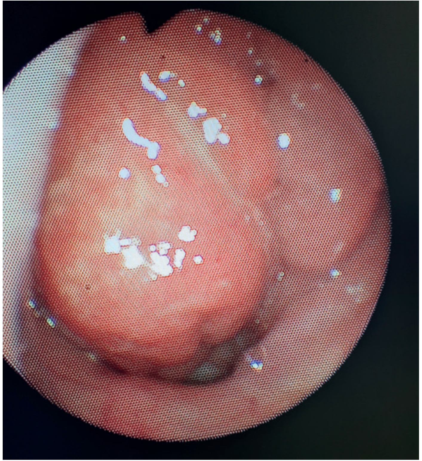
Endoscopic image of adenoid hypertrophy. - Bailey & Love's Short Practice of Surgery, 28th ed.
Clinical Features of Adenoid Hypertrophy
Nasal / Upper Airway Symptoms
- Chronic nasal obstruction - persistent mouth breathing
- Rhinorrhea and post-nasal drip
- Nasal voice (hyponasality / "adenoid voice")
- Snoring and disturbed sleep
- Chronic cough (from post-nasal drip)
Ear Symptoms
- Otitis media with effusion (OME / "glue ear") - adenoid acts as a bacterial reservoir and blocks eustachian tube drainage
- Recurrent acute otitis media (AOM)
- Conductive hearing loss
Sleep-Disordered Breathing
- Obstructive sleep apnoea (OSA) - especially when adenoid hypertrophy coexists with tonsillar hypertrophy
- Irregular snoring with apnoeic pauses and loud inspiratory snorts
- Restless sleep, unusual sleep positions
- Daytime somnolence, behavioural problems, poor school performance
- Long-term: secondary cardiac complications (cor pulmonale, pulmonary hypertension)
Adenoid Facies
Chronic nasal obstruction from childhood produces a characteristic facial appearance:
-
Open-mouth posture (mouth breathing)
-
Crowded/malocclused teeth
-
High-arched palate
-
Underdeveloped nostrils
-
Periorbital edema
-
Long, narrow face
-
KJ Lee's Essential Otolaryngology, p. 1509; Textbook of Family Medicine 9e, p. 1080
Grading of Adenoid Hypertrophy
Based on the degree of choanal obstruction on nasendoscopy (Scott-Brown's grading):
| Grade | Description |
|---|---|
| Grade I | Adenoid tissue filling up to 1/3 of the vertical choanae |
| Grade II | Adenoid tissue filling 1/3 to 2/3 of the choanae |
| Grade III | From 2/3 to nearly complete obstruction |
| Grade IV | Complete choanal obstruction |
The gold standard for assessing adenoid size is nasopharyngoscopy (flexible nasoendoscopy). Mirror examination underestimates choanal occlusion. Palpation is a poor measure. When endoscopy is not tolerated (young children), a lateral soft-tissue neck radiograph is a practical alternative.
- Scott-Brown's Otorhinolaryngology, Vol. 2, p. 331
Diagnosis
- Clinical history and examination - usually sufficient
- Flexible nasopharyngoscopy - gold standard for direct visualisation
- Lateral neck radiograph - shows "pad" of adenoid tissue in the postnasal space (soft-tissue film); practical when endoscopy not tolerated
- Polysomnography (sleep study) - for suspected OSA to quantify apnoea-hypopnoea index (AHI)
- Do NOT routinely use MRI or acoustic rhinomanometry in clinical practice (research tools only)
Role of Adenoids in Chronic Rhinosinusitis (CRS)
The adenoid is in close proximity to the paranasal sinuses and can serve as a bacterial biofilm reservoir. Key findings from Cummings Otolaryngology:
-
88-99% of adenoid mucosal surface in children with CRS is covered by dense biofilm, compared to only 0-6.5% in controls (children with sleep apnoea only)
-
Adenoid bacterial flora mirrors that of the middle meatus in children with chronic/recurrent sinusitis
-
This explains why adenoidectomy improves CRS symptoms even when adenoid size is not the main problem - it is the reservoir function, not just the obstructive size, that matters
-
Cummings Otolaryngology, p. 3784
Management
Conservative (Watchful Waiting)
- Appropriate for mild, well-tolerated symptoms
- Most adenoid hypertrophy will involute with age
Medical Treatment
-
Intranasal corticosteroid sprays - evidence from meta-analyses of RCTs shows reduction in adenoid size and improvements in:
- Middle ear effusion and audiometric thresholds
- Nasal obstruction, rhinorrhoea, cough, snoring, and sleep apnoea
- Trials were judged as moderate quality; further RCTs needed
- Two 2025 systematic reviews (PMIDs 40609250 and 41056645) support intranasal steroids as an effective, safe option for adenoid hypertrophy in children
-
Scott-Brown's Otorhinolaryngology, Vol. 2, p. 6283
Surgical: Adenoidectomy
Indications:
| Indication | Notes |
|---|---|
| OSA (adenotonsillectomy) | Most common current indication; curative in children |
| Adenotonsillar hypertrophy with dysphagia, speech abnormality, dental malocclusion | Adenotonsillectomy |
| Recurrent/chronic AOM or OME requiring repeat tympanostomy tubes | Adenoidectomy regardless of adenoid size (size not the only factor) |
| Nasal obstruction severe/persistent and not responding to conservative management | |
| Suspicion of nasopharyngeal malignancy | Biopsy/removal for diagnosis |
Note: For OME, the adenoid size is not an indication for removal - its role as a bacterial reservoir is the rationale. KJ Lee's, p. 9083: "Adenoidectomy, regardless of adenoidal size, is helpful in children with chronic OM with effusion requiring multiple sets of tubes."
Contraindications / Cautions:
- Cleft palate or submucous cleft palate - risk of velopharyngeal insufficiency (VPI) and hypernasal speech post-operatively; if surgery needed, partial adenoidectomy preserving an inferior ridge is performed
- Bleeding disorders
Surgical Techniques:
| Technique | Notes |
|---|---|
| Blind curettage (traditional) | Most common in UK (79.2%); unpredictable bleeding (~50 mL); poor choanal access |
| Suction diathermy (direct vision) | Preferred; minimal blood loss (~4 mL); excellent haemostasis; allows partial adenoidectomy |
| Microdebrider | 20% faster than curettage; high unit cost |
| Coblation® | Less blood loss, more complete removal; cost limits use unless combined with tonsillectomy |
| KTP laser | Avoid - high risk of nasopharyngeal stenosis |
- Scott-Brown's Otorhinolaryngology, Vol. 2, p. 6296-6323
Peri-operative considerations for OSA:
- Avoid sedative premedications and opiate analgesics post-operatively - they can further depress the child's respiratory drive in a patient already predisposed to apnoea
- Bailey & Love's, p. 9601
Pre-operative investigations:
- Routine investigations not indicated for ASA grade 1-2 children
- Specific testing warranted for: sickle cell disease, thalassaemia, Down syndrome, congenital heart disease
Recent Evidence (2025-2026)
- Two independent 2025 meta-analyses (PMID 40609250 and PMID 41056645) confirm that intranasal corticosteroids are effective and safe for adenoid hypertrophy, reducing both adenoid size and associated symptoms - supporting them as a first-line medical option before surgery.
- A 2025 meta-analysis (PMID 40958230) found a bidirectional association between adenoid hypertrophy and laryngopharyngeal reflux (LPR) - each condition may exacerbate the other, and LPR treatment should be considered in refractory adenoid hypertrophy.
- A 2026 systematic review (PMID 41344444) highlights the growing importance of adenoid biofilms in driving recurrent and chronic ENT disease in children - supporting adenoidectomy as a reservoir-eliminating strategy.
Summary: The adenoid is a single midline lymphoid structure in the nasopharynx, part of Waldeyer's ring, that plays an immune role in early childhood but causes significant clinical disease when hypertrophied. Key consequences are nasal obstruction, OSA, and eustachian tube dysfunction (causing OME and recurrent AOM). It also acts as a bacterial biofilm reservoir contributing to chronic sinusitis. Treatment options include intranasal steroids (increasingly evidence-backed) and adenoidectomy, with surgical technique having moved toward direct-vision methods for better outcomes. In adults, prominent adenoid tissue demands investigation for malignancy or HIV.
This is a shared conversation. Sign in to Orris to start your own chat.