Tracheoesophagela fistula
tracheoesophageal fistula congenital diagnosis management Merck Manual CDC
"tracheoesophageal fistula" AND treatment
Tracheoesophageal fistula (TEF)
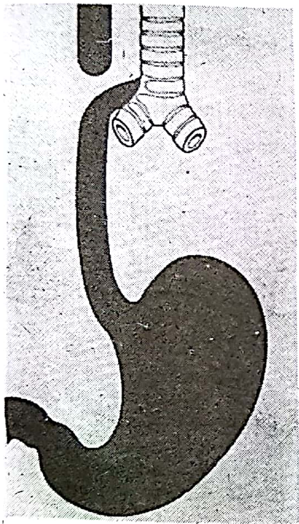
Most common type
Clinical features in a newborn
- Excessive salivation or frothing
- Choking, coughing, cyanosis, or respiratory distress during feeds
- Aspiration pneumonia
- Inability to pass a nasogastric/orogastric tube into the stomach
- Maternal polyhydramnios may be present antenatally
Diagnosis
- Pass a radio-opaque nasogastric tube: in esophageal atresia it stops and coils in the upper esophageal pouch.
- Chest and abdominal X-ray confirms the tube position.
- Gas in the stomach suggests a distal TEF. - Pye's Surgical Handicraft, p. 413
- Echocardiography and renal ultrasound are commonly done because associated anomalies are frequent.
Associations
- Vertebral defects
- Anal atresia
- Cardiac anomalies
- TE tracheoesophageal fistula
- Renal anomalies
- Limb abnormalities
Management
- Keep the infant nil by mouth.
- Continuous suction of the upper pouch to reduce aspiration.
- Position head-up, stabilize airway and breathing, give IV fluids.
- Treat aspiration or sepsis if present.
- Pediatric surgical repair: ligation/division of the fistula and primary esophageal anastomosis when feasible.
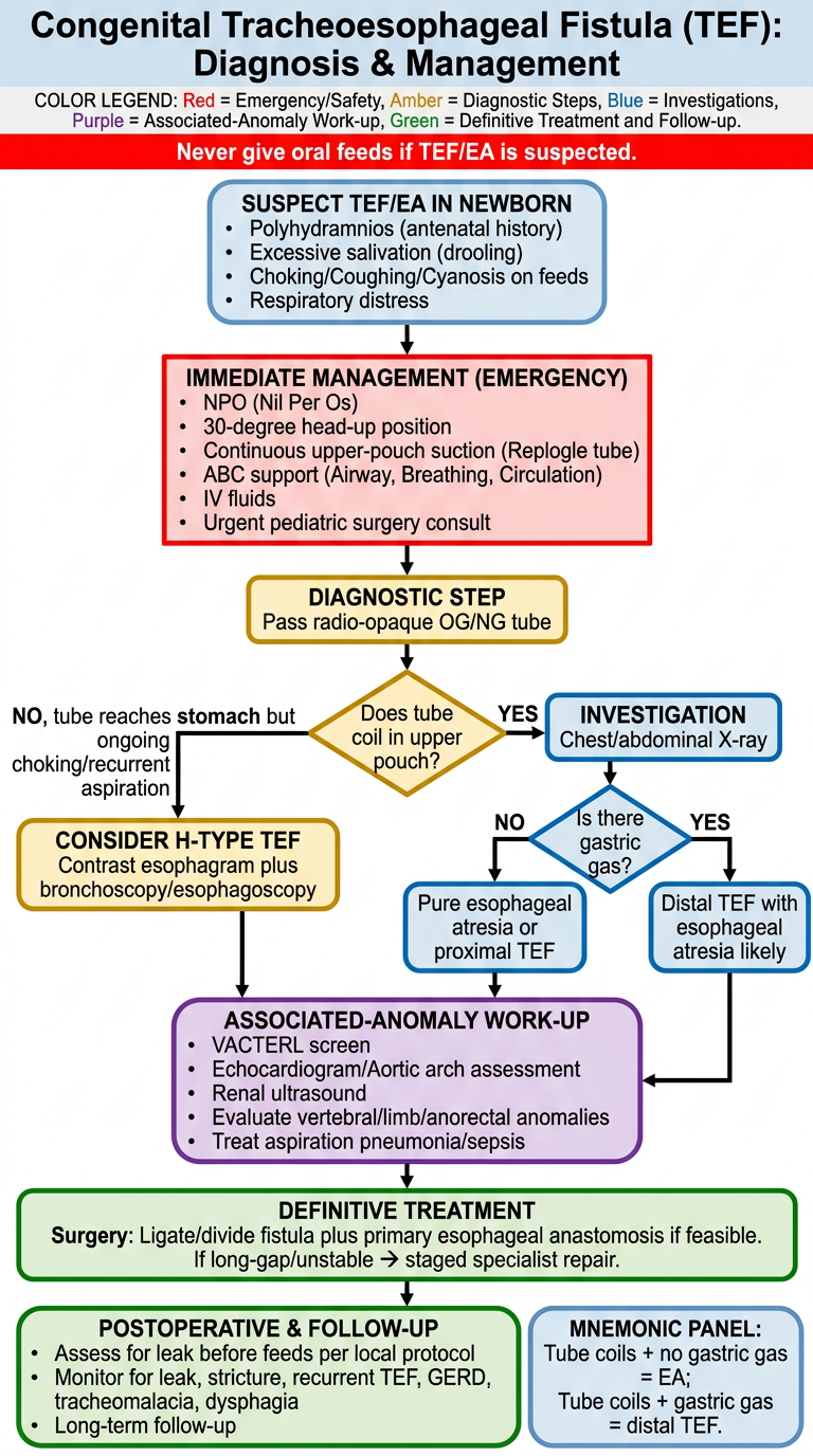
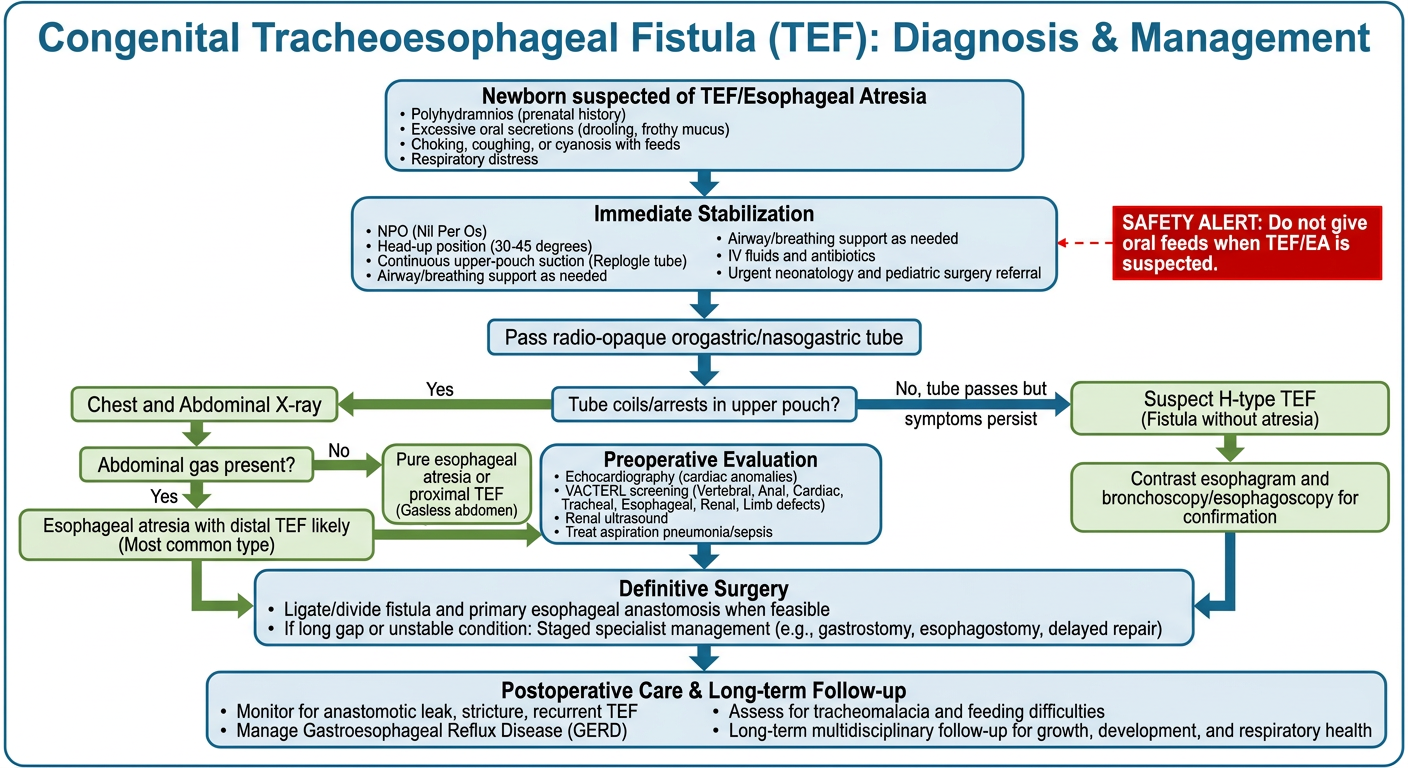
Create a TEF diagnosis and management flowchart
congenital tracheoesophageal fistula diagnosis stabilization management guideline
"tracheoesophageal fistula" AND diagnosis
Congenital TEF with/without esophageal atresia: diagnosis and management flowchart
flowchart TD
    A[Newborn with suspected TEF / esophageal atresia] --> B{Suggestive features?}

    B -->|Yes| C[Polyhydramnios antenatally<br/>Excess oral secretions<br/>Choking, coughing, cyanosis with feeds<br/>Respiratory distress / aspiration]
    B -->|No, but recurrent cough/choking with feeds| H[Consider isolated H-type TEF]

    C --> D[Immediate stabilization]
    D --> D1[NPO - do not feed orally]
    D --> D2[Head-up / reverse Trendelenburg position]
    D --> D3[Continuous suction of upper esophageal pouch]
    D --> D4[Airway and breathing support as required]
    D --> D5[IV fluids, correct hypoglycemia/electrolytes]
    D --> D6[Urgent neonatology and pediatric surgery referral]

    D --> E[Attempt gentle passage of radio-opaque orogastric/nasogastric tube]
    E --> F{Does tube pass into stomach?}

    F -->|No - tube arrests/coils in upper pouch| G[Chest and abdominal X-ray with tube in situ]
    G --> G1{Abdominal gas present?}
    G1 -->|Yes| I[Esophageal atresia with distal TEF likely]
    G1 -->|No| J[Pure esophageal atresia or proximal TEF possible]

    F -->|Yes, but symptoms persist| H
    H --> H1[Contrast esophagram under specialist guidance<br/>and/or bronchoscopy ± esophagoscopy]
    H1 --> H2[Identify H-type TEF and its location]

    I --> K[Preoperative assessment]
    J --> K
    H2 --> K

    K --> K1[Evaluate associated anomalies: VACTERL]
    K --> K2[Echocardiography - cardiac anatomy and aortic arch]
    K --> K3[Renal ultrasound; vertebral, limb and anorectal assessment]
    K --> K4[Treat aspiration pneumonia/sepsis if present]
    K --> K5[Plan airway and surgical approach]

    K --> L{Suitable for primary repair?}
    L -->|Yes| M[Definitive operation:<br/>ligate/divide fistula<br/>primary esophageal anastomosis where applicable]
    L -->|No / long-gap or unstable infant| N[Staged management in specialist center:<br/>control fistula, decompress stomach,<br/>nutritional support, delayed reconstruction]

    M --> O[Postoperative care]
    N --> O
    O --> O1[Ventilatory and hemodynamic support as needed]
    O --> O2[Assess anastomosis before feeds per local surgical protocol]
    O --> O3[Monitor for leak, stricture, recurrent TEF, GERD, tracheomalacia and dysphagia]
    O --> O4[Long-term multidisciplinary follow-up]
Key diagnostic point
Safety note
I want in image
A clear medical educational flowchart titled “Congenital Tracheoesophageal Fistula (TEF): Diagnosis & Management”. Use a clean vertical clinical algorithm with rounded boxes and arrows. Content: 1) Newborn suspected of TEF/Esophageal Atresia: polyhydramnios, excessive secretions, choking/cyanosis with feeds, respiratory distress. 2) Immediate stabilization: NPO, head-up position, continuous upper-pouch suction, airway/breathing support, IV fluids, urgent neonatology and pediatric surgery referral. 3) Pass radio-opaque orogastric/nasogastric tube. Decision: tube coils/arrests in upper pouch? Yes -> chest and abdominal X-ray. Decision: abdominal gas? Yes -> Esophageal atresia with distal TEF likely. No -> pure esophageal atresia/proximal TEF. No, tube passes but symptoms persist -> suspect H-type TEF -> contrast esophagram and bronchoscopy/esophagoscopy. 4) Preoperative evaluation: echocardiography and VACTERL screening, renal ultrasound, treat aspiration/sepsis. 5) Definitive surgery: ligate/divide fistula and primary esophageal anastomosis when feasible; if long gap/unstable, staged specialist management. 6) Postoperative care: monitor for leak, stricture, recurrent TEF, GERD, tracheomalacia and feeding difficulties; long-term follow-up. Include a small red safety callout: “Do not give oral feeds when TEF/EA is suspected.” Ensure all wording is correctly spelled, readable, and self-contained. Do not use images of patients or graphic surgical scenes.
Make a color-coded version for medical students
Color-coded, high-yield medical student infographic flowchart titled “Congenital Tracheoesophageal Fistula (TEF): Diagnosis & Management”. Make all text sharp, large, correctly spelled, and easy to revise from. Vertical flowchart with color legend: red = emergency/safety, amber = diagnostic steps, blue = investigations, purple = associated-anomaly work-up, green = definitive treatment and follow-up. Flow: Suspect TEF/EA in newborn: polyhydramnios, excessive salivation, choking/coughing/cyanosis on feeds, respiratory distress. Red immediate management box: NPO, 30-degree head-up, continuous upper-pouch suction, ABC support, IV fluids, urgent pediatric surgery. Amber: pass radio-opaque OG/NG tube. Decision diamond: Does tube coil in upper pouch? Yes -> blue Chest/abdominal X-ray. Decision: Is there gastric gas? Yes -> distal TEF with esophageal atresia likely. No -> pure esophageal atresia or proximal TEF. No, tube reaches stomach but ongoing choking/recurrent aspiration -> consider H-type TEF -> contrast esophagram plus bronchoscopy/esophagoscopy. Purple: VACTERL screen, echocardiogram/aortic arch, renal ultrasound, evaluate vertebral/limb/anorectal anomalies, treat aspiration pneumonia/sepsis. Green: surgery, ligate/divide fistula plus primary esophageal anastomosis if feasible. If long-gap/unstable -> staged specialist repair. Green postoperative box: assess for leak before feeds per local protocol, monitor leak, stricture, recurrent TEF, GERD, tracheomalacia, dysphagia; long-term follow-up. Add red safety banner: “Never give oral feeds if TEF/EA is suspected.” Include a tiny mnemonic panel: “Tube coils + no gastric gas = EA; tube coils + gastric gas = distal TEF.” No patient photos or graphic operative images.