
Blood supply of long bones with clinical anatomy
blood supply of long bone diagram nutrient artery periosteal
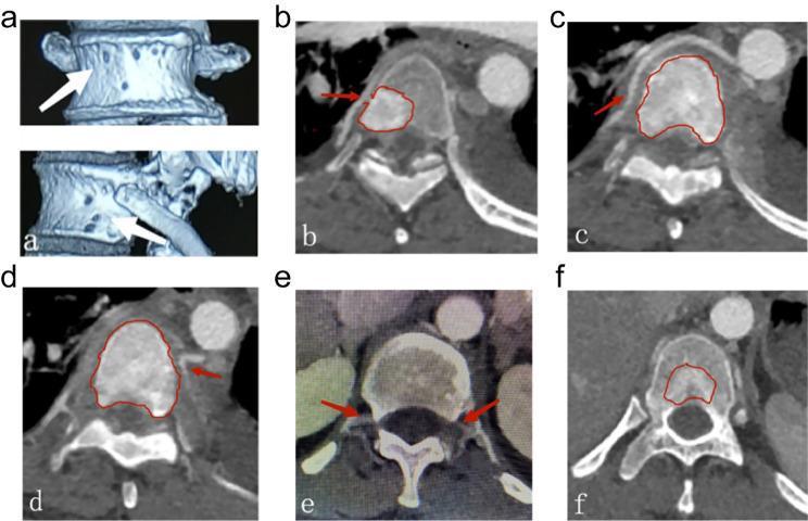
This composite of diagnostic images illustrates the vascular anatomy and tumoral blood supply of the vertebral column. Panel (a) presents 3D reconstructed CT images showing the anterior and lateral vertebral body surfaces, with white arrows indicating the entry points (pores) of nutrient arteries, including periosteal and metaphyseal rete-like branches. Panels (b) through (f) are axial contrast-enhanced CT scans of the thoracic vertebrae. Red arrows in (b) and (c) demonstrate nutrient arteries originating from the right segmental artery, while (d) shows an origin from the left. Panel (e) highlights bilateral segmental and spinal branches providing blood flow to the posterior vertebral body. Red outlines in (b, c, d, f) demarcate hypervascular metastatic spinal malignancies within the vertebral body, illustrating how specific segmental arterial branches provide nourishment to the tumor. These images serve as a clinical guide for understanding vertebral vascular territories (Zone 1 and 2) and planning preoperative arterial embolization to manage intraoperative bleeding in spinal oncology.
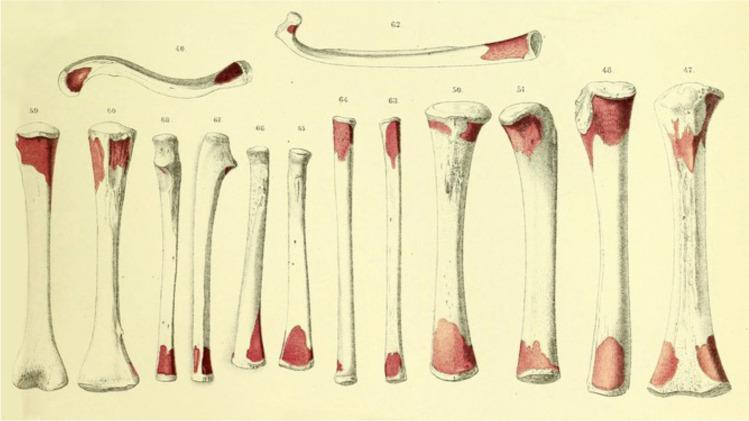
This historical anatomical diagram, credited to Albert Kölliker, illustrates the zones of periosteal resorption on various human long bones, including the femur, tibia, humerus, and clavicle. The illustration depicts thirteen labeled bones (numbered 46–66) showcasing different morphologies, from thick weight-bearing bones to slender and curved structures. The primary educational focus is the visual representation of bone remodeling processes during growth. Distinctive red-shaded areas mark specific regions of bone resorption, which are predominantly localized at the epiphyses and metaphyses. These red zones vary in size and shape, appearing as irregular patches or concentrated caps at the bone ends, while the mid-diaphyses remain white. This diagram historically served to prove the osteogenetic function of the periosteum and the necessity of surface resorption for maintaining appropriate bone shape and proportion during longitudinal and appositional growth. It is a critical visual aid for understanding skeletal development and the cellular activities of osteoclasts in physiological bone modeling.
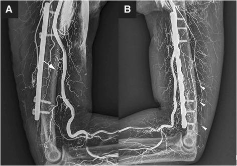
Two-panel plain radiograph (X-ray) depicting vascular perfusion of the humerus and elbow region, utilized for evaluating blood supply integrity following orthopedic surgery. Panel A shows an anteromedial view highlighting the main nutrient artery (white arrow), originating from the brachial artery and entering the mid-distal humeral shaft. Panel B demonstrates the posterior aspect, identifying multiple accessory nutrient arteries (white arrowheads) arising from the profunda brachii artery. Both images display surgical hardware in situ, including a long radiopaque metallic plate and cortical screws fixed to the humeral shaft, likely representing a Minimally Invasive Plate Osteosynthesis (MIPO) procedure. The radiographic contrast outlines the extensive arterial network, including the brachial artery and its branches, alongside the bone anatomy of the humerus, distal humerus, and proximal radius/ulna. This clinical image illustrates the vascular anatomy essential for humeral fracture healing and the impact of surgical fixation on regional perfusion.
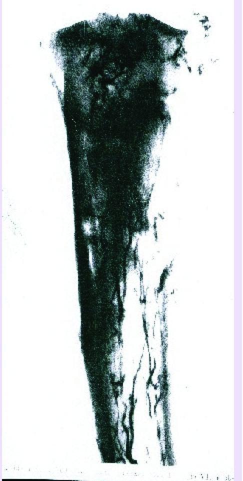
This diagnostic image is an angiogram illustrating the arterial vascularization of the proximal extremity of a human tibia. The image highlights the complex circulatory patterns essential for bone health and regeneration. The diaphyseal region demonstrates an arterial supply primarily derived from the nutrient artery, visible as a prominent, longitudinal vessel within the medullary canal. In contrast, the epiphyseal and metaphyseal regions show a more diffuse and intricate network of finer vessels. These arise from periosteal, capsular, and ligamentous sources, providing collateral circulation to the joint-adjacent structures. The contrast-enhanced vessels appear as dark, branching linear structures against the dense silhouette of the mineralized bone. This visualization is clinically relevant for understanding bone graft integration, the pathophysiology of osteonecrosis, and the healing of complex fractures or pseudarthrosis where vascular integrity is paramount for osteogenesis and remodeling.
osteomyelitis metaphysis hematogenous spread long bone children
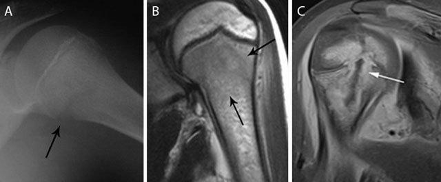
This composite figure illustrates the radiological presentation of acute pediatric hematogenous osteomyelitis in the proximal humerus across different modalities. Panel A is a plain radiograph showing a subtle osteolytic lesion in the metaphysis with early cortical blurring and loss of delineation at the medial aspect (black arrow). Panel B presents a coronal T2-weighted MRI showing hyperintense bone marrow edema (black arrows) primarily localized to the metaphyseal region. Panel C displays a coronal T1-weighted post-gadolinium contrast image with fat saturation, revealing a rim-enhancing intra-osseous abscess (white arrow). This image highlights transphyseal spread, where the infection has crossed the growth plate to involve the epiphysis, a critical finding in pediatric musculoskeletal imaging. These panels collectively demonstrate the evolution of findings from subtle radiographic changes to advanced abscess formation and epiphyseal extension, emphasizing the high sensitivity of MRI for detecting bone marrow changes and identifying surgical targets like rim-enhancing collections.
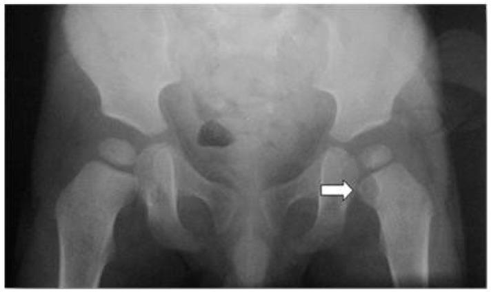
This anterior-posterior (AP) radiograph of a pediatric pelvis and proximal femurs demonstrates a focal osteolytic lesion in the proximal metaphysis of the left femur, marked by a white arrow. The lesion presents as a well-defined, radiolucent area situated just distal to the growth plate (epiphyseal plate). The surrounding cortical bone appears thinned, and the internal architecture of the lesion lacks calcification or an evident matrix. Key anatomical structures visible include the iliac wings, sacroiliac joints, pubic symphysis, and both hip joints. The femoral heads appear well-seated within the acetabula. In a pediatric clinical context involving fever and hip pain, this radiographic finding is highly suggestive of acute hematogenous osteomyelitis or a Brodie's abscess. The educational focus of the image is the identification of early lytic bone changes in pediatric musculoskeletal infection and the anatomical localization of metaphyseal lesions in long bones.
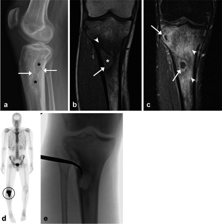
This figure presents a multi-modal imaging series of chronic hematogenous osteomyelitis in the proximal tibia of a 27-year-old male. (a) Lateral radiograph shows irregular lucent areas in the proximal tibia metaphysis (asterisks) with prominent reactive sclerosis (arrows). (b) Coronal T1-weighted MRI demonstrates a well-defined intraosseous abscess (asterisk) exhibiting the 'penumbra sign'—a thin hyperintense rim (arrow) indicative of vascularized granulation tissue—against a background of low-signal reactive sclerosis (arrowhead). (c) Coronal T1 post-contrast fat-saturated MRI reveals peripheral enhancement of the abscesses (arrows) and diffuse, intense enhancement of the adjacent bone marrow (arrowheads), indicating active inflammation. (d) Whole-body bone scan shows a focal area of intense radiopharmaceutical uptake in the right proximal tibia. (e) Intraoperative fluoroscopic image captures the aspiration of purulent material from the osseous pockets. These findings collectively illustrate the classic radiographic and MR features of Brodie’s abscess and chronic osteomyelitis.
Blood Supply of Long Bones - with Clinical Anatomy
Overview
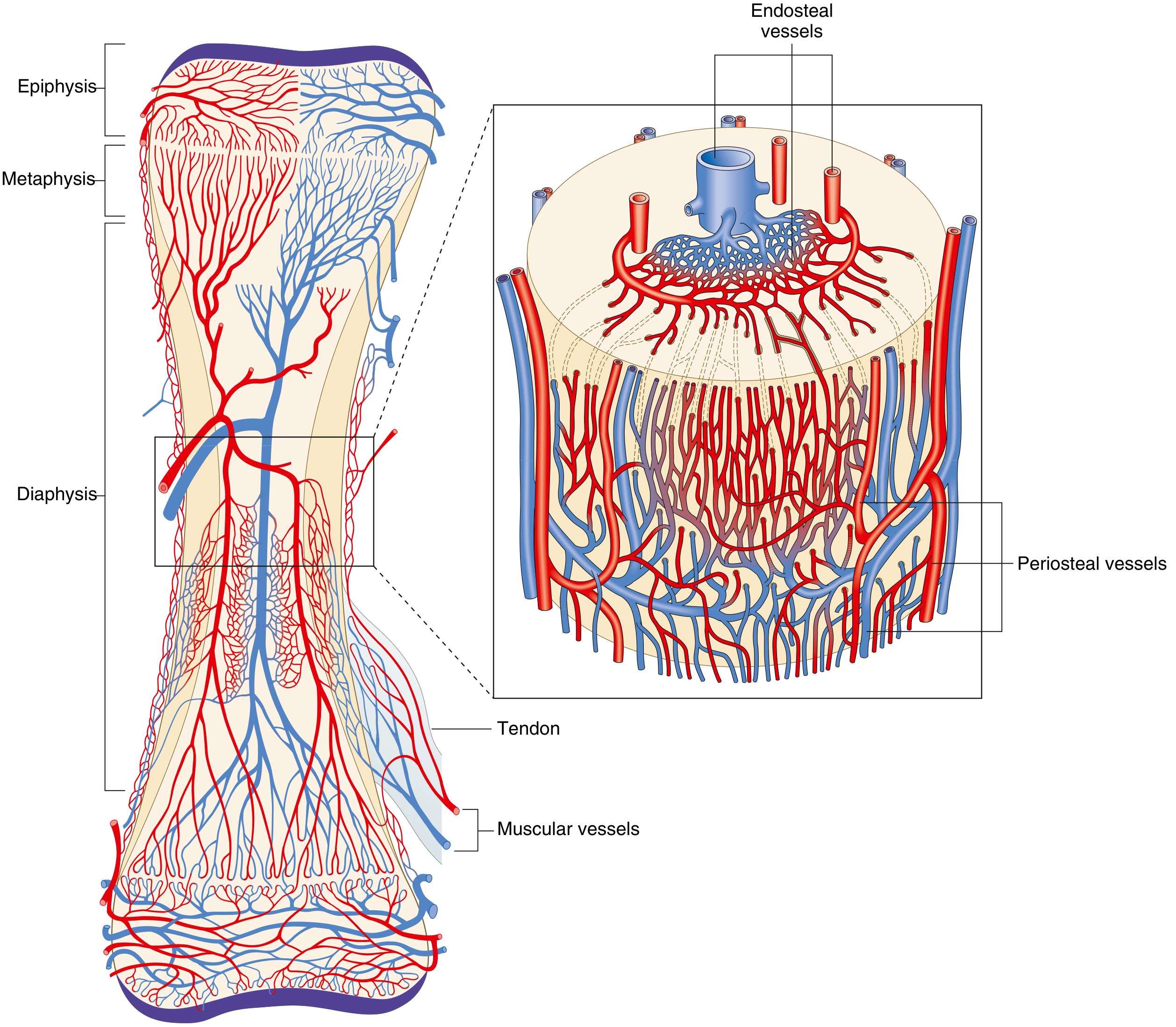
The Three Systems of Blood Supply
1. Nutrient Artery System (Diaphyseal / Medullary System)
- Branches from a named systemic artery (e.g., brachial for humerus, posterior tibial for tibia)
- Enters via the nutrient foramen in the diaphyseal cortex, directed obliquely toward the nearest growing end ("towards the elbow, away from the knee")
- Passes through the nutrient canal into the medullary cavity
- Divides into ascending and descending medullary arteries within the canal
- These branch into arterioles in the endosteal cortex, supplying blood via the Haversian system
- Supplies the inner 2/3 of the mature diaphyseal cortex
- Serves as the primary artery to bone marrow
- High pressure system
- Accounts for 60% of cortical bone vascularization
2. Metaphyseal-Epiphyseal System
- Arises from the periarticular vascular plexus (e.g., geniculate arteries for knee, circumflex humeral arteries for shoulder)
- Multiple small vessels enter through numerous foramina in the metaphysis
- Supplies the metaphysis and epiphysis richly
- Epiphyseal arteries enter through the non-articular bone and supply the bony epiphysis
- Metaphyseal arteries developmentally arise from periosteal vessels incorporated into the metaphysis as bone grows in diameter
- In the immature skeleton, the physis (growth plate) acts as a barrier - epiphyseal and metaphyseal circulations are separate
- After growth plate closure in adults, anastomoses form between metaphyseal and epiphyseal vessels
3. Periosteal System
- Capillaries derived from surrounding muscles, tendons, and fascial attachments
- Supplies the outer 1/3 of the mature diaphyseal cortex
- Blood enters via Volkmann's canals (transverse canals perpendicular to Haversian canals), then into Haversian canals
- Low pressure system
- Less dominant in normal mature bone
- Bone tissue lacks lymphatic vessels - lymphatic drainage occurs only from the periosteum
Diagrams
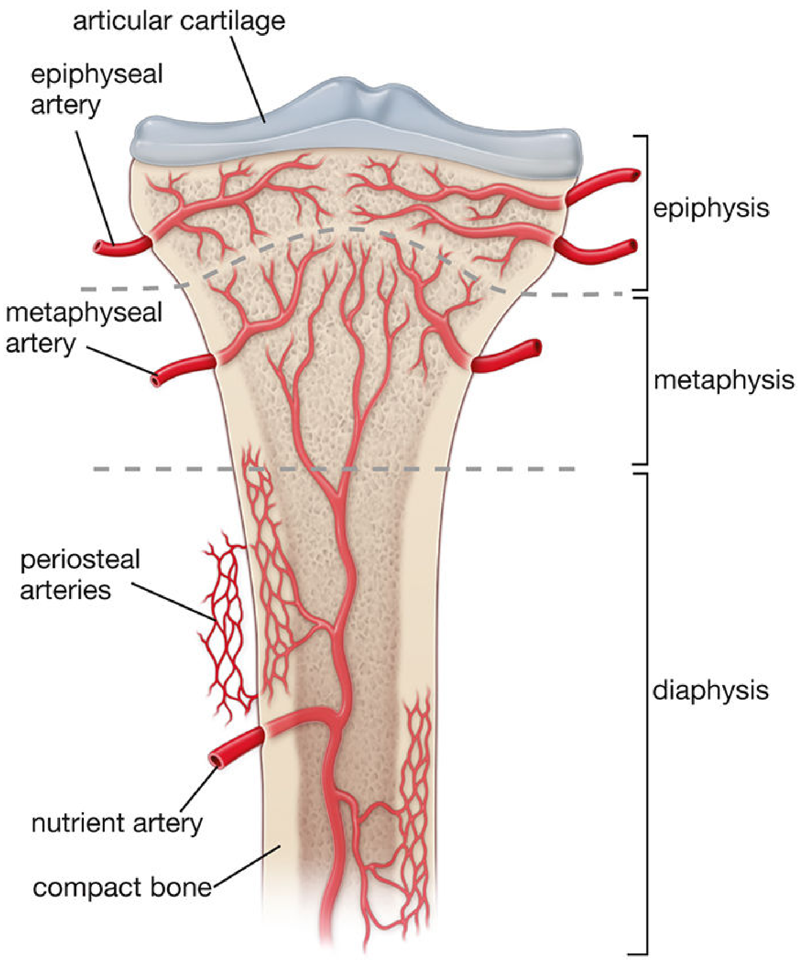
Direction of Blood Flow
| State | Arterial Flow | Venous Flow |
|---|---|---|
| Normal mature bone | Centrifugal (inside → outside) | Centripetal (outside → inside) |
| Fracture / immature bone | Centripetal (periosteal dominates) | Centrifugal |
- In normal bone, the high-pressure nutrient artery pushes blood outward through the cortex; the low-pressure periosteal system provides the outer third
- After fracture disrupting the nutrient artery (or in the immature skeleton), periosteal pressure dominates and flow reverses to centripetal
Regulation of Bone Blood Flow
- Regulated by metabolic, humoral, and autonomic inputs
- Hypoxia, hypercapnia, and sympathectomy all increase arterial bone blood flow
- Vessels within bone have vasoactive receptors (β-adrenergic, muscarinic, thromboxane/prostaglandin)
- The arterial system has greater potential for vasoconstriction than vasodilation
CLINICAL ANATOMY
1. Hematogenous Osteomyelitis - Why the Metaphysis?
- The metaphyseal capillary loops make sharp hairpin turns at the physis
- Blood flow here is slow and sinusoidal - bacteria lodge in the sluggish sinusoidal vessels
- There are few phagocytic cells in this region (relatively immunocompromised zone)
- This creates an ideal environment for bacterial seeding
| Age Group | Spread Pattern | Reason |
|---|---|---|
| Neonates/Infants (<1yr) | Metaphysis → Epiphysis → Joint (septic arthritis common) | Transphyseal vessels still present; physis not a barrier |
| Children (1yr - puberty) | Lateral via Volkmann canals → Subperiosteal abscess | Physis acts as barrier; periosteum loosely attached |
| Adults (post-epiphyseal fusion) | Metaphysis → Epiphysis → Joint (again) | New metaphyseal-epiphyseal anastomoses form after plate closure; periosteum firmly attached (limits subperiosteal abscess) |
2. Avascular Necrosis (AVN / Osteonecrosis)
- Scaphoid - blood supply enters distally through the waist; proximal pole relies on retrograde flow
- Femoral head - retinacular vessels (branches of medial circumflex femoral artery); disrupted by femoral neck fractures or hip dislocation
- Talus - 3 arterial sources, all disrupted in high-energy fracture-dislocations
- Odontoid process - small arterial supply
3. Fracture Healing and Blood Supply
- Bone blood flow is the major determinant of fracture healing
- Initial response = decrease in bone blood flow
- Blood flow increases within hours to days (regional acceleratory phenomenon)
- Peaks at approximately 2 weeks
- Returns to normal in 3-5 months
- Unreamed intramedullary nails preserve endosteal blood supply
- Reamed nails devascularize the inner 50-80% of the cortex and delay revascularization
- Stripping periosteum during plating impairs the periosteal contribution to healing
4. Surgical and Procedural Relevance
- Humeral head: Anterior and posterior circumflex humeral arteries are critical - risk of AVN in 4-part proximal humerus fractures and fracture-dislocations
- Femoral head: Medial circumflex femoral artery (via retinacular vessels) is the dominant supply - disrupted in subcapital neck fractures → AVN of femoral head
- Talar body: Blood supply disrupted in Hawkins type III/IV talar fracture-dislocations → AVN
- Nutrient foramen direction: "Towards the elbow, away from the knee" - oblique entry means the direction of the canal can indicate which physis is more dominant (used in fracture biomechanics interpretation)
Summary Table
| System | Supplies | Pressure | % Cortex |
|---|---|---|---|
| Nutrient artery | Inner 2/3 cortex, marrow | High | ~60% |
| Metaphyseal-epiphyseal | Metaphysis, epiphysis | Moderate | - |
| Periosteal | Outer 1/3 cortex | Low | ~40% |
- Miller's Review of Orthopaedics 9th Ed., p. 25-26
- Histology: A Text and Atlas (Wolters Kluwer), p. 589
- Rosen's Emergency Medicine, p. 2720-2721
- Imaging Anatomy: Bones, Joints, Vessels and Nerves (Thieme), p. 172
For UG ??
Blood Supply of Long Bones - UG Level
Quick Recall: Parts of a Long Bone
- Epiphysis - the ends (covered by articular cartilage)
- Physis (growth plate) - active in children, fused in adults
- Metaphysis - the flared region just below the physis
- Diaphysis - the shaft (the long middle part)
Sources of Blood Supply
| # | Source | Supplies |
|---|---|---|
| 1 | Nutrient artery | Shaft (diaphysis) + bone marrow |
| 2 | Metaphyseal & Epiphyseal arteries | The two ends of the bone |
| 3 | Periosteal arteries | Outer cortex of shaft |
1. Nutrient Artery (Most Important - Exam Favourite)
- 1 or 2 per bone, branch of a nearby named systemic artery
- Enters through the nutrient foramen on the diaphysis
- Travels obliquely through cortical bone via the nutrient canal
- Reaches the medullary cavity and divides into:
- Ascending branch → towards one end
- Descending branch → towards the other end
- These further branch into arterioles supplying the inner 2/3 of the cortex via Haversian canals
Direction of nutrient foramen - Memory Trick
"Towards the elbow, away from the knee"
| Bone | Direction nutrient foramen points |
|---|---|
| Humerus | Towards elbow (downward) |
| Radius | Towards elbow (downward) |
| Ulna | Towards elbow (downward) |
| Femur | Away from knee (upward = towards hip) |
| Tibia | Away from knee (downward = towards ankle) |
| Fibula | Away from knee (downward) |
2. Metaphyseal and Epiphyseal Arteries
- Arise from periarticular arterial plexuses around the joint
- e.g., Geniculate arteries for knee, circumflex humeral arteries for shoulder
- Enter the bone through multiple small foramina in the metaphysis and epiphysis
- Supply cancellous (spongy) bone of the ends richly
3. Periosteal Arteries
- Small vessels from surrounding muscles, tendons, and periosteum
- Enter via Volkmann's canals (transverse channels in cortex)
- Supply only the outer 1/3 of the cortex
- Low pressure - normally the least dominant system
Direction of Blood Flow (High-Yield)
| Situation | Flow Direction | Why |
|---|---|---|
| Normal mature bone | Centrifugal (inside → outside) | High-pressure nutrient artery dominates |
| Fracture / child's bone | Centripetal (outside → inside) | Periosteal system takes over |
Think of it as: normally blood flows from the medulla outward through the cortex. When the main supply (nutrient artery) is cut off (fracture), the backup (periosteal) takes over and flows inward.
Clinical Anatomy - The Exam Connections
A. Hematogenous Osteomyelitis - Why the Metaphysis?
- The metaphyseal capillaries make sharp hairpin bends near the growth plate
- Blood flow is slow and sluggish in the sinusoidal capillaries here
- There are few phagocytic cells in this area
- Bacteria settle here easily → infection starts
| Age | Spread | Reason |
|---|---|---|
| Neonate (<1 yr) | Metaphysis → Epiphysis → Joint | Transphyseal vessels cross the physis |
| Child (1 yr - puberty) | Metaphysis → Subperiosteal abscess | Physis is a barrier; periosteum loosely attached |
| Adult | Metaphysis → Epiphysis → Joint | Physis fused, anastomoses reform; periosteum firmly attached |
Clinical pearl: In children, the metaphysis of the lower femur, upper tibia, upper humerus are most commonly involved (fastest-growing ends = richest blood supply = most bacteria settle there)
B. Avascular Necrosis (AVN) - Bones at Risk
| Bone | Why at Risk | Common Cause |
|---|---|---|
| Femoral head | Retinacular vessels easily torn | Femoral neck fracture, hip dislocation |
| Scaphoid (proximal pole) | Distal-to-proximal retrograde supply | Fracture at waist cuts off proximal pole |
| Talus (body) | 3 entry points all disrupted | High-energy fracture-dislocation |
| Odontoid (C2) | Sparse blood supply from below | Fracture of dens |
Rule: if a bone's blood supply enters from one end only (like proximal scaphoid), a fracture between the entry point and that end = AVN of the isolated fragment.
C. Fracture Healing
- Blood supply is the most important factor in fracture healing
- After fracture: initial drop → then rises within hours (regional acceleratory phenomenon) → peaks ~2 weeks → normalizes in 3-5 months
- Stripping the periosteum during surgery removes the periosteal supply → impairs healing
- Intramedullary nails (unreamed) preserve endosteal supply
- Reamed IM nails destroy inner 50-80% of cortical blood supply temporarily
D. Volkmann's Canal vs. Haversian Canal (1-liner)
- Haversian canals = run longitudinally along the bone, carry the main vessels within osteons
- Volkmann's canals = run transversely, connect Haversian canals to the periosteum and to each other
One-Liner Summary for Quick Revision
A long bone gets blood from: (1) nutrient artery → medulla + inner cortex, (2) metaphyseal/epiphyseal arteries → the ends, (3) periosteal arteries → outer cortex. Normal flow is centrifugal. The metaphysis is the osteomyelitis hotspot due to sluggish sinusoidal flow. AVN happens in bones with end-artery / retrograde supply.