
Exophthalmos radio active iodine vs thyroid surgery which should be preferred
Graves ophthalmopathy radioactive iodine thyroid surgery exophthalmos
PMID: 38686801
Graves ophthalmopathy exophthalmos proptosis
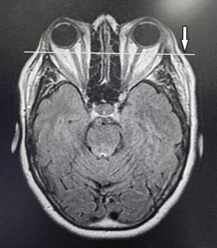
This diagnostic image is an axial view Magnetic Resonance Imaging (MRI) of the head, specifically focusing on the orbits and brain. The scan demonstrates significant bilateral proptosis (exophthalmos), characterized by the anterior displacement of the globes relative to the orbital rims. A horizontal white line and a white arrow on the left side highlight the degree to which the eyeballs protrude beyond the lateral orbital margin. Visible within the retrobulbar space is evidence of extraocular muscle enlargement and orbital fat hypertrophy, typical findings associated with Graves' ophthalmopathy (thyroid-associated orbitopathy). The optic nerves are visible and appear stretched due to the globe displacement. The brain section shows the midbrain, cerebral hemispheres with visible gyri and sulci, and the interhemispheric fissure. This image serves as an educational tool for identifying radiological signs of restrictive orbitopathy and secondary complications like proptosis.
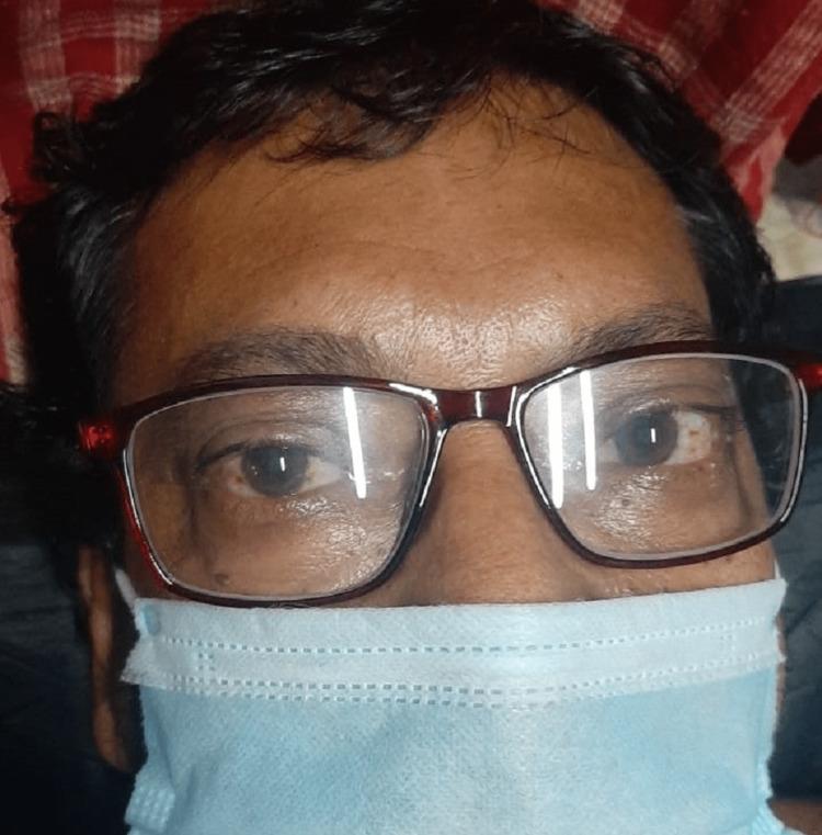
Clinical photograph of a 53-year-old male presenting with bilateral Graves' ophthalmopathy (thyroid eye disease). The image focuses on the periocular region, demonstrating bilateral exophthalmos (proptosis) characterized by the protrusion of the globes from the orbits. Significant scleral show is visible, particularly inferior to the iris, contributing to a characteristic 'staring' expression or Dalrymple's sign. The patient is wearing corrective eyeglasses and a surgical face mask, indicating a clinical setting. This visual illustrates the extrathyroidal manifestations of Graves' disease, an autoimmune condition causing hypertrophy of extraocular muscles and orbital adipose tissue. The image is an educational resource for medical professionals to recognize clinical signs of hyperthyroidism-associated orbitopathy and for calculating Clinical Activity Scores (CAS) in endocrinology and ophthalmology.
Exophthalmos (Graves' Ophthalmopathy): RAI vs. Thyroid Surgery - Which to Prefer?
Why RAI Is Problematic in Exophthalmos
"Radioactive iodine may worsen Graves ophthalmopathy and is contraindicated in patients who have significant Graves ophthalmopathy unless patients receive concomitant high doses of corticosteroid therapy for 4 to 6 weeks after the administration of radioactive iodine."
- Goldman-Cecil Medicine, Table 207-6 & RAI section
"Can worsen Graves' ophthalmopathy (especially in smokers)"
- K.J. Lee's Essential Otolaryngology, Radioactive Iodine Ablation section
- Goldman-Cecil Medicine
Why Surgery Is Preferred
| Feature | RAI | Surgery (Total Thyroidectomy) |
|---|---|---|
| Effect on ophthalmopathy | Worsens GO (especially in smokers) | Does not worsen GO; may improve it |
| Speed of control | 6-8 weeks for TH normalization | Faster normalization |
| GO risk (network meta-analysis) | HR 1.70 vs ATD for developing GO | HR 0.18 vs ATD for GO (lowest risk) |
| Overall mortality | Higher than thyroidectomy | Lowest of all 3 treatments |
| Hypothyroid rate | Up to 80% | Universal (expected and manageable) |
| Cure | ~75-90% after one dose | Definitive cure |
- Faster correction than RAI without the risks of antithyroid drugs
- Lower risk of hypothyroidism compared to RAI in many studies
- Particularly suited when large goiter is also present
The Specific Exception for RAI
The Washington Manual Summary
"Thyroidectomy may be used in patients who refuse RAI therapy, have significant Graves ophthalmopathy, or who relapse or develop side effects with thionamide therapy."
- Washington Manual of Medical Therapeutics
Current Evidence (2024 Network Meta-Analysis)
- Thyroidectomy was associated with significantly lower risk of GO than both ATD (HR 0.31) and RAI (HR 0.18)
- RAI had a higher risk of GO than ATD (HR 1.70)
- Thyroidectomy was also associated with the lowest overall mortality
Practical Summary
| Clinical Scenario | Preferred Treatment |
|---|---|
| Graves disease + significant/active GO | Surgery (total thyroidectomy) - RAI is contraindicated |
| Graves disease + mild/no GO | RAI is generally first-line |
| Graves disease + mild GO + must use RAI | RAI + corticosteroids for 4-6 weeks |
| Graves disease + GO + smoker | Surgery strongly preferred (RAI worsens GO much more in smokers) |
| Pregnant patient with GO | Neither RAI nor surgery acutely - antithyroid drugs first |
- Goldman-Cecil Medicine, 26th ed., Ch. 207 - Radioactive Iodine Therapy & Graves Disease sections
- K.J. Lee's Essential Otolaryngology - Radioactive Iodine Ablation & Surgical Treatment sections
- Washington Manual of Medical Therapeutics - Choice of Definitive Therapy
- Liu X et al. (2024) - Bayesian network meta-analysis, World J Surg, PMID 38686801