
Steven jhonson syndeome
Reading File
Searching sources...
Finding Sources
Searching PubMed
"Stevens-Johnson syndrome"[MeSH Terms] AND management
Now let me read the primary sources - Fitzpatrick's dermatology (most authoritative for SJS) and Goldman-Cecil Medicine:
Reading File
Reading File
Let me now read the main SJS chapter from Fitzpatrick's (Chapter 44) and Goldman-Cecil's full SJS section:
Finding Sources
Reading File
Now let me read the comprehensive Andrews' Diseases of the Skin chapter on SJS/TEN, which is the most detailed:
Reading File
Reading File
Excellent. I now have comprehensive information. Let me read a bit more of Andrews' for the pathogenesis, clinical features, and management details:
Reading File
I now have comprehensive data from multiple authoritative textbooks. Let me also fetch a clinical image of SJS to include in the answer:
Finding Sources
I have enough comprehensive information to write a detailed, well-cited answer now. Let me fetch the TEN image from Goldman-Cecil:
Loading Image
I now have everything needed for a comprehensive, well-illustrated answer.
Stevens-Johnson Syndrome (SJS)
Overview and Classification
Stevens-Johnson syndrome (SJS) and toxic epidermal necrolysis (TEN) represent a spectrum of the same disease - rare, acute, life-threatening mucocutaneous reactions that are nearly always drug-induced. They are classified by the extent of epidermal detachment:
| Category | Epidermal Detachment | Mortality |
|---|---|---|
| SJS | < 10% BSA | ~5% |
| SJS/TEN overlap | 10-30% BSA | ~19% |
| TEN (Lyell syndrome) | > 30% BSA | 15-25% |
(BSA = body surface area)
- Dermatology 2-Volume Set 5e, p. 412
- Goldman-Cecil Medicine, p. 4306
Epidemiology
- Incidence: 1.2-6 per million person-years (SJS); 0.4-1.2 per million (TEN)
- Female:male ratio ~ 1.5:1
- Over 95% of TEN cases and the majority of SJS cases are drug-induced
- HIV infection significantly increases risk (up to 1000-fold)
- Andrews' Diseases of the Skin, p. 342
Causative Drugs
More than 100 drugs have been implicated. The highest-risk drugs include:
Highest risk:
- Allopurinol - currently the most common cause in Europe
- Sulfonamides (TMP-SMX): 1-3 per 100,000
- Anticonvulsants: carbamazepine (14:100,000), lamotrigine (1:1,000 adults), phenytoin, phenobarbital, valproate
- Nevirapine (antiretroviral)
- NSAIDs (especially oxicam class)
Also implicated:
- Aminopenicillins, cephalosporins, quinolones, tetracyclines
- Fansidar (sulfadoxine/pyrimethamine): 10:100,000 - highest incidence of any drug
- Chlormezanone, acetaminophen (particularly in children)
The reaction typically begins 7-21 days after starting the causative drug. For anticonvulsants, risk is greatest during the first 2 months of treatment.
- Andrews' Diseases of the Skin, p. 342
- Goldman-Cecil Medicine, p. 4305
Pharmacogenetics / HLA Associations
Genetic susceptibility plays a major role, especially in specific ethnic groups:
| HLA Allele | Drug | Population |
|---|---|---|
| HLA-B*15:02 | Carbamazepine | Han Chinese, Thai, Malaysian, East Indian |
| HLA-B*15:02 | Lamotrigine | Taiwanese |
| HLA-B*15:02 | Phenytoin | Han Chinese |
| HLA-B*58:01 | Allopurinol | Han Chinese |
| HLA-A*31:01 | Carbamazepine | Europeans |
Clinical implication: HLA typing should be performed in all Asians before starting carbamazepine. The FDA recommends screening for HLA-B*15:02 in patients of Asian ancestry. HLA-B*15:02 is present in 4-15% of affected Asian populations but is extremely rare in Northern Europeans (<0.01%).
- Dermatology 2-Volume Set 5e, p. 412
Pathogenesis
The core mechanism is extensive keratinocyte apoptosis leading to full-thickness epidermal necrosis and separation at the dermal-epidermal junction.
Key mediators:
- Granulysin - a cytotoxic secretory protein released by CD8+ cytotoxic T lymphocytes and NK cells; this is now considered the primary mediator of keratinocyte killing
- Fas-FasL interaction - death receptor-ligand pair triggering apoptosis
- Drug-specific T-cell activation (pharmacogenetic interaction between drug/metabolite and HLA)
- TNF-alpha also contributes to keratinocyte death
The histology shows full-thickness epidermal cell death with a subepidermal split and a lymphohistiocytic infiltrate with eosinophils in the dermis.
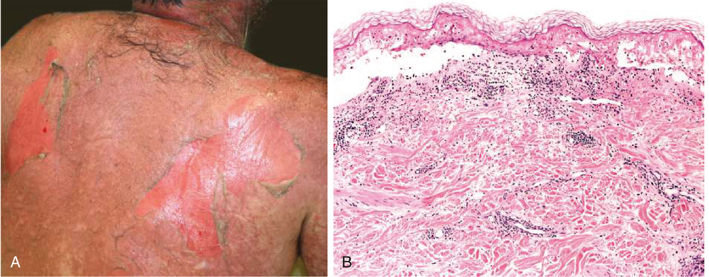
Figure: TEN showing denuded skin (A) and histology with full-thickness epidermal cell death and subepidermal separation (B). From Goldman-Cecil Medicine.
Clinical Features
Prodrome (1-3 days before skin lesions)
- Fever, malaise, sore throat
- Burning/painful eyes
- Painful skin - a key early warning sign
- Upper respiratory tract symptoms
Mucosal Involvement (almost always present)
- Oral/lip mucosa: erosions, hemorrhagic crusting, difficulty eating/swallowing
- Ocular: conjunctivitis, pseudomembrane formation, corneal erosions - can lead to permanent visual impairment
- Genital: erosions, urethritis
- Respiratory: bronchial epithelial sloughing (severe cases)
- Mucous membrane involvement typically precedes or accompanies skin changes
Skin Lesions
- Begin as erythematous macules on the trunk and face
- Evolve to flat, atypical target lesions (not the classic 3-ring targets of EM)
- Nikolsky sign positive: lateral pressure on erythematous skin causes epidermal detachment
- Asboe-Hansen sign: pressure on bullae causes lateral extension
- Blisters coalesce to form large areas of denuded skin
Distribution
- Starts on trunk, spreads to face and proximal extremities
- Palms/soles involved
- Acral surfaces > central in some cases
Systemic Involvement (severe cases/TEN)
- Pulmonary: bronchial sloughing, ARDS
- Hepatic involvement
- Renal impairment
- GI tract involvement
Diagnosis
Clinical diagnosis based on:
- Characteristic skin and mucosal lesions
- Positive Nikolsky sign
- History of drug exposure 7-21 days prior
- Skin biopsy: full-thickness necrosis, subepidermal blister
SCORTEN (Severity of Illness Score for TEN) - calculated at day 1 and day 3:
| Parameter | 1 point each if present |
|---|---|
| Age > 40 years | |
| Malignancy | |
| Heart rate > 120/min | |
| Initial BSA detachment > 10% | |
| BUN > 28 mg/dL (10 mmol/L) | |
| Serum bicarbonate < 20 mEq/L | |
| Serum glucose > 252 mg/dL (14 mmol/L) |
SCORTEN score 0-1 = ~3.2% mortality; score ≥5 = >90% mortality
ALDEN score helps establish causality (drug to SJS/TEN relationship).
Differential Diagnosis
| Condition | Distinguishing Features |
|---|---|
| Staphylococcal scalded skin syndrome (SSSS) | Cleavage is subcorneal (not full thickness); occurs mainly in children; Gram+ staph exotoxin |
| Generalized bullous fixed drug eruption | No systemic involvement; marked hyperpigmentation; previous lesions at same site |
| Drug-induced linear IgA bullous dermatosis | IgA on DIF |
| Erythema multiforme (EM) | True 3-zone targets; acral distribution; usually HSV-triggered; no/minimal mucositis |
| AGEP | Pustular, not bullous; rapid resolution on drug withdrawal |
Management
1. Immediate - Stop the Causative Drug
This is the single most important intervention. Each additional day of continued drug use is associated with increased mortality. Drugs with short half-lives should be stopped first.
2. Supportive Care (mainstay of treatment)
- Transfer to a burn unit or specialized center - outcomes are significantly better
- Fluid and electrolyte replacement (treat like a burn patient)
- Temperature-controlled environment (patients cannot thermoregulate)
- Wound care: non-adhesive dressings, avoid debridement of intact blisters
- Nutritional support: early enteral feeding
- Ophthalmologic evaluation and care - daily, to prevent permanent sequelae (amniotic membrane transplant in acute phase)
- Pain management
- Infection surveillance and treatment (leading cause of death is sepsis)
3. Specific/Adjunctive Therapies
(No single therapy has definitive RCT evidence; options include:)
| Agent | Rationale/Use |
|---|---|
| TNF-alpha inhibitors (etanercept 50 mg SC x1-2 doses) | Blocks TNF-mediated keratinocyte apoptosis; observational data suggests benefit in TEN |
| Cyclosporine (3-5 mg/kg/day) | Inhibits T-cell activation and Fas-FasL pathway; most studied adjunctive agent |
| IVIg (1 g/kg/day x3 days) | May block Fas-FasL; may decrease fever duration; high doses may help |
| Corticosteroids (IV methylprednisolone 1-2 mg/kg or 60 mg q6h short course) | Controversial; may increase infection risk; some benefit in early disease |
- Goldman-Cecil Medicine, p. 4307
- Dermatology 2-Volume Set 5e, p. 413-414
Note: A 2024 systematic review on SJS/TEN from immune checkpoint inhibitors (PMID 39279714) highlights a growing subset of drug-induced cases from oncologic immunotherapy agents, requiring modified management approaches.
Sequelae / Complications
Ocular (most common long-term problem):
- Symblepharon, trichiasis, corneal scarring, dry eye syndrome, visual impairment/blindness
Skin:
- Post-inflammatory hyper/hypopigmentation
- Nail dystrophy or nail loss (anonychia)
- Scarring alopecia
Mucosal:
- Oral/esophageal strictures
- Phimosis, vaginal synechiae
- Bronchiolitis obliterans (rare but severe)
Psychological:
- PTSD, depression, significant reduction in quality of life
Key Points Summary
- SJS/TEN is a spectrum - classified by % BSA epidermal detachment (<10%, 10-30%, >30%)
- Almost always drug-induced in adults; onset 7-21 days after starting the drug
- Top culprits: allopurinol, sulfonamides, carbamazepine, lamotrigine, NSAIDs, nevirapine
- HLA screening (especially HLA-B*15:02) before carbamazepine in Asian patients is mandatory
- Pathogenesis: CD8+ T cell/NK cell-mediated keratinocyte apoptosis via granulysin and Fas-FasL
- Diagnosis: clinical + positive Nikolsky sign + skin biopsy
- SCORTEN predicts mortality
- Treatment: stop the drug immediately + burn unit care + supportive therapy; cyclosporine and etanercept are most promising adjunctive agents
- Ocular sequelae are the most common cause of long-term morbidity
Sources: Andrews' Diseases of the Skin, p. 340-345; Dermatology 2-Volume Set 5e, pp. 412-415; Goldman-Cecil Medicine, pp. 4305-4307; Fitzpatrick's Dermatology Vol 1-2, p. 783
Its effect on esophogus
Searching sources...
Finding Sources
Finding Sources
Reading File
Reading File
Reading File
Now let me also search for any additional details on esophageal stricture management in SJS:
Finding Sources
Searching PubMed
"Stevens-Johnson syndrome"[MeSH Terms] AND "esophagus"[MeSH Terms]
Searching PubMed
"Stevens-Johnson" AND esophageal
Good - I have two highly relevant recent case reports (esophageal bleeding, esophageal epidermoid metaplasia). Let me get their abstracts quickly:
Reading Article
PMID: 39500588
Reading Article
PMID: 39280887
I now have comprehensive, multi-source data to write a focused, well-cited answer.
SJS/TEN: Effects on the Esophagus
The esophagus is the primary gastrointestinal organ affected in SJS/TEN, though involvement beyond the oropharynx is considered uncommon in SJS and more prominent in TEN.
Why the Esophagus Is Affected
The same immune-mediated mechanism driving skin destruction - CD8+ cytotoxic T-lymphocyte attack on epithelial cells - targets the squamous epithelium lining the esophagus. Since the esophagus (especially the upper two-thirds) is lined with non-keratinized stratified squamous epithelium, it is vulnerable to the same Fas-FasL and granulysin-mediated apoptosis that causes epidermal detachment in the skin.
- Yamada's Textbook of Gastroenterology, p. 2313
Acute Phase: Esophageal Manifestations
1. Esophagitis / Mucosal Inflammation
- The first and most common manifestation
- Ranges from mild esophagitis to diffuse ulceration and necrosis
2. Endoscopic Findings
At upper GI endoscopy, the esophagus may show:
- Diffuse erythema and friability
- Whitish plaques - can mimic candida esophagitis (important diagnostic pitfall)
- Necrotic plaques with mucosal sloughing
- Ulcerations along the esophageal mucosa
Biopsy differentiates SJS esophagitis from candidiasis: SJS shows lymphocytic infiltration and epithelial necrosis, not fungal hyphae.
3. Massive Gastrointestinal Bleeding
In severe cases, esophageal mucosal sloughing can lead to life-threatening hemorrhage. A 2024 case report (PMID 39500588) described a patient with SJS who developed massive GI bleeding with haemodynamic instability; endoscopy revealed necrotic plaques and mucosal sloughing throughout the esophagus requiring intensive care, multiple transfusions, and IV dexamethasone.
4. Oropharyngeal/Esophageal Pain
-
Severe oropharyngeal mucositis with erosions and mucosal sloughing
-
Odynophagia (painful swallowing) and dysphagia that may severely limit oral intake
-
Nutritional support (enteral or parenteral) is often required acutely
-
Sleisenger and Fordtran's GI and Liver Disease, p. 381
GI Involvement Beyond the Esophagus
While the esophagus bears the brunt of GI involvement, SJS/TEN can affect other parts of the gut:
| Site | Findings |
|---|---|
| Stomach / Duodenum | Diffuse erythema and friability (even without esophageal involvement) |
| Colon | Appearance resembles severe ulcerative colitis or pseudomembranous colitis; biopsy shows necrosis and lymphocytic infiltrate without crypt abscesses or neutrophils |
| Bowel (extensive TEN) | Mucosal sloughing of large bowel segments - can cause hematemesis, melena, and even intestinal perforation |
The colonic histology pattern resembles graft-versus-host disease - a key distinguishing feature from IBD.
- Sleisenger and Fordtran's GI and Liver Disease, p. 381
Chronic / Long-Term Sequelae
This is clinically important - esophageal complications can persist long after the acute disease resolves.
1. Esophageal Strictures and Webs
- Result from fibrosis and scarring following mucosal healing
- Can be single or multiple
- Cause progressive dysphagia weeks to months after the acute episode
- Esophageal dilation (endoscopic bougie or balloon) is often required for treatment
- Harrison's Principles of Internal Medicine 22E, p. 1872; Yamada's Textbook of Gastroenterology, p. 2313
2. Esophageal Epidermoid Metaplasia (EEM) - Newly Recognized
A 2024 pediatric case report (PMID 39280887) described the first case of esophageal epidermoid metaplasia developing in an 8-year-old girl after SJS with esophageal stricture. EEM is a rare condition in which the normal esophageal squamous epithelium undergoes keratinizing (epidermoid) metaplasia. This is significant because:
- EEM is associated with esophageal squamous cell carcinoma
- The authors recommend close surveillance, biopsy for dysplasia, endoscopic treatment, and TP53 genetic sequencing in pediatric SJS patients with esophageal involvement
Management of Esophageal Involvement
| Phase | Management |
|---|---|
| Acute | NPO or liquid diet; parenteral/enteral nutrition; IV fluids; treat GI bleeding with endoscopy if needed; IV corticosteroids (e.g., dexamethasone) for severe mucosal disease |
| Endoscopic | Upper GI endoscopy for diagnosis and to exclude candidiasis; avoid aggressive intervention on fragile mucosa acutely |
| Long-term | Surveillance for stricture development; serial endoscopic dilation for strictures; biopsy surveillance for dysplasia/metaplasia |
Key Takeaways
- The esophagus is the most commonly affected GI organ in SJS/TEN - other GI sites are rare but described
- Acute findings: esophagitis, diffuse erythema, whitish plaques (mimic candidiasis), mucosal sloughing, and rarely massive hemorrhage
- Colonic involvement histologically mimics GvHD (lymphocytic necrosis, no crypt abscesses)
- Long-term: esophageal strictures and webs requiring dilation are the primary sequela
- Newly recognized: esophageal epidermoid metaplasia post-SJS carries a risk of squamous cell carcinoma - surveillance is warranted
Sources: Yamada's Textbook of Gastroenterology 7e, p. 2313; Sleisenger and Fordtran's GI and Liver Disease, p. 381; Harrison's Principles of Internal Medicine 22E, p. 1872; PMID 39500588 (2024); PMID 39280887 (2024)
This is a shared conversation. Sign in to Orris to start your own chat.