
Define cell injury n explain types n Pathogenisis of reversible cell injury??
reversible cell injury cellular swelling hydropic change pathology
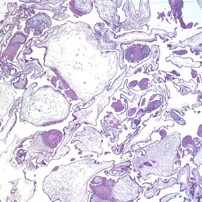
Low-power light microscopy of placental villous tissue from a hydatidiform mole. The slide demonstrates vesicular swelling (hydropic change) of chorionic villi with scalloped margins and diffuse villous edema. Trophoblastic proliferation is evident, appearing as nests and clusters of eosinophilic cytoplasm at the villous surface and within the stroma, consistent with trophoblastic hyperplasia. The severity of edema and hyperplasia varies from villus to villus. Fetal stromal blood vessels are usually absent, a hallmark distinguishing a complete mole from partial mole. The villi lack embryonic tissue, and the overall architecture shows circumferential trophoblastic proliferation around enlarged, hydropic villi. In some regions, hyalinization and cystic changes are visible, reflecting advanced hydropic degeneration. The image corresponds to a gonadotropin-secreting gestational trophoblastic disease with high risk of persistent GTN. Correlates with clinical findings such as markedly elevated hCG and potential uterine enlargement. This morphological pattern supports the differential diagnosis of complete mole versus partial mole or non-neoplastic hydropic changes. The pathology report would emphasize hydropic villi, diffuse trophoblastic hyperplasia, and absence of fetal vessels. This pattern is diagnostic of complete mole on histology and guides clinical management, including suction curettage, serial quantitative hCG monitoring, and pregnancy counseling.
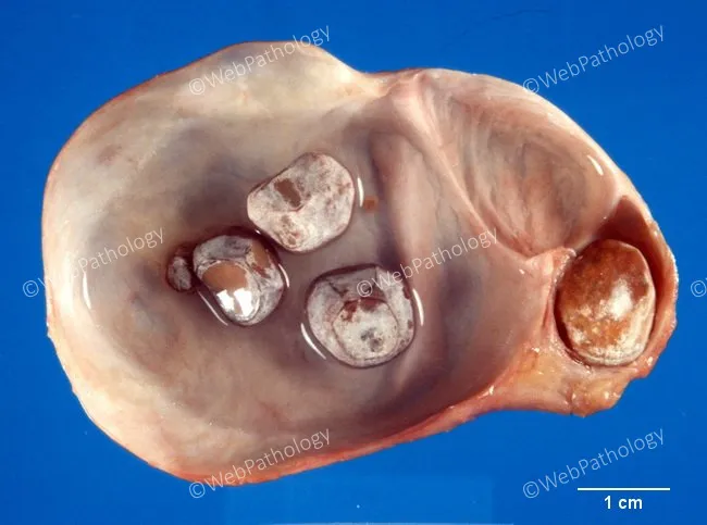
Gross pathology photograph of an opened gallbladder with cholelithiasis demonstrating hydrops due to cystic duct obstruction. The gallbladder is markedly distended with a thin, smooth lining of mucosa. An impacted calculus is seen at the cystic duct near the gallbladder neck on the right edge, with additional calculi within the lumen. Bile stasis and progressive dilation produce hydropic change without frank perforation. In adults, hydrops is most commonly caused by mechanical obstruction of the gallbladder neck or cystic duct by a stone; less commonly by tumor, fibrosis, cystic fibrosis-related thickening, ductal kinking, or external compression by a mass. In children, hydrops is typically acute, reversible, and often infectious; however, this image reflects an adult pattern of obstruction. The mucosa remains relatively smooth, and there is no overt acute cholecystitis shown. Pathophysiologic significance: obstruction leads to increased intraluminal pressure, bile stasis, mucosal atony, and potential secondary inflammation if persistent. Clinically, this pattern supports diagnosis of hydropic gallbladder due to gallstone disease and helps distinguish from inflammatory or neoplastic causes of gallbladder distension. Potential clinical use includes educational reference for surgical pathology, medical student training, radiologic-pathologic correlation, and differential diagnosis of biliary obstruction. Synonyms: gallbladder hydrops, hydropic gallbladder, obstructive cholecystopathy. Clinical relevance.
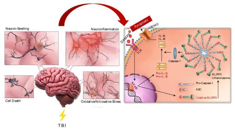
This pathophysiology diagram illustrates the cellular and molecular mechanisms of secondary injury following Traumatic Brain Injury (TBI). The left side features a brain illustration with four inset panels showing pathological processes: neuron swelling (cytotoxic edema), neuroinflammation (characterized by reactive gliosis and microglial activation), cell death (apoptosis/necrosis), and oxidative/nitrosative stress (indicated by mitochondrial dysfunction and ROS accumulation). The right side provides a detailed molecular schema of the NLRP3 inflammasome activation pathway. Damage-Associated Molecular Patterns (DAMPs) are shown interacting with Toll-Like Receptors (TLR), leading to NF-κB nuclear translocation and the synthesis of pro-IL-1β and pro-IL-18. The assembly of the NLRP3 inflammasome from inactive NLRP3, ASC, and pro-caspase-1 is depicted as a circular oligomeric complex. Active caspase-1 then cleaves the pro-cytokines into their mature forms (IL-1β, IL-18) and facilitates gasdermin D (GSDMD) mediated pyroptosis. This diagram summarizes the link between acute mechanical trauma and the chronic inflammatory cascade in neurotrauma.
Cell Injury - Definition, Types, and Pathogenesis of Reversible Cell Injury
Definition of Cell Injury
"Cell injury is the basis of all disease." - Robbins & Kumar Basic Pathology
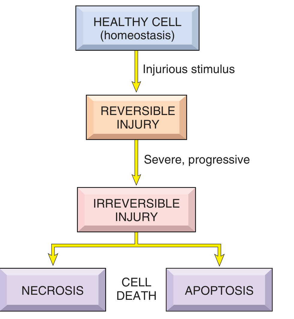
Types of Cell Injury
1. Based on Outcome
| Type | Definition | Outcome |
|---|---|---|
| Reversible | Functional and structural changes that are correctable if the injurious stimulus is removed | Cell recovers fully |
| Irreversible | Cell passes the "point of no return" - injury is too severe or prolonged | Cell death (necrosis or apoptosis) |
2. Based on Causes
Reversible Cell Injury - Definition
Pathogenesis of Reversible Cell Injury
Step-by-Step Pathogenesis
- Hypoxia interferes with mitochondrial oxidative phosphorylation
- Toxins or radiation may directly damage mitochondria
- Result: ATP levels fall
- This energy-dependent pump normally maintains ionic balance
- When ATP fails, the pump stops working
- Na⁺ accumulates inside the cell
- Cl⁻ follows (electrical neutrality)
- Water enters osmotically → cellular swelling (hydropic change)
- Cells switch to anaerobic glycolysis
- Lactic acid accumulates
- Intracellular pH drops
- Acid pH clumps nuclear chromatin
- Na⁺ overload activates the Na⁺/Ca²⁺ exchanger
- Ca²⁺ enters the cell, activating phospholipases, proteases, ATPases, and endonucleases
- Contributes to membrane and organelle damage (still reversible at this stage)
- Reduced ATP and altered pH → polysomes detach from rough ER
- Protein synthesis decreases
- ER dilates, forming intracytoplasmic vacuoles
- Toxic injury disrupts metabolic pathways
- Rapid accumulation of triglyceride-filled lipid vacuoles, especially in the liver
- This is a manifestation of reversible injury
If Stimulus is Removed at This Stage → RECOVERY
Morphological Features of Reversible Cell Injury
A. Cellular Swelling (Hydropic Change / Vacuolar Degeneration)
- Gross: Pallor, increased turgor, increased organ weight (compression of capillaries)
- Light microscopy (LM): Small clear vacuoles in cytoplasm (distended/pinched-off ER segments); eosinophilic (red) cytoplasm due to loss of RNA
- Most common with hypoxia, toxins
B. Fatty Change
- Appearance of lipid vacuoles in cytoplasm
- Principally in organs involved in lipid metabolism (liver)
Ultrastructural Changes (Electron Microscopy)
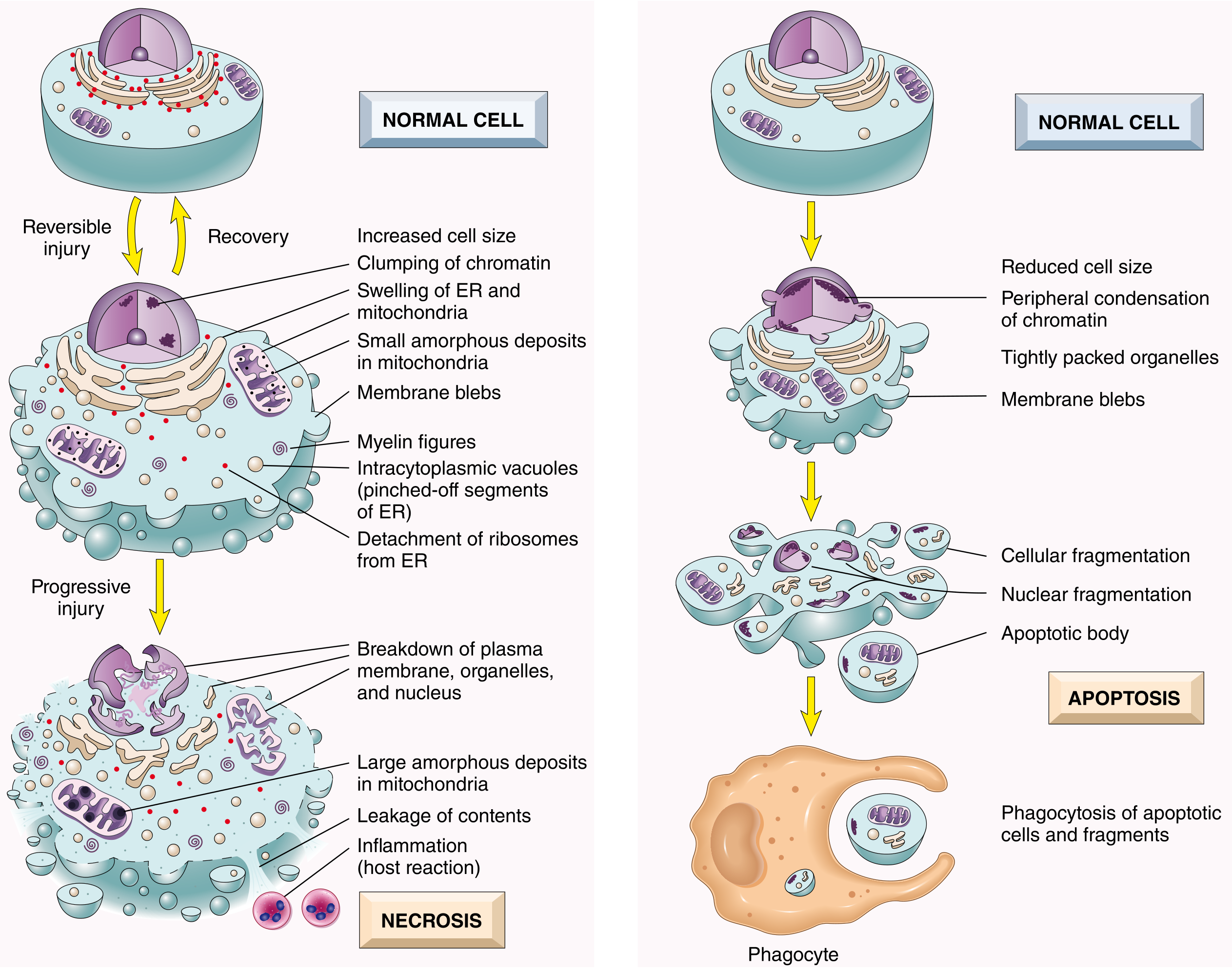
- Plasma membrane - blebbing, blunting, loss of microvilli, loosening of intercellular attachments
- Mitochondria - swelling, appearance of small amorphous densities (calcium-phospholipid deposits)
- Endoplasmic reticulum - dilation with detachment of ribosomes/polysomes, dissociation of polysomes
- Nucleus - clumping and disaggregation of granular and fibrillar elements (chromatin clumping)
- Cytoplasm - accumulation of "myelin figures" (whorl-like phospholipid collections derived from damaged membranes)
Summary: Reversible vs Irreversible - Key Differences
| Feature | Reversible | Irreversible |
|---|---|---|
| Plasma membrane | Blebs (intact) | Disrupted, broken down |
| Mitochondria | Swelling, small amorphous densities | Large amorphous densities, cristae disrupted |
| Cell contents | Retained | Leak out |
| Inflammation | Not triggered | Triggered |
| Outcome | Recovery if stimulus removed | Necrosis or apoptosis |
| Point of no return | Not yet crossed | Crossed |
Key Concept Summary
- Reversible injury hallmarks: cellular swelling + fatty change
- Central mechanism: ATP depletion → Na⁺-K⁺ pump failure → water influx → swelling
- Point of no return is crossed when: plasma membranes are irreversibly damaged, mitochondria show large amorphous densities, and cell contents begin to leak
- If stimulus is removed early: full morphological and functional recovery is possible