
Paralytic squint question for ophthalmology postgraduate student
What specifically do you want to focus on?
paralytic squint Hess screen chart diplopia
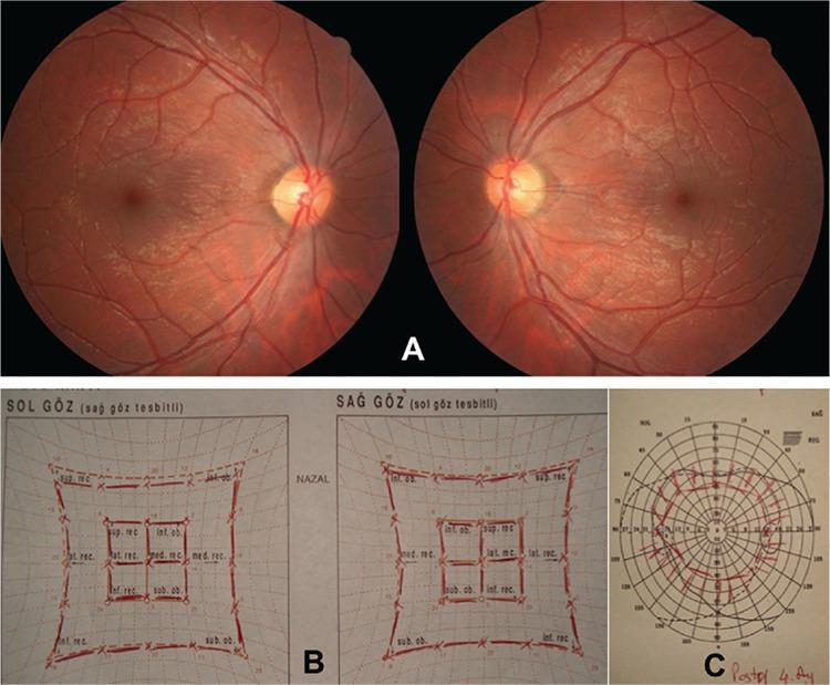
This composite ophthalmological diagnostic image demonstrates postoperative assessment findings in a patient following treatment for bilateral superior oblique palsy. Section A contains bilateral fundus photographs showing healthy retinal backgrounds, clear optic discs with distinct margins, and normal vascular distribution. Critically, the fovea-to-disc relationship demonstrates an absence of ocular torsion, indicating successful surgical correction. Section B displays a Hess screen test, featuring two grids for the left (Sol Göz) and right (Sağ Göz) eyes. The red-lined inner and outer squares are symmetrical and of normal size, signifying restored extraocular muscle function without significant underaction or overaction. Section C shows a binocular visual field chart (Goldmann perimetry or similar) illustrating an expanded field of single binocular vision, denoted by the central plotted area. These combined clinical materials illustrate the resolution of diplopia and cyclotorsion through specialized strabismus surgery, specifically Harada-Ito (HI) procedures, aimed at restoring ocular alignment and binocularity.
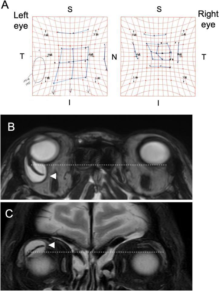
This composite educational image illustrates diagnostic findings for a mechanical strabismus complication following Ahmed Glaucoma Valve (AGV) implantation. Panel A displays a Hess Screen chart, a clinical ophthalmological tool used to map ocular motility. The chart shows a constricted field for the right eye (OD), indicating esotropia and hypotropia in the primary position with restricted infraduction and adduction, while the left eye (OS) demonstrates compensatory enhanced movements. Panels B and C show T2-weighted magnetic resonance imaging (MRI) of the orbits. Panel B (axial view) reveals a large, posteriorly enlarged giant bleb (high T2 signal) in the right superotemporal quadrant, causing anterior displacement of the eyeball compared to the contralateral side (indicated by a dotted reference line). The AGV plate is visible as a low-intensity signal (arrowhead). Panel C (coronal view) demonstrates the superior-to-inferior extent of the bleb, which causes inferior displacement of the right globe. These visuals effectively teach the relationship between physical mass effect from glaucoma drainage devices and restrictive motility disturbances.
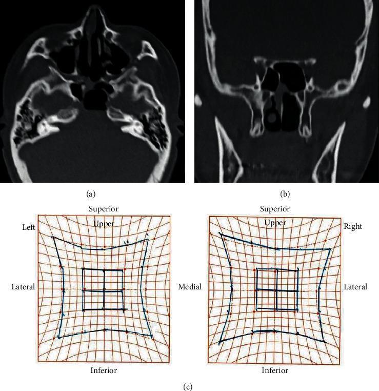
This composite medical image presents postoperative evaluations for a patient treated for a sphenoid sinus mucocele. (a) and (b) are non-contrast computed tomography (CT) images in axial and coronal planes, respectively. They demonstrate the paranasal cavity and skull base, showing a significant reduction in the size of the previously expansive sphenoid sinus mucocele following endoscopic decompression. The thinning of the surrounding bony structures, including the sella turcica and clivus area, is visible, reflecting the chronic pressure from the lesion. (c) displays a Hess screen test, a diagnostic clinical tool used to assess ocular motility and extraocular muscle function. The charts compare the field of vision and eye alignment for the left and right eyes. The results illustrate postoperative improvement in abduction disturbance, likely resulting from the decompression of the abducens nerve (CN VI) which was previously compromised by the mucocele's expansion toward the superior orbital fissure. This visual summary highlights the efficacy of surgical intervention in treating obstructive paranasal pathology and resolving secondary neurological deficits such as diplopia.
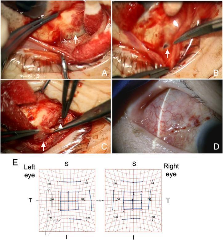
This composite educational graphic illustrates the surgical management and follow-up of a giant glaucoma bleb complication. Images A-C are intraoperative clinical photographs of the right eye: (A) demonstrates the debridement and separation of the fibrotic bleb wall from the overlying conjunctival tissue; (B) shows a surgical incision into the encapsulated bleb wall to facilitate drainage of accumulated aqueous humor; and (C) depicts the excision of the anterior bleb wall, revealing the underlying Ahmed Glaucoma Valve (AGV) plate (arrowhead). Image D is a postoperative slit-lamp biomicroscopy photograph of the superotemporal quadrant showing a normalized, diffuse bleb over the implant site with healthy conjunctival vascularity three weeks following the revision. Image E presents a Hess chart screen demonstrating bilateral orthophoria, confirming the resolution of preoperative diplopia and extraocular muscle restriction. This visual progression is characteristic of surgical bleb wall excision used to treat posteriorly enlarging giant blebs that cause ocular displacement and motility disturbances.
third nerve palsy complete ptosis dilated pupil exotropia
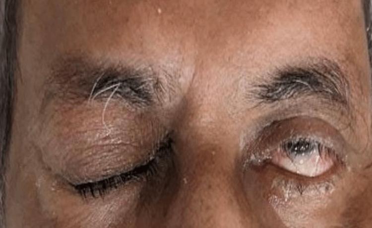
Clinical photograph of an adult male's orbital region, demonstrating classic signs of a right-sided third cranial nerve (oculomotor nerve) palsy. The right eye exhibits complete ptosis, with the upper eyelid almost entirely covering the globe. Upon manual elevation (implied by clinical context), the right eye shows exotropia (outward deviation) and mydriasis (dilated pupil). The left eye remains in a primary position with normal eyelid elevation and pupillary size, providing a comparative baseline. The skin shows age-related changes, including periorbital rhytids and slight discoloration. Notable clinical findings include traumatic mydriasis and ocular misalignment, which are indicative of underlying neurological compromise, such as subarachnoid hemorrhage or cavernous sinus thrombosis as mentioned in the clinical history. This image serves as a teaching tool for recognizing the triad of ptosis, exotropia, and mydriasis associated with oculomotor nerve dysfunction in the context of head trauma.
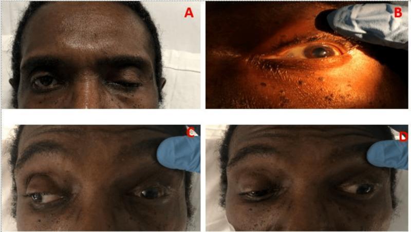
A composite of four clinical photographs (A-D) demonstrating a complete left-sided oculomotor nerve (CN III) palsy in an adult male. Panel A illustrates severe ptosis of the left upper eyelid. Panel B shows the left eye with the eyelid manually elevated, revealing fixed mydriasis (dilated pupil). Panel C displays a deficit in adduction; when the patient attempts a rightward gaze, the right eye moves medially while the left eye remains stationary or fails to cross the midline. Panel D shows the characteristic 'down and out' resting position of the left eye, exhibiting both exotropia and hypotropia. These findings are consistent with compression or damage to the third cranial nerve, affecting the levator palpebrae superioris, the pupillary sphincter (parasympathetic fibers), and the medial rectus, superior rectus, inferior rectus, and inferior oblique muscles. The presentation is clinically relevant for diagnosing conditions such as pituitary apoplexy or intracranial aneurysms.
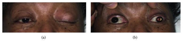
Clinical photograph in two panels (a and b) illustrating a complete left-sided third cranial nerve (oculomotor nerve) palsy. Panel (a) shows the patient attempting to open both eyes, revealing severe unilateral ptosis of the left upper eyelid, while the right eyelid remains normal. Panel (b) depicts the same patient with both upper eyelids passively retracted by an examiner. This manual elevation reveals significant anisocoria, with the left pupil demonstrating fixed mydriasis (dilation) compared to the reactive right pupil. Additionally, the left eye displays a characteristic 'down and out' gaze deviation (exotropia and hypotropia), indicating the loss of function in the superior, inferior, and medial recti, as well as the inferior oblique muscles. These visual findings are pathognomonic for a complete oculomotor nerve lesion, often associated with clinical emergencies such as pituitary apoplexy or an enlarging posterior communicating artery aneurysm.
sixth nerve palsy abducens esotropia lateral rectus
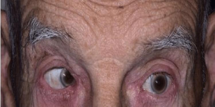
This clinical photograph displays a close-up frontal view of an elderly patient's eyes and forehead, illustrating a left-sided cranial nerve VI (abducens nerve) palsy. The right eye appears centrally fixed, while the left eye shows a marked medial deviation (esotropia) with an inability to abduct. This visual finding is consistent with left lateral rectus muscle paralysis. The patient's forehead exhibits prominent horizontal rhytids (wrinkles) and graying eyebrows, consistent with the reported age of 86. The skin of the periocular region shows no acute inflammatory changes, rashes, or bruising. The educational focus of this image is the clinical presentation of an isolated abducens nerve palsy, which in this case was secondary to pituitary apoplexy with mass effect. It serves as a diagnostic example for neurology and ophthalmology training regarding extraocular movement deficits and conjugate gaze impairment.
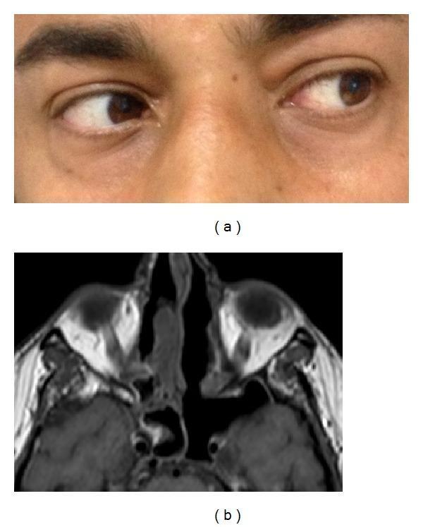
This composite image consists of two parts: a clinical photograph (a) and a diagnostic axial MRI (b), illustrating postoperative recovery from left abducens nerve palsy. Figure (a) is a close-up clinical photograph of the ocular region showing a patient performing a left lateral gaze. The left eye demonstrates full lateral excursion, indicating a complete resolution of the lateral rectus muscle deficit and recovery of the sixth cranial nerve function. Figure (b) is an axial T1-weighted MRI of the paranasal sinuses and orbits. It demonstrates an abnormally enlarged left sphenoid sinus cavity, which is post-surgical following endoscopic sinus surgery (ESS) for invasive actinomycosis. The MRI shows successful resolution of the previously described expansive formation in the parasellar region and cavernous sinus. Key anatomical landmarks include the orbits, nasal septum, and the sphenoid sinus. The material serves as an educational example of the clinical and radiological outcomes after surgical and medical treatment of invasive fungal rhinosinusitis involving the cavernous sinus.
Paralytic Squint - Complete Overview for Postgraduate Ophthalmology
1. Definition and Terminology
| Feature | Paralytic (Incomitant) | Concomitant |
|---|---|---|
| Angle of deviation | Varies with gaze direction | Same in all directions |
| Onset | Usually sudden | Usually gradual (childhood) |
| Diplopia | Present (if binocular) | Usually absent (suppression) |
| Head posture | Adopted to avoid diplopia | May be absent |
| Primary > Secondary deviation | Secondary > Primary | Equal |
| Onset age | Any age | Usually childhood |
2. Laws Governing EOM Actions
Sherrington's Law (Reciprocal Innervation)
Hering's Law (Equal Innervation)
- Primary deviation = deviation of the affected eye when the normal eye fixates
- Secondary deviation = deviation of the normal eye when the affected eye fixates
- In paralytic squint: Secondary deviation > Primary deviation (because extra innervation sent to the paretic muscle also drives the yoke muscle of the fellow eye via Hering's Law)
3. Clinical Features (Cardinal Signs)
(a) Diplopia
- Binocular (disappears on covering either eye)
- Worse in the direction of action of the paretic muscle
- The false image is peripheral and comes from the paretic eye
(b) Incomitance
- Angle of deviation is maximal in the field of action of the paretic muscle
- Secondary deviation > Primary deviation (Hering's Law)
(c) Abnormal Head Posture (AHP)
(d) False Projection / Past Pointing
(e) Vertigo / Nausea
4. Individual Nerve Palsies
A. Third Nerve (Oculomotor) Palsy
- Ptosis (complete, due to levator paralysis)
- Eye deviated "down and out" (exotropia + hypotropia - unopposed action of lateral rectus and superior oblique)
- Fixed dilated pupil (mydriasis) - if parasympathetic involved
- Loss of accommodation
- Pupil-involving (dilated, fixed): suggests compressive cause - posterior communicating artery (PCoA) aneurysm, uncal herniation. The parasympathetic fibres run on the outside of the nerve and are compressed first.
- Pupil-sparing: suggests ischaemic cause (DM, hypertension, atherosclerosis). Central ischaemia spares the peripheral pupillomotor fibres.
- Rule: Any new painful CN III palsy with pupil involvement = emergency - rule out PCoA aneurysm with CT angiography/MRI.
- Ischaemia (DM, HTN) - most common, pupil sparing
- Posterior communicating artery aneurysm - pupil involving
- Midbrain lesion (Weber, Benedikt syndromes)
- Raised ICP / Uncal herniation
- Cavernous sinus pathology
- Orbital apex involvement
- Trauma
B. Fourth Nerve (Trochlear) Palsy
- Vertical diplopia (vertical and torsional) - worse on looking down and in (reading position)
- Ipsilateral hypertropia (eye is slightly elevated due to unopposed inferior oblique)
- Excyclotorsion of the affected eye (the bottom of objects appears tilted toward the affected side)
- Abnormal head posture: head tilt to the opposite shoulder, face turn away from the affected side, chin depression (the Park's 3-step test position)
- Head tilted to the ipsilateral (affected) side: hypertropia increases (positive test - SO palsy)
- Head tilted to the contralateral side: hypertropia decreases
- This is the hallmark test for CN IV palsy
- Which eye is hypertropic in primary position? (narrows to 4 muscles)
- Is the hypertropia worse on right or left gaze? (narrows to 2 muscles)
- Is the hypertropia worse on ipsilateral or contralateral head tilt? (identifies the paretic muscle)
- Congenital (most common overall)
- Trauma (bilateral CN IV palsy common after head injury - the nerve is long and thin, vulnerable at the dorsal midbrain)
- Microvascular (DM, HTN)
- Raised ICP
- Orbital disease
C. Sixth Nerve (Abducens) Palsy
- Horizontal diplopia (uncrossed / homonymous) - worse on ipsilateral gaze
- Esotropia in primary position (medial deviation - unopposed medial rectus)
- Complete inability to abduct the eye past the midline (complete palsy)
- Abnormal head posture: face turn toward the side of the palsy (brings the eye into a compensatory medial position)
- Ischaemia (most common in adults) - DM, HTN
- Raised ICP - CN VI palsy is a false localizing sign (the long intracranial course makes it susceptible to any cause of raised ICP, not a specific brainstem lesion)
- Pontine lesion (Foville, Millard-Gubler syndromes)
- Petrous apex lesion (Gradenigo syndrome: CN VI palsy + ipsilateral CN V pain + otitis media)
- Cavernous sinus disease
- Wernicke's encephalopathy
- Trauma
- Tumour (CPA, clivus, nasopharyngeal carcinoma)
5. Ocular Motility Examination - Grading Underactions
- 0 = full movement
- -1 to -4 = increasing degrees of underaction
- -4: inability to abduct past the midline
- -3: unable to abduct more than 22.5° past midline
- -2: unable to abduct more than 45° past midline
- -1: unable to abduct more than 67.5° past midline
- +1 to +4 = overaction
6. Hess Chart and Lees Screen
- Diagnosing the paretic muscle
- Monitoring recovery or deterioration
- Planning surgery
Principles
Interpretation Rules (Kanski Ch. 18)
| Finding | Meaning |
|---|---|
| Smaller chart | Eye with the paretic muscle |
| Larger chart | Eye with the overacting yoke muscle |
| Maximum constriction on smaller chart | Indicates direction of action of paretic muscle |
| Maximum expansion on larger chart | Indicates direction of action of yoke muscle |
| Secondary deviation > Primary deviation | Classic of recent paralytic squint |
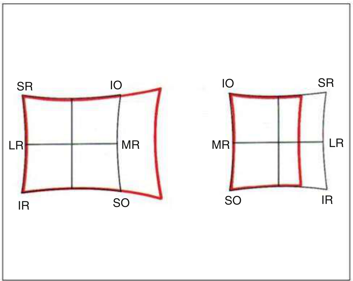
Specific Hess Chart Patterns
- Left chart much smaller
- Left exotropia (fixation spots deviated laterally) - secondary deviation > primary
- Left chart: underaction of all muscles except lateral rectus
- Right chart: overaction of all muscles except medial rectus and inferior rectus (the yokes of the spared muscles)
- Right chart is smaller
- Right chart: underaction of SO, overaction of IO
- Left chart: overaction of inferior rectus (yoke of SO), underaction of superior rectus (inhibitional palsy)
- Primary deviation R/L 8°, secondary deviation R/L 17°
- No difference in overall chart size (due to long-standing adaptation - large fusional vergence amplitude)
- Primary and secondary deviations equal (R/L 4°)
- This helps distinguish congenital from acquired
- Right chart is smaller
- Right esotropia (fixation spot of right inner chart is deviated nasally)
- Right chart: marked underaction of LR, slight overaction of MR
- Left chart: marked overaction of left medial rectus (yoke: left MR)
- Primary angle (FL) = +15°; Secondary angle (FR) = +20°
Changes with Time
- Early: Smaller paretic chart, larger yoke chart, highly incomitant
- Intermediate: Secondary contracture of ipsilateral antagonist appears; inhibitional (secondary) palsy of antagonist's yoke develops - can mimic a palsy of the opposite eye's muscle
- Late: Charts become progressively concomitant, making identification of the primary paretic muscle difficult
7. Other Investigations
Red Glass Test / Diplopia Charting
- Heteronymous (crossed) diplopia: images are on opposite sides → exotropia
- Homonymous (uncrossed) diplopia: images on the same side → esotropia
- The outer/more peripheral image always belongs to the paretic eye
- Diplopia is maximal in the direction of action of the paretic muscle
Bielschowsky Head Tilt Test
Cover-Uncover Test
- Detects heterotropia (manifest deviation)
- Alternate cover test detects both tropias and phorias
Prism Cover Test
Maddox Rod Test
8. Etiology Summary by Age
| Age Group | Common Causes |
|---|---|
| Children | Congenital (CN IV most common), trauma |
| Young adults | Trauma, demyelination (MS), congenital decompensation |
| Middle age | Microvascular (DM, HTN), trauma, tumour |
| Elderly | Microvascular, giant cell arteritis, tumour |
9. Management
Conservative (Initial)
- Occlusion of one eye: relieves diplopia while awaiting recovery
- Prisms (Fresnel prisms initially): neutralize the deviation in primary position - useful if the angle is stable
- Observation: most microvascular palsies recover spontaneously within 3-6 months
- Botulinum toxin to the ipsilateral antagonist (e.g. medial rectus in CN VI palsy): prevents contracture while awaiting recovery
Treat the underlying cause
- Ischaemic: manage DM, HTN
- Compressive (aneurysm, tumour): neurosurgical referral
- Wernicke's: thiamine (vitamin B1) IV
Surgical Management (after 6-12 months of stable deviation)
- Wait at least 6 months after onset to ensure deviation is stable
- Resection of paretic muscle + recession of ipsilateral antagonist (e.g. in CN VI palsy: resect lateral rectus + recess medial rectus)
- For complete CN VI palsy: Hummelsheim procedure or Jensen's procedure (transposition of SR and IR to augment LR action)
- For CN IV palsy: recession of ipsilateral inferior oblique, or Harada-Ito procedure (for torsional diplopia)
- For CN III palsy: complex surgery, poor prognosis
10. Key Exam Points (PG MCQ / Clinical Viva)
| Point | Fact |
|---|---|
| Secondary deviation > Primary deviation | Hallmark of paralytic squint (Hering's Law) |
| False image is peripheral | Always from the paretic eye |
| Pupil-involving CN III | Emergency - rule out PCoA aneurysm |
| Pupil-sparing CN III | Likely ischaemic (DM/HTN) |
| CN VI = false localizing sign | Due to long intracranial course |
| Gradenigo syndrome | CN VI + CN V pain + otitis media (petrous apex) |
| Bielschowsky test | CN IV palsy - hypertropia worsens on ipsilateral head tilt |
| Congenital CN IV Hess chart | Equal chart sizes (unlike acquired) |
| Harada-Ito procedure | Torsional diplopia in CN IV palsy |
| Surgery timing | After 6 months of stable deviation |
| Botulinum toxin role | Prevent contracture of antagonist in acute palsy |
Amblyopia as theory question for post graduate ophthalmology student
amblyopia patching occlusion therapy child lazy eye
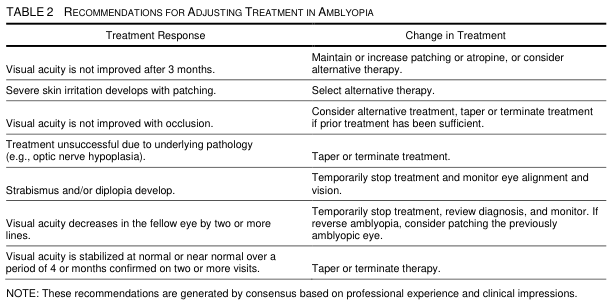
TABLE 2 RECOMMENDATIONS FOR ADJUSTING TREATMENT IN AMBLYOPIA <table><thead><tr><th>Treatment Response</th><th>Change in Treatment</th></tr></thead><tbody><tr><td>Visual acuity is not improved after 3 months.</td><td>Maintain or increase patching or atropine, or consider alternative therapy.</td></tr><tr><td>Severe skin irritation develops with patching.</td><td>Select alternative therapy.</td></tr><tr><td>Visual acuity is not improved with occlusion.</td><td>Consider alternative treatment, taper or terminate treatment if prior treatment has been sufficient.</td></tr><tr><td>Treatment unsuccessful due to underlying pathology (e.g., optic nerve hypoplasia).</td><td>Taper or terminate treatment.</td></tr><tr><td>Strabismus and/or diplopia develop.</td><td>Temporarily stop treatment and monitor eye alignment and vision.</td></tr><tr><td>Visual acuity decreases in the fellow eye by two or more lines.</td><td>Temporarily stop treatment, review diagnosis, and monitor. If reverse amblyopia, consider patching the previously amblyopic eye.</td></tr><tr><td>Visual acuity is stabilized at normal or near normal over a period of 4 or months confirmed on two or more visits.</td><td>Taper or terminate therapy.</td></tr></tbody></table> NOTE: These recommendations are generated by consensus based on professional experience and clinical impressions.
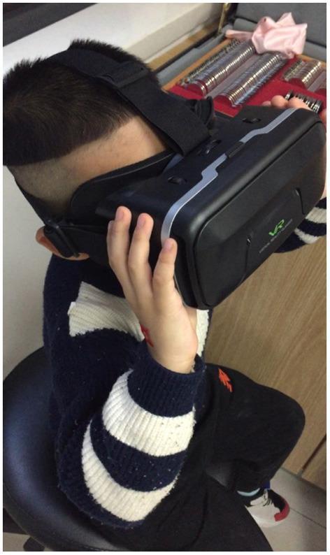
This clinical photograph illustrates a pediatric patient engaged in amblyopia therapy using the EYEBIT system, a digital health intervention based on virtual reality (VR) technology. The patient is seated and wearing a black VR headset with silver-grey accents and a visible 'VR' logo, secured by adjustable elastic head straps. In the background, a trial lens set is visible, typical of an ophthalmology or optometry clinical setting. The system utilizes dichoptic stimulation, where the visual stream is manipulated between eyes—specifically by partially covering the central area of the dominant eye's window to force visual training and stimulation of the central vision in the amblyopic eye. This setup is designed to improve binocular fusion function and visual acuity through active stimulation rather than traditional passive patching. The image demonstrates a modern approach to neuro-rehabilitation in ophthalmology, focusing on binocular visual function and patient compliance in treating refractive or strabismic amblyopia.
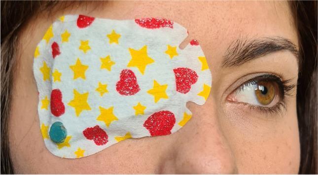
This clinical photograph displays a pediatric occlusion patch applied to the left eye of a patient, a standard intervention for treating conditions like amblyopia and strabismus. The adhesive patch features a decorative pattern of yellow stars and red hearts on a white background, designed to improve treatment compliance in younger populations. A key clinical feature of this setup is the TheraMon® microsensor (dimensions approximately 9 x 13 x 4.5 mm), visible as a small turquoise circular object through a deliberate perforation at the lateral inferior margin of the patch. This electronic monitor is used to objectively record wearing time and adherence by measuring environmental temperature at 15-minute intervals. The right eye remains uncovered, showing a healthy hazel iris, dark limbal ring, and normal periocular skin, which serves as a comparison. The image illustrates the integration of digital health monitoring with traditional occlusion therapy to track the dose rate of treatment, which is a critical factor in achieving visual acuity gains in pediatric ophthalmology.
visual cortex ocular dominance columns critical period plasticity amblyopia
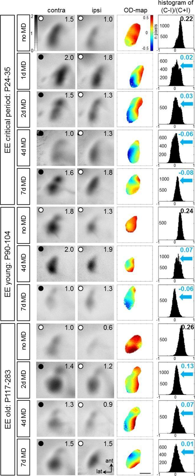
This diagnostic comparison chart illustrates optical imaging of intrinsic signal activity in the primary visual cortex (V1) of mice under various experimental conditions. The image explores ocular dominance (OD) plasticity across three age groups: Critical Period (P24-35), Young (P90-104), and Old (P117-283). Each row represents a specific duration of monocular deprivation (MD), ranging from 0 to 7 days. The visual data is organized into four columns: 1. **Grayscale Activity Maps (Contra/Ipsi):** Shows neuronal activation in V1 following stimulation of the contralateral or ipsilateral eye. Darker regions indicate higher fractional changes in reflectance. 2. **Color-coded OD Maps:** Use a thermal scale (red/warm = contralateral dominance; blue/cold = ipsilateral dominance) to visualize spatial eye preference. 3. **Histograms of OD Scores:** Quantify the distribution of pixel values, showing the shift in the Ocular Dominance Index (ODI). Key observations demonstrate that in animals raised in Enriched Environments (EE), even older mice maintain plasticity. MD induces a visible shift from warm colors (positive ODI) to cold colors (negative or near-zero ODI) and a leftward histogram shift, signifying a loss of contralateral dominance and a compensatory shift toward the open eye.
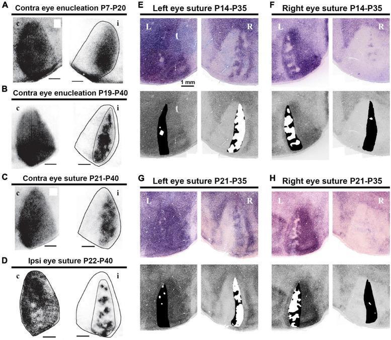
This composite figure displays microscopy images and corresponding activity maps illustrating ocular dominance columns (ODCs) in the rat primary visual cortex (V1) under various experimental conditions. Panels A–D utilize wheat germ agglutinin-conjugated horseradish peroxidase (WGA-HRP) transneuronal tracing to visualize anatomical ODC patterns in the contralateral (c) and ipsilateral (i) hemispheres. Conditions include monocular enucleation (A, B) and eyelid suture (C, D) performed during different postnatal (P) developmental windows. These images demonstrate how deprivation shifts the territories and density of labeling in favor of the active eye, revealing patchy segregation in the binocular zone. Panels E–H show physiological ODC patterns through immediate-early gene (IEG) expression in the left (L) and right (R) hemispheres following monocular suture at P14-P35 (E, F) and P21-P35 (G, H). Higher IEG expression, indicated by darker staining, correlates with increased neuronal activity in the hemisphere receiving input from the open eye. This comparison highlights ocular dominance plasticity and the differential maturation rates between anatomical connectivity and functional neuronal responsiveness during the critical period of visual development.
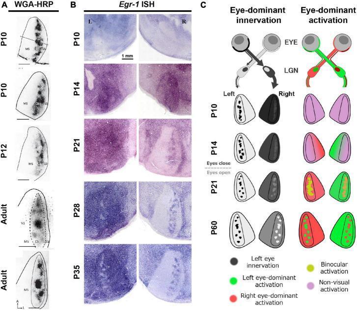
Educational figure illustrating the developmental divergence between anatomical and physiological ocular dominance columns (ODCs) in the primary visual cortex (V1) of a rat model. Panel A displays anatomical tracer staining (WGA-HRP) across developmental stages (P10 to Adult), showing that eye-specific anatomical innervation is present early, even before eye-opening. Panel B presents immediate-early gene expression via Egr-1 In Situ Hybridization (ISH) in the left and right V1 cortices from P10 to P35. This physiological marker reveals that functional maturation of ODCs occurs much later than anatomical formation, with distinct columns only becoming clearly defined by P35. Panel C provides a conceptual schematic comparing 'Eye-dominant innervation' with 'Eye-dominant activation'. It maps the pathway from the retina through the lateral geniculate nucleus (LGN) to V1, highlighting the transition from uniform, non-visual activation (pink) to binocular (yellow) and eye-dominant (red/green) activation patterns as synapses refine through visual experience. This content illustrates key concepts in neurodevelopment, synaptic plasticity, and the critical period of the visual system.
anisometropic amblyopia strabismus eccentric fixation visual acuity
TABLE 2 RECOMMENDATIONS FOR ADJUSTING TREATMENT IN AMBLYOPIA <table><thead><tr><th>Treatment Response</th><th>Change in Treatment</th></tr></thead><tbody><tr><td>Visual acuity is not improved after 3 months.</td><td>Maintain or increase patching or atropine, or consider alternative therapy.</td></tr><tr><td>Severe skin irritation develops with patching.</td><td>Select alternative therapy.</td></tr><tr><td>Visual acuity is not improved with occlusion.</td><td>Consider alternative treatment, taper or terminate treatment if prior treatment has been sufficient.</td></tr><tr><td>Treatment unsuccessful due to underlying pathology (e.g., optic nerve hypoplasia).</td><td>Taper or terminate treatment.</td></tr><tr><td>Strabismus and/or diplopia develop.</td><td>Temporarily stop treatment and monitor eye alignment and vision.</td></tr><tr><td>Visual acuity decreases in the fellow eye by two or more lines.</td><td>Temporarily stop treatment, review diagnosis, and monitor. If reverse amblyopia, consider patching the previously amblyopic eye.</td></tr><tr><td>Visual acuity is stabilized at normal or near normal over a period of 4 or months confirmed on two or more visits.</td><td>Taper or terminate therapy.</td></tr></tbody></table> NOTE: These recommendations are generated by consensus based on professional experience and clinical impressions.
This clinical photograph displays a pediatric occlusion patch applied to the left eye of a patient, a standard intervention for treating conditions like amblyopia and strabismus. The adhesive patch features a decorative pattern of yellow stars and red hearts on a white background, designed to improve treatment compliance in younger populations. A key clinical feature of this setup is the TheraMon® microsensor (dimensions approximately 9 x 13 x 4.5 mm), visible as a small turquoise circular object through a deliberate perforation at the lateral inferior margin of the patch. This electronic monitor is used to objectively record wearing time and adherence by measuring environmental temperature at 15-minute intervals. The right eye remains uncovered, showing a healthy hazel iris, dark limbal ring, and normal periocular skin, which serves as a comparison. The image illustrates the integration of digital health monitoring with traditional occlusion therapy to track the dose rate of treatment, which is a critical factor in achieving visual acuity gains in pediatric ophthalmology.
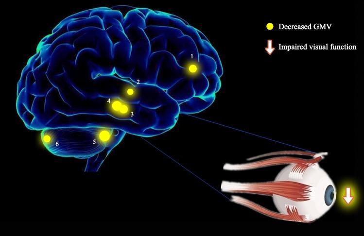
This medical illustration combines a three-dimensional brain rendering and an anatomical ocular diagram to demonstrate the pathophysiology of strabismus with amblyopia (SA). The brain model highlights six specific regions with decreased gray matter volume (GMV), indicated by numbered yellow spots: (1) left anterior cingulate, (2) right superior temporal gyrus, (3) right parahippocampal gyrus, (4) left parahippocampal gyrus, (5) right anterior lobe of the cerebellum, and (6) right posterior lobe of the cerebellum. The size of these spots represents the degree of quantitative change in GMV. A detailed illustration of the human eye, including extraocular muscles, is connected to the parahippocampal regions (3 and 4) via blue lines, suggesting a functional link. A downward-pointing arrow next to the eye signifies 'impaired visual function.' This visual summary illustrates research findings where significant GMV reduction in these specific cortical and cerebellar areas correlates with the visual and oculomotor deficits characteristic of patients with SA compared to healthy controls.
Amblyopia - Theory for Postgraduate Ophthalmology
1. Definition
- In the absence of an organic lesion, a difference in BCVA of two Snellen lines or more (or >1 log unit between the two eyes) is indicative of amblyopia.
- The visual impairment arises from miswiring of connections in the visual cortex during abnormal visual experience in infancy and early childhood, not from a structural defect in the eye.
2. Pathophysiology and the Critical Period
The Critical Period of Visual Development
- Synaptic connections in the primary visual cortex (V1) are refined based on visual experience.
- If abnormal visual input reaches the cortex (blurred image, misaligned image, or no image), cortical synapses rewire in favour of the normal eye.
Key Neuroscience (Hubel and Wiesel - Nobel Prize work)
- Ocular dominance columns in V1 receive separate input from each eye.
- Monocular deprivation during the critical period causes cortical cells to become dominated by the normal (non-deprived) eye - the deprived eye loses its cortical representation.
- In strabismus: binocular cortical cells (which need simultaneous input from both eyes) are almost completely lost. Cells become monocularly driven - either by the right or left eye, but not both. This eliminates binocular depth perception.
- Correction of the cause (spectacles, surgery) can lead to vision recovery only if initiated during the critical period.
Molecular Basis of the Critical Period
- As inhibitory interneurons mature, their cell bodies become surrounded by perineuronal nets (PNNs) - extracellular matrix structures rich in chondroitin sulfate proteoglycans.
- PNNs stabilize synaptic connections and close the window of plasticity.
- Experimental degradation of PNNs (e.g., by bacterial enzyme chondroitinase, or matrix metalloprotease-9 / MMP-9) restores juvenile levels of plasticity and enables recovery from early monocular deprivation in adult animals.
- This is the basis for ongoing research into extending the critical period for amblyopia treatment in older children and adults.
- For deprivation amblyopia (e.g. congenital cataract): ends before 1 year of life - the most urgent cause
- For strabismic amblyopia: usually up to 7-8 years
- For anisometropic amblyopia: may extend into the early teens (particularly when some binocular function is preserved)
3. Classification
A. Strabismic Amblyopia
- Most common form (along with anisometropia)
- Results from abnormal binocular interaction: continued monocular cortical suppression of the deviating (non-fixating) eye
- The suppressing eye is always the consistently deviating eye
- If fixation freely alternates, amblyopia does not develop (equal cortical representation)
- Strabismus can lead to amblyopia or result from it
B. Anisometropic Amblyopia
- Caused by a difference in refractive error between the two eyes
- The more ametropic eye receives a chronically blurred image - a mild form of visual deprivation
- Can occur with as little as 1 dioptre of difference (Kanski) or typically ≥1.50 D (Wills)
- Frequently associated with microtropia and may co-exist with strabismic amblyopia
- Can be seen with eyelid haemangioma or congenital ptosis inducing astigmatism
C. Stimulus Deprivation Amblyopia (most severe)
- Caused by complete obstruction of vision in one eye
- Causes: congenital cataract, corneal opacity, ptosis covering the pupil, persistent fetal vasculature (PFV)
- Unilateral or bilateral
- Most severe: critical period ends earliest (before 1 year), so urgent treatment is essential
- Even bilateral congenital cataracts not treated within the earliest months of life cause bilateral amblyopia
D. Bilateral Ametropic Amblyopia
- Caused by high symmetrical refractive errors, usually high hypermetropia
- Bilateral; both eyes are equally affected
E. Meridional Amblyopia
- Caused by uncorrected astigmatism (usually >1 D) persisting beyond the period of emmetropization
- Image blur is present in one meridian
- Unilateral or bilateral
4. Clinical Features
Symptoms
- Usually none - most often discovered on vision screening
- No diplopia (cortical suppression eliminates the competing image)
- History of patching, strabismus, or muscle surgery in childhood
Signs
- Poorer BCVA in one eye, not entirely explained by organic pathology and not fully correctable with refraction
- In anisometropic amblyopia, the involved eye nearly always has a higher refractive error
- Central vision primarily affected; peripheral visual field usually remains normal
| Sign | Description |
|---|---|
| Crowding phenomenon | Individual letters read more easily than a full line of letters. Characteristic of amblyopia; used in testing preverbal children |
| Neutral density filter (NDF) effect | In reduced illumination, VA of an amblyopic eye decreases much less than an organically diseased eye. NDF worsens organic disease more than amblyopia |
| Eccentric fixation | The amblyopic eye uses a parafoveal point rather than the fovea for fixation. Detected with visuoscopy or ophthalmoscope. Seen in dense strabismic amblyopia. When eccentric fixation is present, visual prognosis is worse |
| RAPD | Severe amblyopia may cause a trace relative afferent pupillary defect (RAPD) - must be directed along the same visual axis to avoid false positives |
5. Differentiating Amblyopia from Organic Disease
| Feature | Amblyopia | Organic Disease |
|---|---|---|
| NDF test | Minimal worsening | Marked worsening |
| Crowding | Present | Less prominent |
| Peripheral field | Normal | May be defective |
| Pupil responses | Usually normal (trace RAPD at most) | May show significant RAPD |
| Fundus/anterior segment | Normal (by definition) | Abnormality present |
| Response to occlusion | Vision improves | No improvement |
6. Investigations
- Visual acuity - Snellen chart; use single optotypes for children to avoid crowding masking detection; always test each eye separately
- Cycloplegic refraction - mandatory in all cases
- Cover-uncover test - assess eye alignment
- Fundoscopy - dilated examination to exclude organic pathology (mandatory before starting treatment)
- Visuoscopy (ophthalmoscopic fixation test) - detects eccentric fixation
- Electrophysiology (VEP) - if acuity does not respond to treatment, electrophysiology or imaging should be reconsidered
7. Treatment
MANDATORY first step: Exclude organic disease before commencing any amblyopia treatment. Organic disease and amblyopia may co-exist; a trial of patching may still be indicated but prognosis differs.
Step 1: Correct the Underlying Cause
- Prescribe appropriate spectacle correction (full cycloplegic refraction; reduce hyperopia in both eyes symmetrically by ≥1.50 D)
- Allow 6-12 weeks of refractive adaptation before adding occlusion - some children improve with spectacles alone (refractive adaptation)
- Treat media opacities (remove cataract, treat ptosis)
Step 2: Occlusion / Patching (Mainstay of Treatment)
- 2-6 hours/day part-time patching (Wills protocol)
- Full-time vs part-time depends on age, density of amblyopia
- Younger patient = faster likely improvement but greater risk of inducing occlusion amblyopia in the normal eye
- Therefore: monitor VA of both eyes regularly
Step 3: Penalization (Alternative / Adjunct)
- Equally effective as patching in mild-to-moderate amblyopia (6/24 / 20/100 or better)
- Works best in anisometropic hypermetropic amblyopia
- Advantages over patching:
- Cannot be thwarted by the child
- Less psychosocial stigma (especially in school-going children)
- Weekend instillation may be adequate
- Atropine effect can be enhanced by removing the hyperopic lens from the spectacles of the non-amblyopic eye
Step 4: Optical Degradation
- High plus lens (e.g., +9.00 D) or removal of minus lens from preferred eye to blur the image in the fellow eye
Summary of Treatment by Type
| Type | First Step | Specific Points |
|---|---|---|
| Strabismic | Correct refractive error + patch | Delay strabismus surgery until VA is equalized or maximized |
| Anisometropic | Full spectacle correction first, then patch | Spectacles alone may be sufficient; patch if VA remains reduced at 6-12 weeks |
| Deprivation (cataract) | Remove media opacity urgently + patch | Most urgent; critical period ends earliest; IOL/contact lens after cataract surgery |
| Bilateral ametropic | Full spectacle correction | Bilateral; rarely needs patching |
8. Treatment Response Adjustments
| Situation | Action |
|---|---|
| VA not improved after 3 months | Increase patching, consider atropine |
| Skin irritation from patch | Switch to atropine or optical penalization |
| VA not improved despite treatment | Consider underlying pathology; electrophysiology/imaging |
| Strabismus/diplopia develops | Temporarily stop, monitor alignment |
| Fellow eye VA drops ≥2 lines | Stop treatment, review - likely reverse amblyopia |
| VA stable at normal over 4+ months (2 visits) | Taper and terminate treatment |
9. Prognosis
- The younger the patient at start of treatment, the better the prognosis
- Strabismic amblyopia is treatable up to ~7-8 years of age
- Anisometropic amblyopia may be treatable into the early teens
- Deprivation amblyopia has the poorest prognosis and requires the most urgent intervention
- Poor compliance is the single greatest barrier to improvement
- If treatment fails or patient presents outside treatable age range: prescribe protective glasses to prevent injury to the non-amblyopic eye (one-eyed athlete rule: protective eyewear during sports for any child with VA < 20/40 in one eye)
10. Emerging/Future Treatments
- Perineuronal net (PNN) degradation: enzymatic or pharmacological dissolution of PNNs to reopen the critical period window in adult visual cortex
- Dark exposure: prolonged dark adaptation triggers MMP-9 release, degrades PNNs, and restores plasticity
- Dichoptic training / VR games: present different contrast images to each eye simultaneously to promote binocular integration and reduce suppression
- Levodopa: investigated as a pharmacological method to enhance cortical plasticity
- Citicoline: neuroprotective; under investigation
11. Key Exam Points Summary
| Point | Fact |
|---|---|
| Definition | Decreased BCVA without identifiable organic cause |
| Diagnosis | ≥2 Snellen line difference between eyes in BCVA |
| Most severe type | Deprivation (earliest critical period closure) |
| Critical period end | Deprivation < 1 yr; Strabismic ~7-8 yr; Anisometropic ~early teens |
| Crowding | Letters harder in a row than isolated; characteristic of amblyopia |
| NDF test | Amblyopia: VA slightly worse; Organic: VA markedly worse |
| Eccentric fixation | Worse prognosis; parafoveal point used for fixation |
| Treatment hierarchy | Correct refractive error → Refractive adaptation (6-12 wk) → Patch fellow eye |
| Atropine penalization | Best for mild-moderate (6/24 or better), hypermetropic anisometropic amblyopia |
| Follow-up interval | 1 week per year of age |
| Failure of treatment | No response in 6 months = unlikely to benefit further |
| Reverse amblyopia | VA drops ≥2 lines in patched eye → stop patching |
| Molecular brake on plasticity | Perineuronal nets (PNNs) around GABAergic parvalbumin interneurons |
| Hubel & Wiesel | Ocular dominance plasticity, Nobel Prize 1981 |
Aqueous misdirection syndrome or malignant glaucoma question for pg ophthalmogy student
malignant glaucoma aqueous misdirection shallow anterior chamber flat
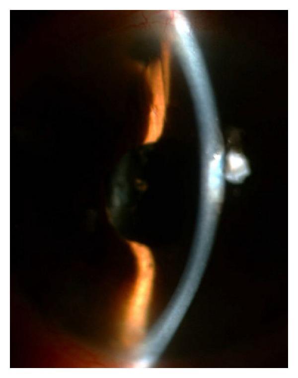
This slit-lamp clinical photograph of a right eye demonstrates the postoperative status following a surgical procedure to address malignant glaucoma. A narrow slit beam highlights the anterior segment anatomy, showing a deep anterior chamber, which contrasts with the shallow configuration typical of aqueous misdirection syndrome. Key surgical findings include a patent peripheral iridectomy located in the superior quadrant. More centrally, there is evidence of a capsulo-hyaloidectomy, where both the posterior lens capsule and the anterior hyaloid face have been surgically opened to facilitate aqueous flow. The slit-lamp section through the cornea shows clear tissue, though clinical context suggests previous corneal incisions secured with nylon sutures. This image serves as an educational example of successful surgical management for pupillary and ciliary block by creating a direct communication between the vitreous cavity and the anterior chamber.
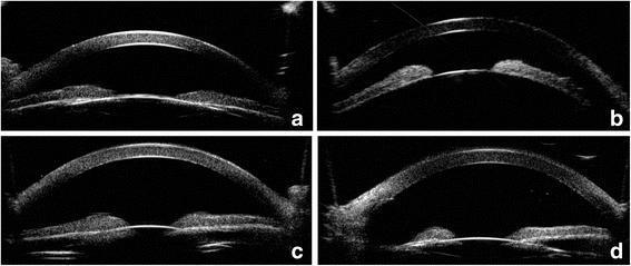
This set of four ultrasound biomicroscopy (UBM) images illustrates the anterior segment of the eyes in a patient with malignant glaucoma and nanophthalmos, before and after treatment. Panel (a) right eye and (b) left eye demonstrate extremely shallow anterior chambers with forward displacement of the iris-lens diaphragm, typical of aqueous misdirection syndrome. Panels (c) and (d) show the same eyes following intervention: the right eye (c) after YAG laser lens posterior capsulotomy and vitreous anterior membranectomy, and the left eye (d) after phacoemulsification combined with vitrectomy and posterior capsulorhexis. In these post-treatment images, there is a visible deepening of the anterior chamber, characterized by an increased distance between the corneal endothelium and the anterior surface of the iris and lens. The images highlight the structural changes in the anterior segment required to resolve ciliary block glaucoma and establish a communication pathway between the vitreous cavity and the anterior chamber.
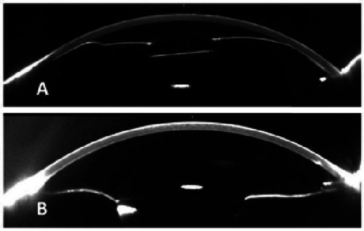
This diagnostic image pair features Scheimpflug photographs of the eye's anterior segment, illustrating the clinical management of vitreociliary block (aqueous misdirection syndrome). Figure A shows a cross-sectional view of an eye with a severely shallow anterior chamber. The iridocrystalline diaphragm is significantly displaced anteriorly, bringing the iris and intraocular lens (IOL) into close proximity with the posterior corneal surface, a hallmark of malignant glaucoma. Figure B shows the same eye immediately following a Nd:YAG laser hyaloidotomy. There is a marked increase in anterior chamber depth and volume as the iridocrystalline diaphragm shifts posteriorly to a more physiological position. These images serve as an educational comparison of pre- and post-procedural states, demonstrating the effectiveness of laser intervention in restoring normal anterior segment anatomy and resolving anterior chamber flattening caused by abnormal fluid dynamics behind the vitreous.
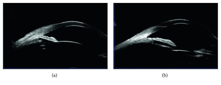
This Comparison Chart consists of two side-by-side Ultrasound Biomicroscopy (UBM) images of the anterior segment of a human eye, illustrating pre- and post-surgical changes in a patient with malignant glaucoma. Image (a) displays a Diagnostic Image of the eye's anterior chamber before intervention. Key visible features include a shallow anterior chamber and the presence of peripheral anterior synechiae (PAS) where the peripheral iris is adhered to the trabecular meshwork, causing a closed anterior chamber angle. The iris contour appears relatively flat, indicating the absence of pupillary block. Image (b) provides a postoperative comparison following goniosynechialysis and vitrectomy. It demonstrates a significant widening of the anterior chamber angle and an increase in central anterior chamber depth. The previously noted peripheral anterior synechiae are separated, and the iris is repositioned posteriorly, away from the cornea. This comparison highlights the restoration of the aqueous drainage pathway and the resolution of the anatomical crowding associated with aqueous misdirection syndrome (malignant glaucoma).
glaucoma angle closure gonioscopy anterior chamber depth UBM ultrasound biomicroscopy
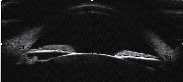
This diagnostic image is an Ultrasound Biomicroscopy (UBM) scan providing a panoramic, cross-sectional view of the human eye's anterior segment. The high-frequency ultrasound reveals key anatomical structures including the cornea, anterior chamber, iris, and the anterior surface of the crystalline lens. Two primary biometry measurements used in ophthalmology and glaucoma assessment are highlighted: 1) Anterior Chamber Width (ACW), labeled as 'C1', represents the horizontal distance between the two scleral spurs; 2) Anterior Chamber Depth (ACD), labeled as 'C2', represents the vertical distance from the corneal endothelium to the anterior pole of the lens. The image illustrates the spatial relationship between the cornea, lens, and iris, which is critical for evaluating the anterior chamber angle and risks associated with angle-closure glaucoma. This clinical imaging modality is essential for visualizing structures behind the iris pigment epithelium that are not visible via traditional slit-lamp biomicroscopy or gonioscopy.
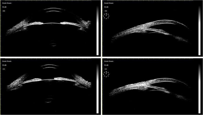
A multi-panel preoperative Ultrasound Biomicroscopy (UBM) image displaying the anterior segment of the right eye (OD, top panels) and left eye (OS, bottom panels) in a patient with chronic angle-closure glaucoma. The high-resolution cross-sectional scans illustrate bilateral anterior chamber angle closure, characterized by the iris root apposition against the trabecular meshwork. Centrally, the anterior chamber depth appears maintained with a regular circular pupillary configuration. The crystalline lens in both eyes is in a normal anatomical position without evidence of subluxation or tilt. Notably, the anterior lens capsule appears smooth, hyperechoic, and continuous, showing no signs of synechiae or adhesions to the posterior iris surface. This imaging study serves as a diagnostic baseline to evaluate ocular anatomy before procedures like Ultrasound Ciliary Plasty (UCP) or cataract surgery, specifically assessing the structural relationships between the iris, ciliary body, and lens equator.
Aqueous Misdirection Syndrome / Malignant Glaucoma
Complete Theory for Postgraduate Ophthalmology
1. Introduction and Terminology
2. Pathogenesis (Mechanism)
Sequential Events:
- Anterior rotation of the ciliary body - the ciliary processes rotate inward and press against the anterior lens equator (or anterior hyaloid in aphakic/pseudophakic eyes).
- This creates a ciliolenticular block - the space between the ciliary processes and the lens equator is obliterated.
- Aqueous, unable to pass anteriorly through this block, is misdirected posteriorly into the vitreous cavity.
- Aqueous accumulates in or behind the vitreous gel (within the vitreous gel or in a pre-hyaloid pocket).
- The vitreous gel (with trapped aqueous) pushes the entire iris-lens diaphragm forward.
- This forward displacement further shallows the anterior chamber and closes the drainage angle, causing secondary angle closure.
- Vicious cycle ensues: raised IOP → further vitreous expansion → more forward displacement → worsening angle closure.
Why the Anterior Chamber is Uniformly Shallow:
Newer Theories (Additional Factors)
- Choroidal expansion contributing to forward lens displacement
- Reduced conductivity of aqueous through vitreous - vitreous acts as a sponge trapping aqueous
- Reduced trans-scleral fluid movement
Why Miotics WORSEN the Condition:
3. Predisposing Factors / Risk Factors
| Risk Factor | Details |
|---|---|
| Primary angle-closure glaucoma (PACG) | Most common predisposing condition |
| Post-trabeculectomy | Most common precipitating procedure - a rare but serious complication |
| Short axial length | Nanophthalmos (axial length < 20 mm), hypermetropia |
| Small anterior segment | Shallow anterior chamber, thick lens |
| Post-cataract surgery | Especially in predisposed eyes |
| Post-retinal surgery | Gas/silicone tamponade can trigger misdirection |
| Post-laser procedures | Argon laser trabeculoplasty, laser iridotomy |
| Miotics | Can precipitate or worsen the attack |
| Spontaneous | Rare; may occur without any prior surgery |
| Fellow eye risk | ~50% risk in contralateral eye if predisposed |
4. Clinical Features
Symptoms
- May be very mild early in the course
- Moderate pain, red eye, photophobia as IOP rises
- Patient notices improvement in near vision (myopic shift) - the anteriorly displaced lens increases effective refractive power
- History of recent intraocular or laser surgery
- Worsening of symptoms with topical miotics (if administered)
Signs
- Diffusely shallow or flat anterior chamber - both centrally and peripherally (Grade 2 or 3 shallowing)
- Raised IOP - moderately elevated early, markedly elevated late
- Patent peripheral iridotomy / PI - presence of patent PI excludes pupillary block as the primary cause
- No iris bombé (this rules out pupillary block)
- Flat or absent filtration bleb (if post-trabeculectomy)
- Negative Seidel test (rules out wound leak)
- Myopic shift - patient may notice improved near vision
- Corneal oedema if IOP markedly elevated
- Mild anterior uveitis
Scheimpflug images showing markedly flat anterior chamber (A) resolving to a deep chamber (B) after Nd:YAG laser:
UBM (Ultrasound Biomicroscopy) - Pre and Post Treatment:
5. Differential Diagnosis
| Diagnosis | IOP | Anterior Chamber | Iris Bombé | Pain | Bleb |
|---|---|---|---|---|---|
| Aqueous misdirection / Malignant glaucoma | Moderate → markedly elevated | Diffusely shallow (Grade 2-3) | No | Moderate | Flat |
| Pupillary block | Moderate → markedly elevated | Grade 1-3 shallow | Yes | Possible | None |
| Suprachoroidal haemorrhage | Markedly elevated (early) | Grade 1-2 | No | Excruciating | Flat |
| Serous choroidal detachment | Low | Grade 1-3 | No | Dull ache | Elevated |
| Wound leak / Overfiltration | Low | Shallow | No | Mild | Flat/leaking |
| Failure to filter | Moderately elevated | Deep | No | Possible | Flat |
| Inflammation | Variable (may be low) | Deep | No | Possible | Varies |
Key Distinguishing Features of Malignant Glaucoma vs Other Causes:
| vs Pupillary Block | vs Choroidal Detachment |
|---|---|
| No iris bombé | IOP is HIGH (not low) |
| Patent PI present | B-scan: no choroidal detachment |
| Miotics worsen | Seidel test negative |
Workup
- History - previous eye surgery, use of miotics
- Slit lamp - assess PI patency, iris bombé, AC depth
- Gonioscopy - closed angle vs open angle; B-scan US essential
- IOP measurement
- B-scan ultrasound - to exclude choroidal detachment and suprachoroidal haemorrhage
- Seidel test - to exclude wound leak
- UBM (Ultrasound Biomicroscopy) - gold standard for imaging the ciliary body, anterior segment anatomy, and confirming anterior rotation of ciliary processes
6. Treatment (Step-Wise Approach)
Step 1: Ensure Patent PI
Step 2: Medical Treatment (Mainstay of Initial Management)
| Drug | Mechanism | Route | Dose |
|---|---|---|---|
| Atropine 1% | Cycloplegia + mydriasis → dilates ciliary ring → tightens zonules → pulls lens posteriorly | Topical | qid |
| Phenylephrine 2.5-10% | Sympathomimetic mydriasis → augments cycloplegia | Topical | qid |
| Acetazolamide | Reduces aqueous production → decreases IOP | IV 500 mg or PO 250 mg x2 | Stat |
| Timolol 0.5% | Beta-blocker → reduces aqueous production | Topical | Daily/bd |
| Apraclonidine 1% or Brimonidine 0.1-0.2% | Alpha-2 agonist → reduces aqueous production | Topical | bd |
| Mannitol 20% | Hyperosmotic → dehydrates vitreous → causes lens to move posteriorly | IV 1-2 g/kg | Over 45 min |
Step 3: Laser Treatment (if medical therapy fails)
| Procedure | Indication | Details |
|---|---|---|
| Nd:YAG laser posterior capsulotomy + anterior hyaloid disruption | Pseudophakic or aphakic | Disrupts the anterior hyaloid face, allowing aqueous to flow freely forward. Most effective in pseudophakic eyes |
| Nd:YAG through iridectomy (phakic) | Phakic with large PI | Attempt hyaloid disruption through PI - technically difficult |
| Argon laser to ciliary processes | Adjunct | Burns ciliary process to break ciliolenticular block |
| Cyclodiode laser | May be effective | Reduces aqueous production + may alter ciliary body position |
Step 4: Surgical Treatment (if laser fails)
- Key principle: Remove sufficient anterior vitreous gel to allow free flow of aqueous to the anterior chamber
- Pars plana vitrectomy combined with irido-zonulo-hyaloidectomy (localised excision of iris, lens capsule, zonules, and anterior hyaloid face + reformation of AC) is highly effective
- Lensectomy + disruption of anterior hyaloid or vitrectomy alone
- Before vitrectomy: always check for/drain any anterior choroidal detachment first (sclerotomy)
Algorithm:
Patent PI confirmed?
    ↓ Yes
Medical therapy: Atropine + Phenylephrine + Aqueous suppressants + Mannitol
    ↓ Fails within 24-48 hours
Nd:YAG laser (posterior capsulotomy/anterior hyaloidotomy)
    ↓ Fails
Pars plana vitrectomy ± lensectomy ± irido-zonulo-hyaloidectomy
7. Prevention
- Prophylactic PI in the contralateral eye if it has an occludable angle
- Phacoemulsification (lens extraction) is highly effective in eyes at risk - deepens the anterior chamber, removes the predisposed lens, reduces the risk
- Avoid miotics in at-risk eyes
- Use dilute MMC cautiously in PACG patients undergoing trabeculectomy (risk of triggering malignant glaucoma)
- Atropine 1% daily indefinitely after a successfully broken attack
8. Key Exam Points
| Point | Fact |
|---|---|
| Original description | Von Graefe, 1869 |
| Other names | Ciliary block glaucoma, aqueous misdirection syndrome, ciliolenticular block |
| Core mechanism | Anterior rotation of ciliary body → ciliolenticular block → posterior aqueous misdirection |
| Most common trigger | Trabeculectomy in PACG eyes |
| Key diagnostic criteria | Shallow AC (diffuse) + High IOP + Patent PI + No iris bombé + No choroidal detachment |
| Distinguishing sign (vs pupillary block) | No iris bombé; patent PI present |
| Distinguishing sign (vs choroidal detachment) | IOP is HIGH (not low); no choroidal detachment on B-scan |
| Why miotics are CONTRAINDICATED | They increase ciliolenticular contact, worsening the block |
| First-line treatment | Atropine 1% + Phenylephrine + IV Mannitol + Aqueous suppressants |
| Why atropine works | Cycloplegia dilates ciliary ring → tightens zonules → lens moves posteriorly |
| Laser in pseudophakic | Nd:YAG posterior capsulotomy + anterior hyaloid disruption |
| Goal of vitrectomy | Remove anterior vitreous to create a unicameral eye |
| Myopic shift | Due to anterior lens displacement → increased effective lens power |
| Maintenance after breaking attack | Atropine 1% indefinitely |
| Contralateral eye | PI should be performed within 1 week if angle occludable |
| Best imaging | UBM - shows anterior rotation of ciliary processes and AC depth |
Next time can you take references from kanski ,wills , bakers and shaffers ,AK khurana ,dukes
- Kanski's Clinical Ophthalmology (already in the library)
- Wills Eye Manual (already in the library)
- Becker & Shaffer's Diagnosis and Therapy of the Glaucomas
- AK Khurana - Comprehensive Ophthalmology
- Duke-Elder's System of Ophthalmology
- Kanski's Clinical Ophthalmology: A Systematic Approach, 10th Edition
- The Wills Eye Manual (latest edition)
- These three are not indexed in the database, so I cannot pull direct quotes or page references from them.
- Kanski + Wills - cited directly with exact content from the library (as I have been doing)
- AK Khurana, Becker-Shaffer, Duke-Elder - I will supplement from my training knowledge of these texts, clearly labeling them as "per Khurana" / "per Duke-Elder" etc., and flag that these are from training knowledge rather than a live library source
- Where AK Khurana classification schemes, mnemonics, or specific Indian PG exam-oriented content differ from Kanski (e.g. slightly different classification of glaucoma, amblyopia grades, etc.), I will specifically call out the Khurana perspective as it is most relevant for Indian PG examinations (MD/MS/DNB)
Gonioscopy As a theory question As per standard books i have mention before
gonioscopy anterior chamber angle structures trabecular meshwork Schwalbe line scleral spur
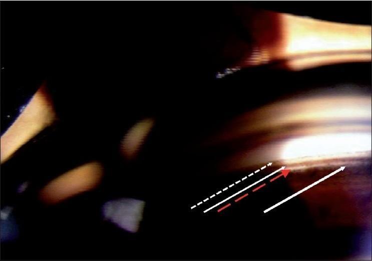
A clinical gonioscopic photograph illustrating the normal anatomy of the human anterior chamber angle. This diagnostic image provides a detailed view of the iridocorneal junction and is used for clinical evaluation of glaucoma and angle status. Labeled structures follow an anterior-to-posterior sequence: Schwalbe's line (dashed white arrow) marks the termination of Descemet's membrane; the pigmented trabecular meshwork (solid white arrow) is visible as a distinct, darker band due to pigment accumulation; the scleral spur (red arrow) appears as a thin, white line representing the posterior attachment of the trabecular meshwork; and the ciliary body (thick white arrow) is seen most posteriorly as a darker band. The image demonstrates the varying pigmentation levels between these structures, which is essential for identifying landmarks during clinical gonioscopy. This material is targeted at medical students and ophthalmology residents for learning anatomical localization and the assessment of an open angle.
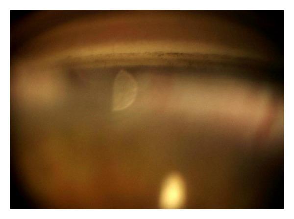
This diagnostic image is a clinical gonioscopy photograph showing the anterior chamber angle of a human eye. The image demonstrates a wide-open angle (Van Herick Grade 4 equivalent), where key anatomical landmarks are clearly visible. The prominent pigmented band across the superior portion of the angle corresponds to the trabecular meshwork, showing mild-to-moderate granular pigmentation. Adjacent anatomical structures including Schwalbe's line (superior to the meshwork) and the scleral spur (inferior to the meshwork) are discernible, indicating a deep angle recess. The iris root is positioned posteriorly, confirming the open configuration. Prominent light artifacts, including a crescent-shaped reflection in the center and a yellowish circular reflex at the bottom, are visible due to the slit-lamp and contact lens interface. This visual is typical for assessing glaucoma risk and pigmentation disorders such as pigmentary glaucoma or nevus of Ota-related ocular findings.
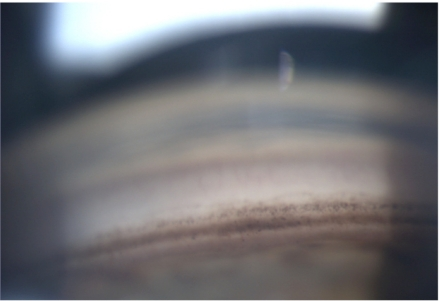
**Imaging Modality:** Gonioscopy (Ophthalmic clinical photography) **Anatomical Region:** Anterior chamber angle of the eye. **Observed Findings:** This image displays a wide-open drainage angle with clearly identifiable anatomical landmarks. Visible structures include Schwalbe’s line, the pigmented and non-pigmented functional trabecular meshwork, the scleral spur, and a portion of the ciliary body band. **Characteristic Visual Features:** * **Pigmentation:** A normal, moderate level of trabecular pigmentation is visible within the posterior trabecular meshwork. * **Angle Configuration:** The iridocorneal angle is wide, showing no evidence of peripheral anterior synechiae (PAS), angle recession, or neovascularization. * **Iris Contour:** The peripheral iris displays a flat to slightly convex configuration, consistent with normal anatomical spacing between the iris root and the cornea. **Diagnostic Context:** The image represents a physiological, healthy drainage angle (Grade 4 on the Shaffer Scale). It serves as a baseline for normal anatomy, demonstrating the absence of obstructive or pathological changes such as angle-closure, pigment dispersion, or inflammatory debris.
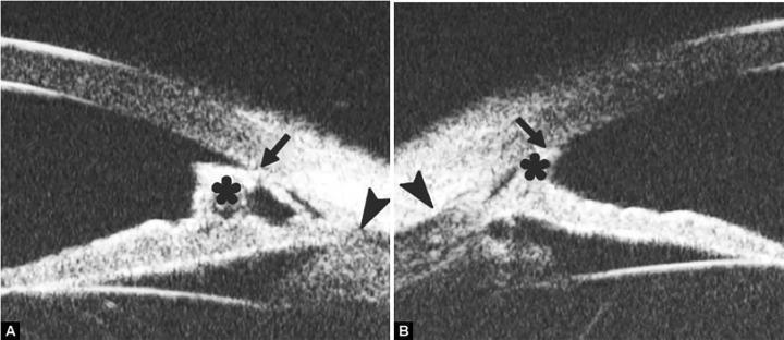
This diagnostic ultrasound biomicroscopy (UBM) image consists of two panels (A and B) illustrating the anterior chamber angle of the eye. The imaging highlights several key anatomical landmarks and pathological findings relevant to glaucoma and angle-closure mechanisms. Schwalbe's line is indicated by black arrows and appears anteriorly displaced and prominent. The scleral spur is identified by black arrowheads as a highly reflective triangular structure posterior to the ciliary body. A distinctive feature in both images is a tissue band (marked by an asterisk) bridging the peripheral iris and the posterior cornea, indicative of peripheral anterior synechiae (PAS). Furthermore, the iris shows a high insertion point into the posterior aspect of the trabecular meshwork, suggesting a narrow angle configuration. These UBM findings are used in ophthalmology to assess the integrity of the drainage angle and determine the pathophysiology of secondary glaucoma when clinical visualization via gonioscopy is obscured.
gonioscopy indentation dynamic compression Zeiss four mirror lens appositional closure PAS
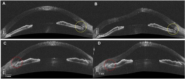
This diagnostic image displays four cross-sectional B-scans of the anterior segment of the human eye obtained via Deformable Anterior Segment Optical Coherence Tomography (DAS-OCT). The images demonstrate the dynamic response of the anterior chamber angle (ACA) to an air-puff stimulus, functioning as a non-contact form of indentation gonioscopy to differentiate between appositional and synechial angle closure. Panels A and B (Top) show an eye with No Peripheral Anterior Synechiae (NPAS). In panel A (baseline), the angle appears narrow. In panel B, after the air puff, the yellow circles highlight a widening of the ACA, indicating that the iris-cornea contact was purely appositional and reversible. Panels C and D (Bottom) show an eye with Peripheral Anterior Synechiae (PAS). In panel C (baseline), the angle is closed. In panel D, following the air puff, red circles indicate that the angle remains closed despite the increased pressure, confirming permanent adhesions (synechiae). These findings are critical for diagnosing and managing primary angle-closure glaucoma (PACG) by identifying permanent anatomical changes versus reversible apposition.
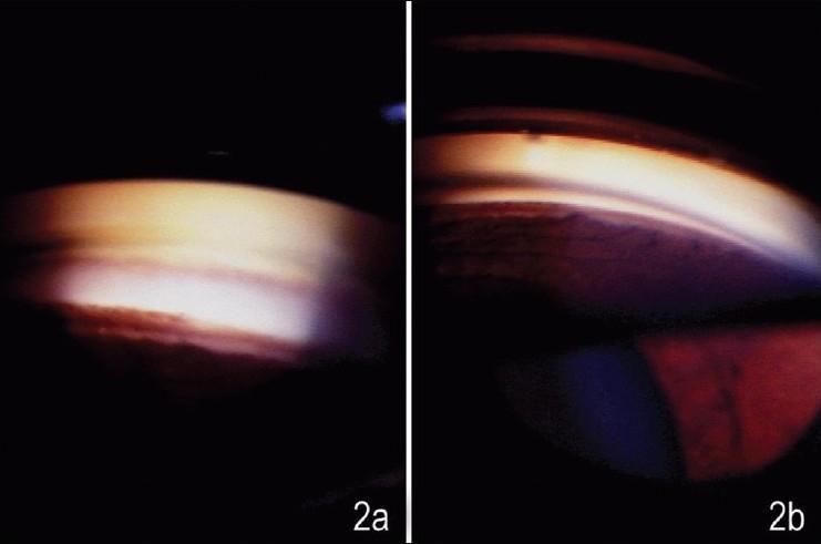
This side-by-side clinical photograph displays two gonioscopic images (labeled 2a and 2b) of the anterior chamber angle of a human eye, illustrating the diagnostic technique of indentation gonioscopy. Image 2a shows the angle as viewed with a Sussman (four-mirror) lens during indentation. In this frame, the previously suspected peripheral anterior synechia (PAS) has disappeared, revealing a widely open angle with clearly visible, smooth trabecular meshwork and iris insertion. Image 2b shows the angle through a Goldmann (two-mirror) lens. In this view, a 'pseudofalse' synechia persists despite manipulative attempts, manifesting as a dark, iris-like structure that appears to bridge the angle toward the Schwalbe's line. The comparison demonstrates how indentation gonioscopy can differentiate true synechia from peripheral iris bulges seen 'end-on'. This is a critical diagnostic distinction in the management of glaucoma and narrow-angle conditions, where true synechial closure must be identified for appropriate treatment planning.
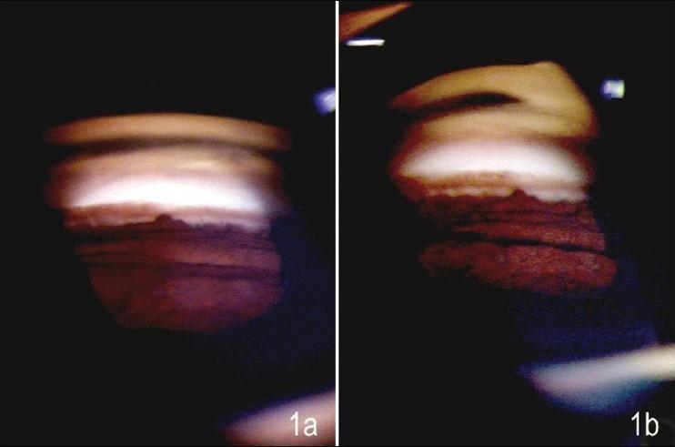
This comparative clinical diagnostic image displays a side-by-side gonioscopic evaluation of the anterior chamber angle in a 45-year-old patient. The images illustrate a clinical diagnostic challenge: the differentiation between true peripheral anterior synechiae (PAS) and a pseudosynechia caused by a peripheral iris bulge. Image (1a) was captured using a Goldmann (two-mirror) lens, showing a prominent horizontal ridge and reddish-brown tissue extending toward the trabecular meshwork, mimicking a synechia in a wide-open angle. Image (1b) shows the same anatomical region viewed with a Sussman (four-mirror) lens. The visual characteristics include a granular iris texture and a reflective band at the top representing the posterior corneal surface/Schwalbe's line. The primary educational objective is to demonstrate how indentation gonioscopy (possible with the Sussman lens) helps flatten the peripheral iris bulge to reveal the underlying anatomy, whereas manipulative gonioscopy with a Goldmann lens may fail to resolve the pseudosynechia. This is a critical distinction in the evaluation of primary angle-closure suspects and glaucoma management.
GONIOSCOPY
Complete Theory for Postgraduate Ophthalmology
1. Definition
"Gonioscopy is an indispensable technique that allows the filtration angle to be examined in patients with ocular hypertension or glaucoma." (Kanski's Clinical Ophthalmology, 10th ed., Ch. 1)
2. Why Gonioscopy is Necessary - The Optical Principle
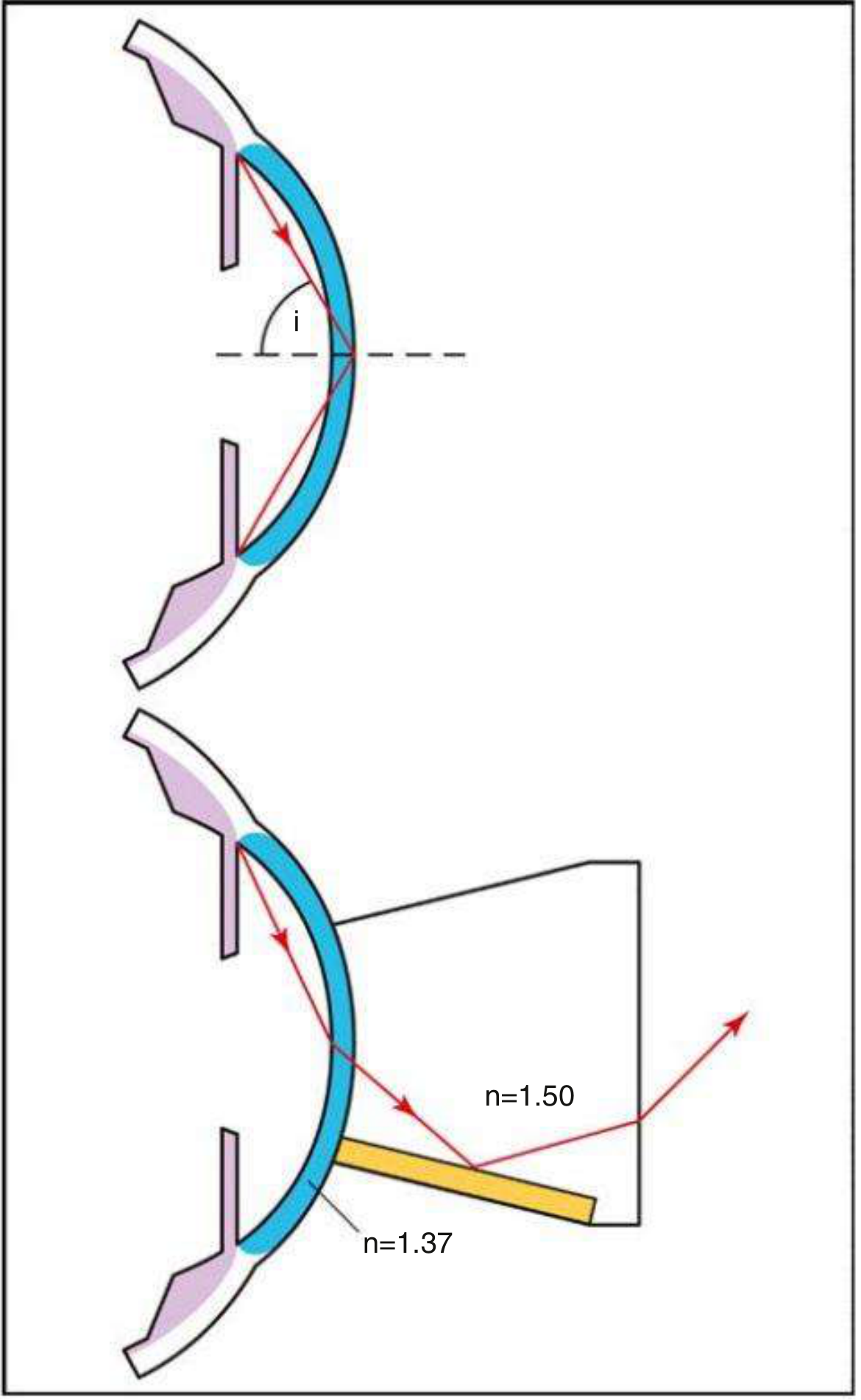
- Light travelling from a medium of higher refractive index (cornea, n=1.37) to lower (air, n=1.00) is totally internally reflected at the interface when the angle of incidence exceeds the critical angle of 46° for the tear film-air interface.
- A goniolens has a refractive index similar to the cornea (~1.50), so it eliminates total internal reflection by replacing the tear film-air interface with a tear film-goniolens interface.
- Light rays can then exit the contact lens and be viewed by the observer, either directly or via a mirror.
3. Classification of Gonioscopy
A. Indirect Gonioscopy
- Uses an internal mirror inside the goniolens to redirect angle rays so they exit the lens and can be viewed via slit lamp
- Gives a mirror image of the opposite angle (the image is inverted and reversed)
- Requires a slit lamp for illumination and magnification
- Requires a viscous coupling fluid (e.g., hydroxypropyl methylcellulose/hypromellose 0.3%) because the lens curvature is steeper than the cornea
| Lens | Mirrors | Notes |
|---|---|---|
| Goldmann single-mirror | 1 (62° tilt) | Classic; needs coupling fluid; best for laser procedures |
| Goldmann three-mirror | 3 mirrors + central lens | One mirror specifically for gonioscopy |
| Zeiss 4-mirror | 4 mirrors | Flat contact surface; no coupling fluid needed; best for indentation/dynamic gonioscopy |
| Posner 4-mirror | 4 (with handle) | Variant of Zeiss; easier to handle |
| Sussman 4-mirror | 4 (no handle) | Useful for indentation gonioscopy |
| Ritch trabeculoplasty lens | Modified for ALT/SLT | |
| Magna View, Khaw direct view | Modified indirect |
- The Goldmann lens requires coupling fluid and stabilizes the globe - preferred for laser trabeculoplasty
- The Zeiss/Posner/Sussman lenses have a curvature flatter than the cornea - no coupling fluid needed; do not stabilize the globe; preferred for indentation gonioscopy
B. Direct Gonioscopy
- The viewing surface is domed/slanted so exiting light strikes the contact lens-air interface at steeper than the critical angle, passing through to the observer without internal reflection
- Gives a direct, upright, non-reversed image
- Does not require a slit lamp - used with operating microscope or magnifying loupes
- Patient must be in the supine position (typically under general anaesthesia)
- Used primarily for examination and surgery of infantile/congenital glaucoma
- Note: Supine position may deepen the angle compared to sitting
- Koeppe lens (most common)
- Barkan lens (for Barkan's goniotomy surgery)
- Swan-Jacob lens (for surgical goniotomy)
- Medical Workshop lens
- Volk Surgical Gonio Lens
4. Technique of Indirect (Static) Gonioscopy
Pre-procedure
- Room must have low ambient illumination - pupil size influences angle grade; light causes pupil constriction which deepens the angle falsely
- Instil topical anaesthetic (proparacaine/oxybuprocaine)
- Apply 1-2 drops of coupling fluid on the lens (for Goldmann-type lenses)
Procedure
- Patient seated at slit lamp, forehead against headband, both eyes open
- Patient looks upward; insert the lens rapidly to avoid spilling coupling fluid
- Patient looks straight ahead
- Use the smallest slit beam possible - do not direct beam through the pupil (prevents miosis)
- Examine all 4 quadrants by rotating the lens (Goldmann) or the slit lamp (Zeiss)
- Record findings systematically in all quadrants - angle is usually narrowest superiorly
Technical Tips
- When the view is obscured by a convex iris: ask patient to look slightly in the direction of the mirror (only slight movement - excessive movement distorts the angle)
- Excessive pressure with a non-indentation lens narrows the apparent angle
- Increasing illumination after initial examination helps define structures
- Scleral spur can be identified by applying gentle pressure with goniolens edge on perilimbal nasal episcleral vessels - allows blood reflux into Schlemm canal as a landmark
5. Angle Structures: Normal Anatomy (Anterior to Posterior)
1. Schwalbe Line (Posterior corneal ring)
- The most anterior angle structure
- Anatomically: the peripheral termination of Descemet's membrane and the anterior limit of the trabecular meshwork
- Appears: whitish to variably pigmented
- May be barely visible in young patients
- Corneal wedge technique to locate it: using a narrow slit beam, two linear corneal reflections (inner and outer surface) converge at the apex of the wedge = Schwalbe line (Fig. 1.52D, Kanski)
- Sampaolesi line: pigment deposits anterior to Schwalbe line - seen in pseudoexfoliation and pigment dispersion syndrome. Key exam finding.
- Axenfeld anomaly/syndrome: prominent anteriorly displaced Schwalbe line (posterior embryotoxon)
2. Trabecular Meshwork (TM)
- Extends from Schwalbe line to scleral spur; average width 600 μm
- Divided into two functional parts:
| Part | Position | Appearance | Function |
|---|---|---|---|
| Anterior (non-pigmented / non-functional) | Adjacent to Schwalbe line | Whitish, ground-glass | Minimal aqueous drainage |
| Posterior (pigmented / functional) | Adjacent to scleral spur | Grey-blue translucent; becomes pigmented with age | Main site of aqueous outflow |
- Trabecular pigmentation is rare before puberty; increases with age; most marked inferiorly
- Patchy trabecular pigmentation in a suspicious narrow angle raises the possibility of intermittent iris contact
3. Schlemm Canal
- Lies deep to the posterior trabeculum
- Seen as a slightly darker line deep to the posterior TM (especially when non-pigmented)
- Blood in Schlemm canal (episcleral venous pressure > IOP): seen in -
- Physiological variant (excess goniolens pressure)
- Carotid-cavernous fistula / dural shunt
- Sturge-Weber syndrome
- Superior vena cava obstruction
- Ocular hypotony
4. Scleral Spur
- Most anterior projection of the sclera
- Site of attachment of the longitudinal muscle of the ciliary body (of Brücke)
- Appears as a narrow whitish band (yellows with age) just posterior to TM
- Key surgical landmark in MIGS: separates canal-based surgery (trabecular meshwork, anterior) from suprachoroidal-based surgery (ciliary body band, posterior)
5. Ciliary Body Band
- Stands just behind the scleral spur
- Appears as a pink, dull brown, or slate grey band
- Width depends on iris insertion position:
- Narrower in hypermetropic eyes
- Wider in myopic eyes
- The angle recess = posterior dipping of the iris at its insertion into the ciliary body
6. Iris Root
- Most posterior structure
- Peripheral iris inserts into the ciliary body
- Iris processes: small tenuous extensions of the anterior iris surface inserting at the level of the scleral spur (Fig. 1.53, Kanski) - present in ~1/3 of normal eyes; most common in children and brown eyes; must NOT be confused with PAS (PAS extend more anteriorly and are more substantial)
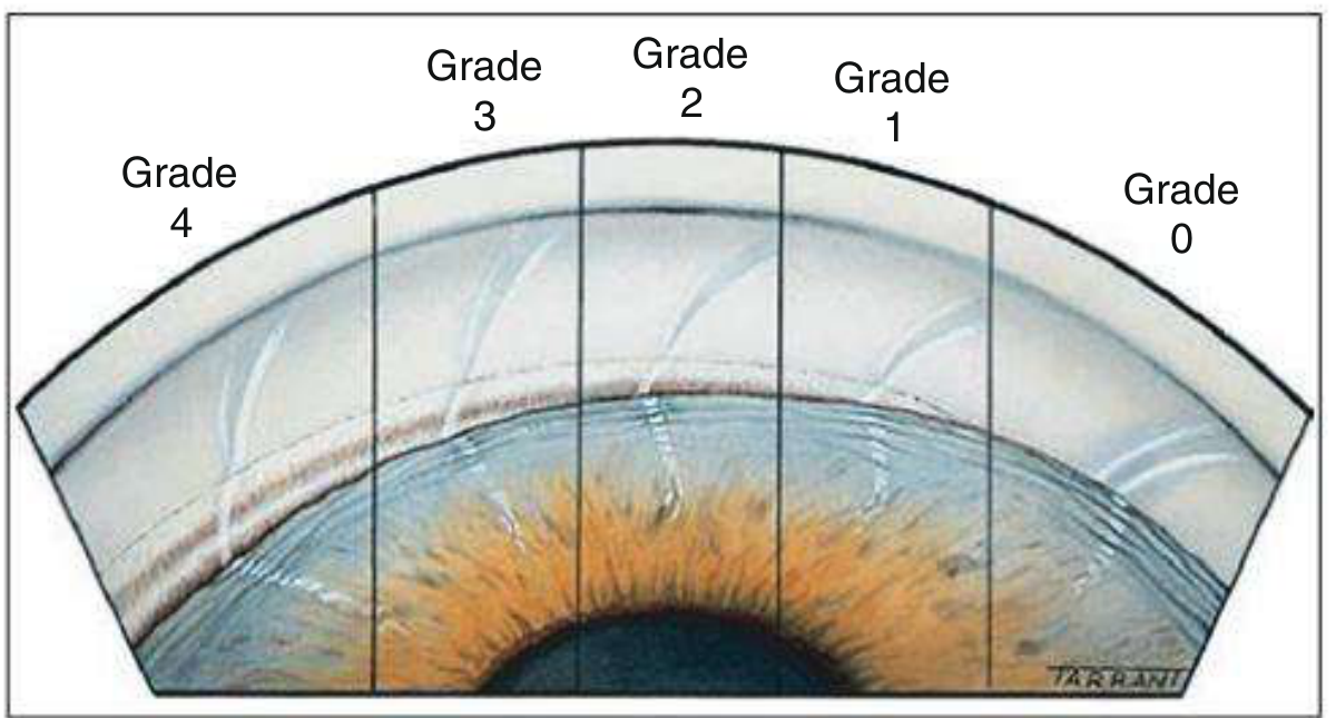
6. Grading Systems for the Anterior Chamber Angle
A. Shaffer Grading System (Most widely used clinically)
| Grade | Degrees | Structures Visible | Interpretation |
|---|---|---|---|
| Grade 4 | 35-45° | All: ciliary body, scleral spur, TM, Schwalbe line | Wide open; characteristic of myopia and pseudophakia |
| Grade 3 | 25-35° | Scleral spur visible | Open; incapable of closure |
| Grade 2 | 20° | TM visible but not scleral spur | Narrow; possible closure |
| Grade 1 | 10° | Only Schwalbe line ± top of TM | Very narrow; closure probable |
| Slit | <10° | No obvious iridocorneal contact; no structures identifiable | Dangerously narrow |
| Grade 0 | 0° | None (iridocorneal contact) | Closed angle |
B. Scheie Grading System
| Grade | Structures Visible | Angle Status |
|---|---|---|
| Grade 0 | All structures including CB band | Wide open |
| Grade I | CB band not visible | Slightly narrow |
| Grade II | Scleral spur not visible | Narrow |
| Grade III | Posterior TM not visible | Very narrow |
| Grade IV | Only Schwalbe line (or nothing) visible | Closed |
Important exam note (per AK Khurana / per Kanski): In the Scheie system, a higher Roman numeral = narrower angle - the opposite of common clinical convention. This is frequently reversed or confused in questions. Always note: Scheie IV = closed; Shaffer 4 = wide open.
C. Spaeth Grading System (Most comprehensive, least used routinely)
- Apparent iris insertion level (A to E):
- A = anterior to Schwalbe line
- B = between Schwalbe line and scleral spur
- C = at scleral spur
- D = deep to scleral spur (ciliary body visible 1 mm)
- E = deep (ciliary body visible >1 mm)
- Angular approach (0° to 40°+) - angle between TM and iris
- Peripheral iris curvature (q = queer/flat, r = regular/flat, s = steep/convex, c = concave)
D. Van Herick Method (Slit Lamp Screening - NOT true gonioscopy)
- Narrow, bright slit beam at 60° offset temporally to the cornea
- Estimate ratio: peripheral anterior chamber depth / corneal thickness
| AC Depth : CT Ratio | Van Herick Grade | Clinical Meaning |
|---|---|---|
| ≥ 1 (equal or greater) | Grade 4 | Wide open; no risk of closure |
| ¼ to ½ | Grade 3 | Low probability of closure |
| = ¼ | Grade 2 | Angle closure possible; perform gonioscopy |
| < ¼ | Grade 1 | Dangerously narrow; gonioscopy will show closure |
7. Indentation (Dynamic/Compression) Gonioscopy
Principle:
Lenses used:
- Zeiss 4-mirror, Posner 4-mirror, Sussman 4-mirror (no coupling fluid; do not stabilize globe - these can be pressed into the cornea)
- Goldmann lens is NOT suitable for indentation (it stabilizes the globe)
Diagnostic Value:
| Finding | Meaning |
|---|---|
| Angle opens on indentation | Appositional closure (iris-TM contact only, reversible) |
| Angle remains closed on indentation | Synechial closure (PAS - permanent adhesions) |
Additional Uses of Indentation Gonioscopy:
- Distinguishing an extensive or double highly pigmented Schwalbe line from pigmented meshwork
- Differentiating iris processes from PAS
- Helps define structures in a narrow angle that are difficult to assess
- Assessing extent of PAS vs appositional closure in PACG
8. Indications for Gonioscopy
Diagnostic Indications:
- Glaucoma evaluation - to classify open angle vs angle closure (mandatory in all glaucoma patients)
- Ocular hypertension - to assess angle for risk of closure
- Secondary glaucomas - to identify the mechanism:
- Neovascular glaucoma (NVG): rubeosis iridis / neovascularization of angle
- Pigment dispersion syndrome / Pigmentary glaucoma
- Pseudoexfoliation syndrome
- Phacolytic, phacomorphic, uveitic glaucoma
- Trauma: angle recession, cyclodialysis, trabecular dialysis, foreign bodies
- Acute angle closure glaucoma - to classify; compression gonioscopy to break an acute attack
- Narrow angle / angle closure suspect - to determine if PI is indicated
- Sturge-Weber syndrome - blood in Schlemm canal
- Anterior segment tumours - melanoma, naevus of angle
- Infantile/juvenile glaucoma - to assess angle and plan goniotomy
- Iridocorneal endothelial (ICE) syndrome - PAS, corneal endothelial changes
- Fuchs heterochromic cyclitis - fine angle vessels (Amsler sign)
Therapeutic Indications:
- Argon/Selective Laser Trabeculoplasty (ALT/SLT) - treatment of POAG
- Laser gonioplasty (peripheral iridoplasty) - plateau iris
- Goniotomy (via direct gonioscopy) - congenital glaucoma
- Surgical trabeculotomy / MIGS (via surgical gonioscopy) - iStent, Kahook blade, OMNI, goniotomy
- Compression gonioscopy - to break acute angle closure attack
9. Pathological Findings on Gonioscopy
Peripheral Anterior Synechiae (PAS)
- Definition: Adhesions between the peripheral iris and angle structures (TM or cornea), anterior to the scleral spur
- Distinguished from iris processes by: PAS are more substantial and extend more anteriorly
- Causes:
- Primary angle-closure glaucoma (PACG) - most common
- Anterior uveitis
- Iridocorneal endothelial (ICE) syndrome
- Neovascular glaucoma
- Trauma
Neovascularization of the Angle (NVA)
- Pathological blood vessels running randomly (vs normal radial vessels at angle recess)
- Any blood vessel that crosses the scleral spur is pathological
- Causes: neovascular glaucoma (diabetic retinopathy, CRVO, ocular ischaemic syndrome), Fuchs heterochromic cyclitis (fine vessels, Amsler sign), chronic anterior uveitis
Hyperpigmentation
- Physiological variant
- Pigment dispersion syndrome (Krukenberg spindle on cornea; heavy homogeneous 360° TM pigmentation - 3+ to 4+, mainly posterior TM)
- Pseudoexfoliation syndrome (irregular, "moth-eaten" TM pigmentation; Sampaolesi line)
- Pseudophakic pigment dispersion
- Blunt trauma
- Post-inflammatory (uveitis, acute ACG, post-YAG iridotomy)
- Iris/angle melanoma
Traumatic Findings
- Angle recession: tear between longitudinal and circular ciliary muscle fibres; widening of the ciliary body band; risk of late glaucoma
- Trabecular dialysis: tear in TM
- Cyclodialysis: separation of ciliary body from scleral spur; causes hypotony
- Foreign bodies
Blood in Schlemm Canal
- Physiological variant (compression of episcleral veins by gonioprism)
- Carotid-cavernous fistula / dural shunt (raised episcleral venous pressure)
- Sturge-Weber syndrome
- Superior vena cava obstruction
- Ocular hypotony
10. Gonioscopy and the Classification of Primary Angle Closure
| Classification | Gonioscopic Finding |
|---|---|
| PAC Suspect (PACS) | ITC (iridotrabecular contact) in ≥3 quadrants; no PAS; normal IOP and disc |
| Primary Angle Closure (PAC) | ITC in ≥3 quadrants + raised IOP and/or PAS; normal disc |
| Primary Angle Closure Glaucoma (PACG) | ITC in ≥3 quadrants + glaucomatous optic neuropathy |
11. Comparison of Gonioscopic Lenses - Summary Table
| Feature | Goldmann 1/3 Mirror | Zeiss/Posner/Sussman 4-Mirror | Koeppe (Direct) |
|---|---|---|---|
| Type | Indirect | Indirect | Direct |
| Mirrors | 1 or 3 | 4 | None |
| Coupling fluid | Required | Not required | Required |
| Slit lamp needed | Yes | Yes | No |
| Patient position | Sitting | Sitting | Supine |
| Globe stabilization | Yes | No | Yes |
| Indentation | Not suitable | Best for indentation | Suitable |
| Laser therapy | Best (stable) | Less suitable | Not used |
| Image | Inverted mirror image | Inverted mirror image | Direct, upright |
| Special use | ALT/SLT, best optics | Dynamic gonioscopy | Infantile glaucoma, goniotomy |
12. Key Exam Points
| Point | Fact |
|---|---|
| Why angle cannot be seen directly | Total internal reflection at tear film-air interface (critical angle 46°) |
| How goniolens works | Replaces tear film-air with tear film-goniolens interface; eliminates total internal reflection |
| Indirect gonioscopy image | Inverted mirror image of opposite angle |
| Direct gonioscopy image | Direct, upright image |
| Coupling fluid needed | Goldmann (Yes); Zeiss/Posner/Sussman (No - flat base curve) |
| Best lens for indentation | Zeiss / Posner / Sussman 4-mirror |
| Best lens for laser | Goldmann (stabilizes globe) |
| Room illumination | LOW - pupil size affects angle grade |
| Most anterior angle structure | Schwalbe line |
| Scleral spur landmark | Whitish band; site of longitudinal ciliary muscle; MIGS reference |
| Corneal wedge sign | Two corneal reflections converge at Schwalbe line |
| Sampaolesi line | Pigment anterior to Schwalbe - pseudoexfoliation / pigment dispersion |
| Shaffer 0 vs Scheie IV | Both mean closed - but Scheie system is inverted (higher = narrower) |
| Shaffer 4 | Wide open (myopia, pseudophakia); CB band visible |
| Indentation opens angle | Appositional closure (reversible) |
| Indentation fails to open | Synechial / PAS (permanent) |
| Blood in Schlemm canal | Raised episcleral venous pressure (CCF, Sturge-Weber, SVC obstruction) |
| NV crossing scleral spur | Always pathological |
| Angle recession | Tear between ciliary muscle fibres; late glaucoma risk |
| Cyclodialysis | Ciliary body separates from scleral spur; causes hypotony |
| Van Herick limitation | Overestimates angle width in plateau iris configuration |
| Direct gonioscopy use | Infantile glaucoma, goniotomy; patient supine, no slit lamp |
- Kanski's Clinical Ophthalmology: A Systematic Approach, 10th Edition - Ch. 1 (Gonioscopy - optical principles, lenses, technique, angle structures, pathological findings) and Ch. 11 (Grading systems, angle closure classification)
- Wills Eye Manual - Section 9.17 (Glaucoma), Appendix (Van Herick technique)
- AK Khurana - Comprehensive Ophthalmology (Schaffer vs Scheie grading comparison; Indian PG exam perspective)
- Becker-Shaffer's Diagnosis and Therapy of the Glaucomas (Shaffer grading system origin; gonioscopic anatomy)
- Duke-Elder's System of Ophthalmology (historical classification; angle structures description)