
Central line insertion marking point
central line insertion landmark marking point internal jugular subclavian femoral
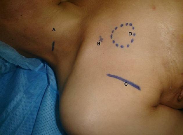
This clinical photograph displays preoperative surgical skin markings on a patient's right neck and upper chest area, identifying anatomical landmarks for central venous access and port-a-cath placement. The image features four distinct blue ink markings labeled A through D: 'A' is a vertical line on the lateral neck corresponding to the internal jugular vein; 'B' is a cross-mark (plus sign) at the infraclavicular region representing the subclavian vein entry point; 'C' is a horizontal curved line near the deltopectoral groove indicating the cephalic vein dissection site; and 'D' is a dotted circular outline on the superior-lateral chest wall marking the planned subcutaneous pocket for the implantable chamber. These markings illustrate the topographic anatomy and surgical planning for various venous access techniques, including percutaneous puncture and surgical cut-down. The content is relevant for surgical oncology, anesthesia, and interventional radiology training regarding the placement of long-term vascular access devices.
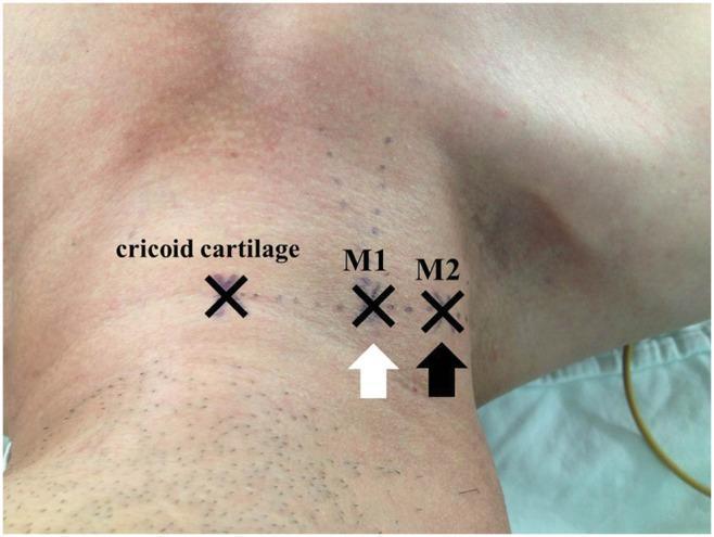
A clinical photograph of the right lateral neck of a patient in a supine, head-down position, demonstrating surface anatomical landmarks for internal jugular vein (IJV) catheterization. Three 'X' markings are visible along a horizontal line at the level of the cricoid cartilage. The leftmost mark indicates the cricoid cartilage itself. The central mark, labeled 'M1' and indicated by a white arrow, represents the apex of the triangle formed by the sternal and clavicular heads of the sternocleidomastoid muscle. The lateral mark, labeled 'M2' and indicated by a black arrow, represents an alternative landmark identified by observing respiratory jugular venodilation and manual palpation of the IJV. The image illustrates the spatial relationship between traditional surface landmarks and dynamic physiological markers (venodilation) used to improve the accuracy of central venous access. Subtle skin elevations near M2 correspond to underlying venous distension, providing clinical guidance for needle insertion in the absence of ultrasound.
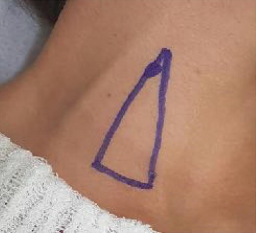
This clinical photograph displays a close-up view of a patient's neck in the dorsal decubitus position, rotated contralaterally to expose the right cervical region. A blue ink marking delineates a triangle representing Sedillot's triangle, an anatomical landmark used for internal jugular vein (IJV) cannulation via the traditional technique (TT). The base of the triangle is situated inferiorly near the clavicle, while the two ascending sides correspond to the sternal and clavicular heads of the sternocleidomastoid muscle. The apex of the triangle is positioned superiorly where these two muscle heads converge. A specific circular mark near the apex indicates the intended puncture point for vascular access, which is clinically localized lateral to the palpated common carotid artery (CCA) pulse. The image serves as an educational guide for identifying superficial anatomical landmarks necessary for central venous catheterization and highlights the surface anatomy used in blind or ultrasound-guided vascular procedures within the field of anesthesiology and critical care.
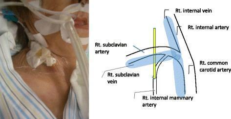
This procedural content consists of a side-by-side clinical photograph and an anatomical diagram illustrating a central venous catheter (CVC) malposition. The left panel is a clinical photograph of a patient's right neck and supraclavicular region, showing an intubated patient with a CVC dressing and medical tubing. The right panel is a schematic diagram of the right-sided vascular anatomy in the neck and upper chest. Labeled structures include the right subclavian artery and vein, right internal jugular vein (labeled as 'Rt. internal vein'), right common carotid artery, and the right internal mammary artery (IMA). A yellow vertical line represents a catheter inserted via a supraclavicular approach. The diagram demonstrates a potential mechanism of iatrogenic injury where the catheter, intended for the subclavian vein, is instead directly inserted into the more deeply seated right internal mammary artery due to a proximal insertion site. This visual serves as an educational tool for anesthesiology and vascular medicine to demonstrate the anatomical risks and potential complications of landmark-guided central venous access.
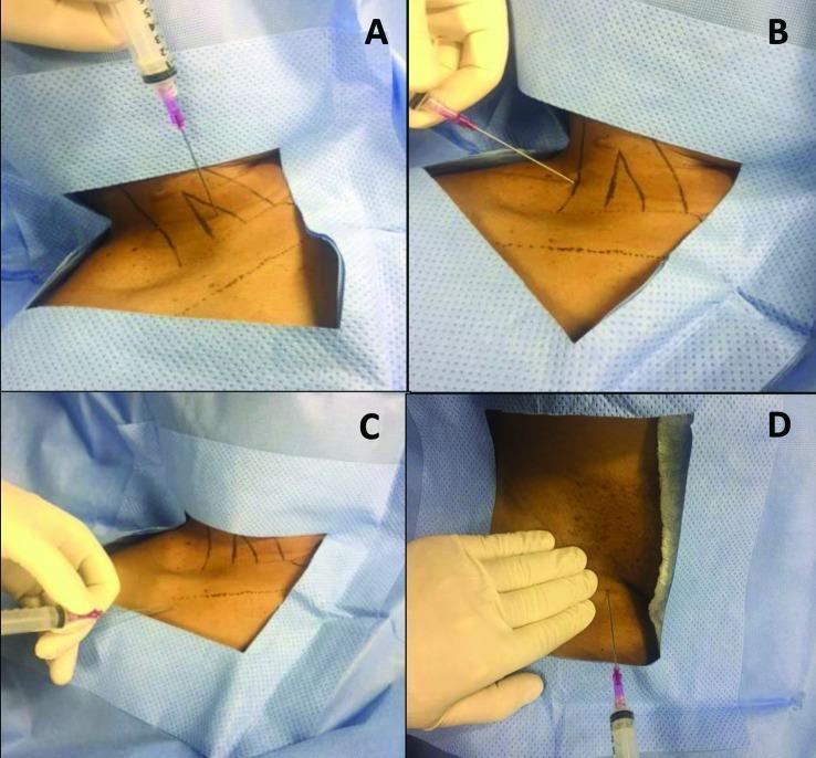
A clinical photograph comprising four panels (A, B, C, D) demonstrating needle puncture techniques for deep vein access during central venous catheterization. Each panel shows a patient prepared with a sterile blue drape and a clinician wearing white sterile gloves using a 10 mL syringe containing pink-tinged fluid. Panel A illustrates an anterior approach to the internal jugular vein (IJV), with the needle inserted at the apex of the triangle formed by the sternal and clavicular heads of the sternocleidomastoid muscle. Panel B shows a posterior approach to the IJV, with the needle entering lateral and posterior to the sternocleidomastoid muscle. Panel C depicts infraclavicular puncture of the subclavian vein, with landmarks indicating insertion between the middle and lateral thirds of the clavicle. Panel D shows a femoral vein puncture in the groin region, medial to the palpated femoral artery pulse. Anatomical landmarks and projected pathways are indicated by black skin markings. These images illustrate common vascular access points used for implanting long-term medical devices like portacaths.
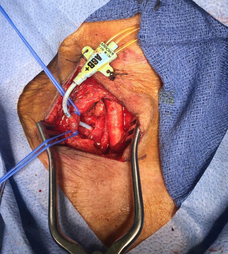
This clinical photograph captures an intraoperative view of a surgical neck exploration performed to address a malpositioned central venous catheter. The image shows a dissected right-sided surgical field with the internal jugular vein (IJV) retracted and controlled using blue vessel loops. A white multi-lumen central line (labeled 'ARROW 7 FR') is visibly traversing through both walls of the IJV. The catheter then tracks deeper into the anatomical space, posterior to the clavicular region, where it is shown piercing the superior aspect of the right subclavian artery. Self-retaining retractors provide exposure of the vascular structures and surrounding soft tissue. This visual serves as a critical educational example of a procedural complication: inadvertent arterial cannulation and through-and-through venous injury during central line placement. It illustrates the complex anatomical relationship between the internal jugular vein and the underlying subclavian artery in the root of the neck.
Create a visual landmarks guide (PDF or poster) for central line insertion sites