
Soft palate image
Reading File
This is a Visual query - image retrieval is the priority. Let me search for soft palate images in the medical library.
Finding Sources
Finding Sources
Let me read the key anatomy sections and look for images.
Reading File
Reading File
Reading File
I found an image URL. Let me fetch it along with looking for more images from other sections.
Loading Image
Reading File
Reading File
Excellent! Found multiple great images. Let me fetch the two additional anatomy images in parallel.
Loading Image
Loading Image
Here are high-quality anatomical images of the soft palate from authoritative medical textbooks:
Soft Palate - Anatomy Images
1. Schematic Overview: Soft Palate & Pharyngeal Regions
(Gray's Anatomy for Students)
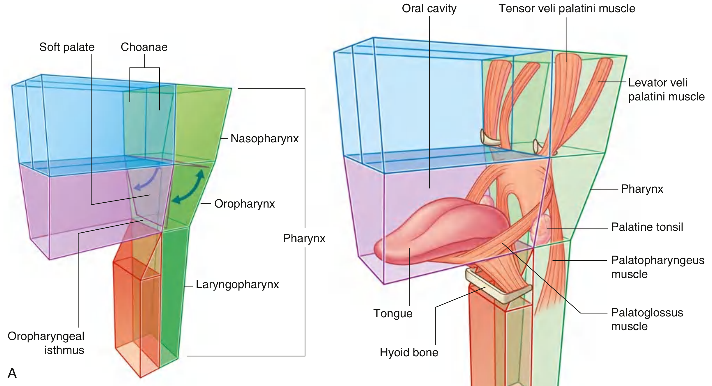
Fig. A shows the three pharyngeal regions (nasopharynx, oropharynx, laryngopharynx) with the soft palate acting as a valve at the superior boundary. Fig. B shows the muscles of the soft palate: tensor veli palatini, levator veli palatini, palatopharyngeus, and palatoglossus, along with the palatine tonsil and hyoid bone.
2. Clinical/Endoscopic View of the Soft Palate
(Gray's Anatomy for Students)
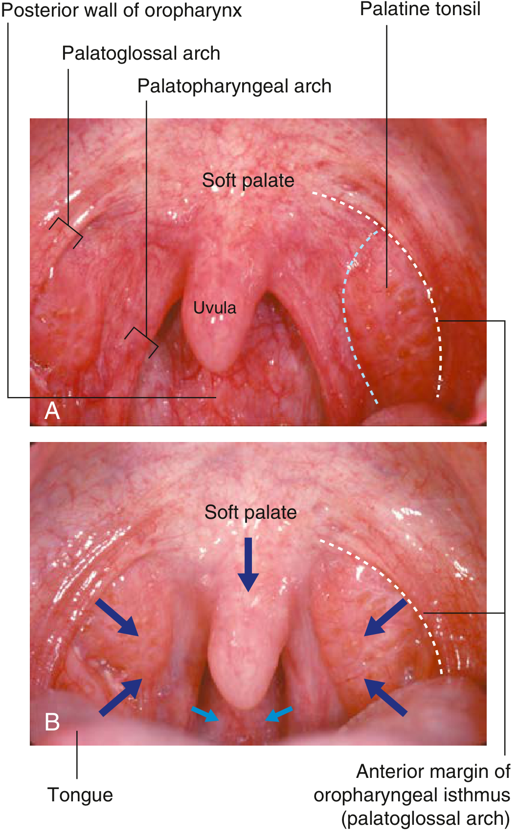
Fig. A is a direct intraoral photograph showing the soft palate, uvula, palatoglossal arch, palatopharyngeal arch, palatine tonsil, and posterior oropharyngeal wall. Fig. B shows the closure of the oropharyngeal isthmus with arrows indicating the direction of muscle movement. This is the view seen during clinical examination ("say ahh").
3. Surgical/Vascular Anatomy of the Soft Palate
(Cummings Otolaryngology Head and Neck Surgery)
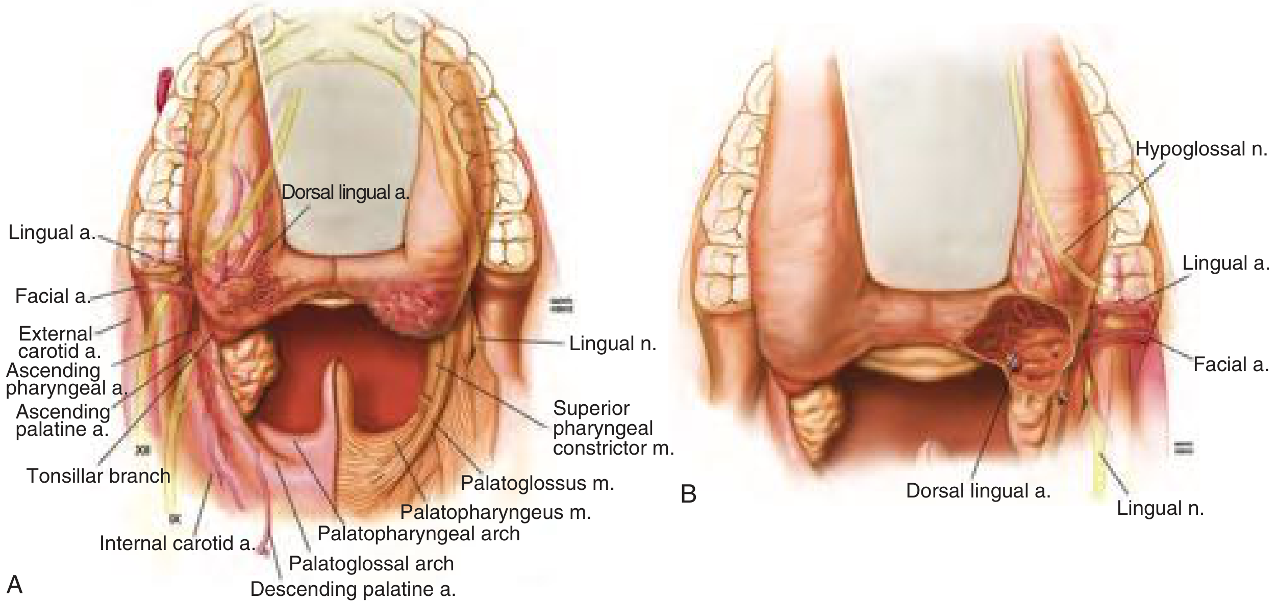
Transoral surgical perspective showing the blood supply and musculature of the oropharynx including the soft palate. Key structures labeled: ascending palatine artery, descending palatine artery, palatoglossus and palatopharyngeus muscles, palatoglossal and palatopharyngeal arches, superior pharyngeal constrictor, tonsillar branches, and key neurovascular relations.
Key Anatomy Summary
| Feature | Detail |
|---|---|
| Location | Posterior continuation of the hard palate; free posterior margin |
| Function | Valvular - elevates to close nasopharynx (swallowing); depresses to close oropharyngeal isthmus |
| Muscles (5) | Tensor veli palatini (CN V3), Levator veli palatini (CN X), Palatoglossus (CN X), Palatopharyngeus (CN X), Musculus uvulae (CN X) |
| Blood supply | Lesser palatine artery (main), ascending palatine branch of facial artery |
| Sensory nerve | Glossopharyngeal nerve (CN IX) + lesser palatine nerve |
| Motor nerve | Vagus nerve (CN X) via pharyngeal plexus - except tensor veli palatini (CN V3) |
| Clinical test | Levator veli palatini tested by saying "ahh" - palate deviates away from the abnormal side |
Sources: Gray's Anatomy for Students; Cummings Otolaryngology Head and Neck Surgery
This is a shared conversation. Sign in to Orris to start your own chat.