
generate most important high yield MCQs for batterjee medical college exams as i have less time to review so generate absolute must know MCQs with pictures/diagram
cd /home/daytona/workspace/attachments/f06032e9-1039-4542-acd4-9238ac5716ab && python3 -c " import pdfplumber files = [ 'Anatomy Of Intercostal space- 2025-2026.pdf', 'Anatomy Of Mediastinum -Part I- 2025-2026.pdf', 'Anatomy Of Mediastinum -Part II- 2025-2026.pdf' ] for fname in files: print(f'\n\n========== {fname} ==========\n') with pdfplumber.open(fname) as pdf: for i, page in enumerate(pdf.pages): text = page.extract_text() if text: print(f'--- Page {i+1} ---') print(text[:3000]) " 2>&1 | head -500
cd /home/daytona/workspace/attachments/f06032e9-1039-4542-acd4-9238ac5716ab && python3 -c " import pdfplumber files = [ 'Anatomy Of Mediastinum -Part I- 2025-2026.pdf', 'Anatomy Of Mediastinum -Part II- 2025-2026.pdf' ] for fname in files: print(f'\n\n========== {fname} ==========\n') with pdfplumber.open(fname) as pdf: for i, page in enumerate(pdf.pages): if i < 20: continue # skip already read pages text = page.extract_text() if text: print(f'--- Page {i+1} ---') print(text[:3000]) " 2>&1
cd /home/daytona/workspace/attachments/f06032e9-1039-4542-acd4-9388ac5716ab && python3 -c " import pdfplumber fname = 'Anatomy Of Mediastinum -Part II- 2025-2026.pdf' print(f'\n\n========== {fname} ==========\n') with pdfplumber.open('/home/daytona/workspace/attachments/f06032e9-1039-4542-acd4-9238ac5716ab/' + fname) as pdf: for i, page in enumerate(pdf.pages): if i >= 20: break text = page.extract_text() if text: print(f'--- Page {i+1} ---') print(text[:3000]) " 2>&1
python3 -c " import pdfplumber fname = '/home/daytona/workspace/attachments/f06032e9-1039-4542-acd4-9238ac5716ab/Anatomy Of Mediastinum -Part II- 2025-2026.pdf' with pdfplumber.open(fname) as pdf: for i, page in enumerate(pdf.pages): if i >= 20: break text = page.extract_text() if text: print(f'--- Page {i+1} ---') print(text[:2000]) " 2>&1
intercostal space neurovascular bundle muscles layers anatomy
mediastinum subdivisions boundaries anatomy diagram
thoracic duct course lymphatic drainage anatomy
azygos venous system hemiazygos veins posterior intercostal
esophagus constrictions relations thoracic anatomy diagram
🏥 High-Yield Anatomy MCQs — Batterjee Medical College
Intercostal Space · Mediastinum Part I & II · 2025–2026
🫁 SECTION 1: INTERCOSTAL SPACE
Q1 — Neurovascular Plane
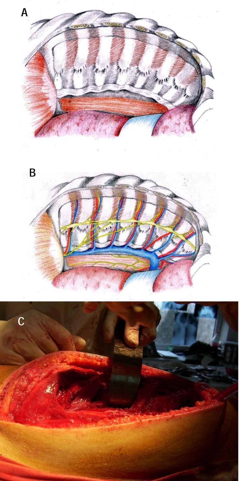
- A) External intercostal and internal intercostal
- B) Internal intercostal and innermost intercostal ✅
- C) Innermost intercostal and endothoracic fascia
- D) Endothoracic fascia and parietal pleura
Answer: B — The intercostal nerve and vessels (VAN) run between the internal intercostal and the innermost intercostal muscles. This is the single most tested fact about the intercostal space.
Q2 — Intercostal Muscle Fiber Directions
- A) External intercostal
- B) Internal intercostal ✅
- C) Subcostalis
- D) Sternocostalis
Answer: B — External intercostal: downward, forward, medially. Internal intercostal: downward, backward, laterally. These are opposite directions — classic MCQ trap.
Q3 — Where Muscles Are Replaced by Membranes
- A) The lateral border of sternum
- B) The midaxillary line
- C) The junction of rib with its costal cartilage ✅
- D) The angle of the rib
Answer: C — External intercostal → replaced anteriorly at rib-cartilage junction → membrane extends to lateral sternum.
Internal intercostal → replaced posteriorly at angle of rib → internal (posterior) intercostal membrane.
Q4 — Sternocostalis (Transversus Thoracis)
- A) 1st to 4th costal cartilages
- B) 2nd to 6th costal cartilages ✅
- C) 4th to 8th costal cartilages
- D) 1st to 3rd ribs
Answer: B — Origin: back of lower 1/3 of sternum + xiphoid. Insertion: 2nd–6th costal cartilages. Fibers ascend superolaterally.
Q5 — Respiratory Function
- A) Internal intercostals only
- B) External intercostals only ✅
- C) Innermost intercostals only
- D) Sternocostalis only
Answer: B — External intercostals = elevate ribs = inspiration.
Internal, innermost, subcostalis, and sternocostalis = depress ribs = expiration.
Q6 — Order of Structures in Costal Groove (VAN)
- A) Artery, Nerve, Vein
- B) Nerve, Artery, Vein
- C) Vein, Artery, Nerve ✅
- D) Artery, Vein, Nerve
Answer: C — V-A-N from top to bottom (Vein, Artery, Nerve). The nerve lies lowest, which is why needles in thoracocentesis are inserted along the upper border of the rib below to avoid injuring VAN.
Q7 — Intercostal Nerve Classification
- A) T1–T3
- B) T3–T6 ✅
- C) T7–T11
- D) T1–T2
Answer: B — T3–T6 are typical (same course and same branches). T1 is atypical (contributes to brachial plexus). T2 is atypical (intercostobrachial nerve). T7–T11 are atypical (pierce diaphragm to supply anterior abdominal wall).
Q8 — Intercostobrachial Nerve
- A) Medial side of forearm
- B) Floor of axilla + upper medial arm ✅
- C) Posterior shoulder
- D) Lateral chest wall
Answer: B — T2's lateral cutaneous branch is undivided and forms the intercostobrachial nerve → supplies floor of axilla and upper medial arm.
Q9 — Posterior Intercostal Arteries — Origin
- A) Directly from the descending thoracic aorta
- B) The superior intercostal artery (from the costocervical trunk of subclavian) ✅
- C) The internal thoracic artery
- D) The musculophrenic artery
Answer: B — Spaces 1 and 2: from superior intercostal artery (branch of costocervical trunk of subclavian artery).
Spaces 3–11: directly from descending thoracic aorta.
Q10 — Internal Thoracic Artery
- A) Musculophrenic and inferior epigastric arteries
- B) Musculophrenic and superior epigastric arteries ✅
- C) Superior epigastric and deep circumflex iliac arteries
- D) Inferior epigastric and lumbar arteries
Answer: B — Internal thoracic artery arises from 1st part of subclavian artery and terminates at 6th ICS → musculophrenic artery + superior epigastric artery.
Q11 — Anterior Intercostal Arteries — Coverage
- A) 11 spaces; from internal thoracic
- B) 9 spaces; from musculophrenic artery ✅
- C) 9 spaces; from superior epigastric artery
- D) 6 spaces; from internal thoracic only
Answer: B — 9 pairs (upper 9 spaces). Spaces 1–6: from internal thoracic. Spaces 7, 8, 9: from musculophrenic artery. Lower 2 spaces (10, 11): no anterior intercostal arteries.
Q12 — Posterior Intercostal Veins (RIGHT side)
- A) The azygos vein directly
- B) The right superior intercostal vein → arch of azygos ✅
- C) The right brachiocephalic vein
- D) The superior hemiazygos vein
Answer: B — Right side: 1st → right brachiocephalic vein. 2nd + 3rd → form right superior intercostal vein → arch of azygos. 4th–11th → directly into azygos vein.
Q13 — Clinical: Thoracocentesis / Tube Thoracostomy (MUST KNOW)
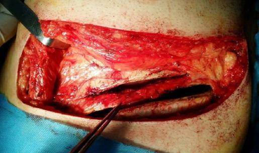
- A) The lower border of the rib above ✅ — No wait:
- A) The lower border of the upper rib
- B) The upper border of the lower rib ✅
- C) The middle of the intercostal space
- D) Along the midaxillary line, midway between ribs
Answer: B — Insert along the upper border of the rib below to avoid the neurovascular bundle (VAN = Vein, Artery, Nerve) that runs in the costal groove under the lower border of the rib above.
Q14 — Case: Stab Wound (From Lecture)
- A) Pectoralis major, intercostal muscles, right lung
- B) Intercostal muscles, internal thoracic artery, right atrium ✅
- C) Serratus anterior, pleura, SVC
- D) Pectoralis minor, diaphragm, right liver lobe
Answer: B — At 2 cm medial to midclavicular line in 4th ICS = near the sternal edge → internal thoracic artery runs here (1 cm lateral to sternum). Right atrium lies behind this region. Pectoralis major overlies the area anteriorly.
🫀 SECTION 2: MEDIASTINUM — PART I (Divisions & Contents)
Q15 — The Dividing Plane
- A) Manubriosternal joint (sternal angle) to lower border of T4 ✅
- B) Xiphoid to T12
- C) Sternal angle to T6
- D) Clavicle to T1
Answer: A — The imaginary plane extends from the sternal angle (angle of Louis) → lower border of T4 vertebra. This is the single most important mediastinal boundary landmark.
Q16 — Superior Mediastinum: Arteries
- A) Right common carotid, right subclavian, left common carotid
- B) Brachiocephalic, left common carotid, left subclavian ✅
- C) Brachiocephalic, left vertebral, left subclavian
- D) Left common carotid, left subclavian, right vertebral
Answer: B — From right to left: 1) Brachiocephalic artery (divides into right common carotid + right subclavian), 2) Left common carotid, 3) Left subclavian.
Q17 — Superior Mediastinum: Nerves
- A) Right recurrent laryngeal nerve
- B) Left recurrent laryngeal nerve ✅
- C) Left vagus nerve
- D) Left phrenic nerve
Answer: B — The left recurrent laryngeal nerve branches from the left vagus in the superior mediastinum, loops under the arch of aorta (ligamentum arteriosum), and ascends to the larynx. The right RLN loops under the right subclavian artery at the root of neck.
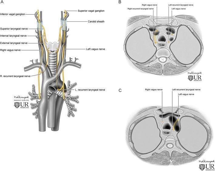
Q18 — Middle Mediastinum Contents
- A) Esophagus
- B) Thoracic duct
- C) Deep cardiac plexus ✅
- D) Thoracic sympathetic chain
Answer: C — Middle mediastinum contains: heart + pericardium, ascending aorta, pulmonary trunk, SVC (lower ½), IVC (1.25 cm), 4 pulmonary veins, azygos termination, right & left main bronchi, phrenic nerves (on sides of pericardium), deep cardiac plexus, inferior tracheobronchial LN.
Q19 — Anterior Mediastinum Contents (Simple but Tested)
- A) Heart and great vessels
- B) Trachea and esophagus
- C) Sternopericardial ligaments, thymus remnants, few lymph nodes ✅
- D) Descending aorta and thoracic duct
Answer: C — Anterior mediastinum is a small space between the sternum and pericardium. Its contents are minimal: sternopericardial ligaments, remains of thymus, few lymph nodes.
Q20 — Posterior Mediastinum Contents
- A) Descending thoracic aorta
- B) Azygos, hemiazygos veins
- C) Ascending aorta ✅
- D) Thoracic duct
Answer: C — Ascending aorta is in the middle mediastinum. The posterior mediastinum contains: descending thoracic aorta, esophagus, thoracic duct, azygos/hemiazygos veins, vagus nerves + sympathetic chains, posterior mediastinal lymph nodes.
Q21 — Mediastinal Syndrome (Clinical Must-Know)
- A) Aortic aneurysm
- B) Mediastinal syndrome due to tumor compression ✅
- C) Pulmonary embolism
- D) Cardiac tamponade
Answer: B — Mediastinal syndrome: tumor compresses:
- SVC → upper body vein engorgement
- Esophagus → dysphagia
- Trachea → dyspnea
- Left recurrent laryngeal nerve → dysphonia/hoarseness
- Phrenic nerve → hemidiaphragm paralysis
Q22 — Thymus Location
- A) Anterior mediastinum only
- B) Superior mediastinum only
- C) Both superior AND anterior mediastinum ✅
- D) Middle mediastinum
Answer: C — Thymus extends from 4th costal cartilage up to the lower poles of thyroid gland → spans both superior and anterior mediastinum.
Q23 — Case: Esophageal Carcinoma at T10 (From Lecture)
- A) Thoracic duct
- B) Vagus nerve ✅
- C) IVC
- D) Aorta
Answer: B — Esophagus passes through the diaphragm at T10 with the vagus nerves (as the esophageal plexus → anterior + posterior gastric nerves). Remember the rule: T8 = IVC, T10 = esophagus + vagus, T12 = aorta + thoracic duct + azygos.
🔬 SECTION 3: MEDIASTINUM — PART II (Esophagus, Thoracic Duct, Thymus)
Q24 — Esophageal Constrictions (4 Sites — VERY HIGH YIELD)
- A) 6 inches from incisor teeth
- B) 9 inches from incisor teeth ✅
- C) 11 inches from incisor teeth
- D) 16 inches from incisor teeth
Answer: B — The 4 constrictions of the esophagus:
- Pharyngo-esophageal junction → 6 inches (C6, narrowest part of alimentary canal except appendix)
- Crossed by aortic arch → 9 inches
- Crossed by left main bronchus → 11 inches
- Diaphragm → 16 inches (T10)
Q25 — Esophagus: Anterior Relations (Critical for Surgery)
- A) Thoracic aorta
- B) Trachea
- C) Left atrium ✅
- D) Right ventricle
Answer: C — Anterior relations of thoracic esophagus include trachea, left recurrent laryngeal nerve, left main bronchus, and left atrium (posterior to pericardium). Enlarged left atrium (e.g., mitral stenosis) → shallow depression on the front of esophagus on barium swallow.
Q26 — Esophageal Relations Diagram
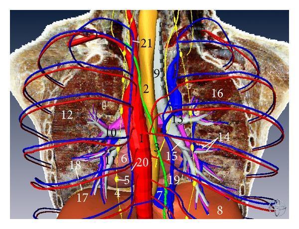
- A) Left side
- B) Anterior
- C) Right side ✅
- D) Posterior
Answer: C — Azygos vein lies on the right side of the esophagus (and arches over the right main bronchus to drain into SVC). The descending thoracic aorta is to the left of the esophagus (T5–T7), then passes posterior (T8–T10).
Q27 — Esophageal Blood Supply (by segment)
- A) Inferior thyroid artery
- B) Descending thoracic aorta branches
- C) Left gastric artery ✅
- D) Right gastric artery
Answer: C — Segmental supply:
- Cervical: inferior thyroid artery
- Thoracic: esophageal branches of descending aorta + bronchial arteries
- Abdominal: left gastric artery (branch of celiac trunk)
Q28 — Esophageal Varices (Portal Hypertension)
- A) Azygos vein → SVC (systemic)
- B) Left gastric vein → portal vein (portal) anastomoses with azygos/hemiazygos (systemic) ✅
- C) Internal thoracic vein → brachiocephalic (systemic)
- D) Inferior thyroid vein → brachiocephalic
Answer: B — Abdominal esophagus drains via left gastric vein → portal vein. In portal hypertension, communications with the azygos/hemiazygos (systemic) dilate → varices → risk of fatal hematemesis.
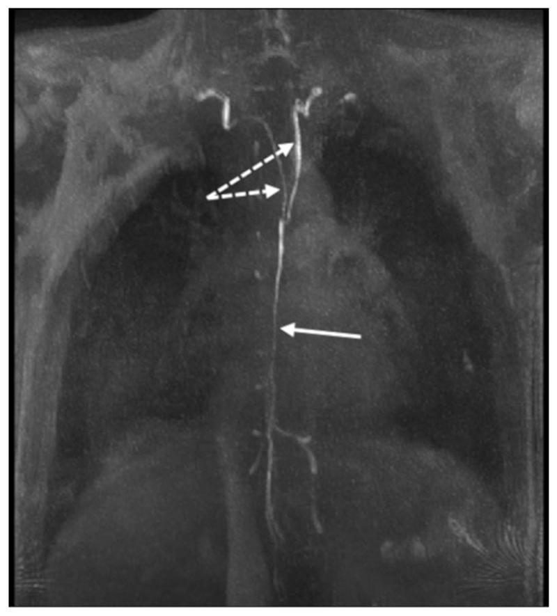
Q29 — Thoracic Duct: Course
- A) T12
- B) T8
- C) T5 ✅
- D) T2
Answer: C — Thoracic duct: enters thorax at T12 (aortic opening), ascends on right side through posterior mediastinum, at T5 it crosses to the left, then ascends in superior mediastinum on the left side of esophagus → ends in angle between left subclavian + left internal jugular veins at C7.
Q30 — Thoracic Duct: What It Drains
- A) Both lower limbs
- B) Left side of thorax
- C) Right upper limb ✅
- D) Left upper limb
Answer: C — The thoracic duct drains: both lower limbs, abdominal cavity, left side of thorax, left upper limb, left side of head & neck.
The right upper limb + right thorax + right head/neck drain via the right lymphatic duct → right subclavian vein.
Q31 — Thoracic Duct: Relations at Aortic Opening
- A) Aorta
- B) Esophagus
- C) Azygos vein ✅
- D) Hemiazygos vein
Answer: C — At the aortic opening: right = azygos vein, left = aorta. (Mnemonic: "Azygos is on the Right, Aorta on the Left" — just like in the chest generally.)
Q32 — Azygos Venous System
- A) Superior hemiazygos vein
- B) Inferior hemiazygos vein ✅
- C) Left brachiocephalic vein
- D) Left superior intercostal vein
Answer: B — Left side drainage:
- 1st → left brachiocephalic vein
- 2nd + 3rd → left superior intercostal vein → left brachiocephalic
- 4th–8th → superior hemiazygos vein
- 9th–11th → inferior hemiazygos vein (Both hemiazygos veins cross to drain into the azygos vein on the right)
Q33 — Esophageal Nerve Supply
- A) Esophageal plexus (vagus)
- B) Recurrent laryngeal nerves ✅
- C) Cervical sympathetic chain
- D) Deep cardiac plexus
Answer: B — Upper esophagus: parasympathetic via recurrent laryngeal nerves. Lower esophagus: via esophageal plexus (formed mainly by vagi). Near the diaphragm, vagal fibers form anterior and posterior gastric nerves entering the abdomen through the esophageal hiatus.
Q34 — Thymus Blood Supply
- A) Internal thoracic + superior thyroid
- B) Internal thoracic + inferior thyroid ✅
- C) Musculophrenic + inferior thyroid
- D) Brachiocephalic + internal thoracic
Answer: B — Internal thoracic artery + inferior thyroid artery. The thymus does NOT receive afferent lymphatics.
Q35 — Diaphragm Openings — THE MOST TESTED TABLE
- A) Esophagus
- B) Thoracic duct
- C) Inferior vena cava ✅
- D) Aorta
Answer: C — The golden rule for diaphragm openings:
| Level | Opening | Structures |
|---|---|---|
| T8 | Caval opening (central tendon) | IVC + right phrenic nerve |
| T10 | Esophageal hiatus | Esophagus + vagus nerves |
| T12 | Aortic hiatus | Aorta + thoracic duct + azygos vein |
Q36 — Tracheo-Esophageal Fistula
- A) Compression of esophagus by aortic arch
- B) Improper separation of trachea from esophagus during development ✅
- C) Achalasia
- D) Portal hypertension
Answer: B — TEF is a congenital anomaly due to failure of proper separation of the respiratory diverticulum from the foregut during embryological development.
Q37 — Left Superior Intercostal Vein
- A) Arch of azygos vein
- B) Superior hemiazygos vein
- C) Left brachiocephalic vein ✅
- D) Inferior hemiazygos vein
Answer: C — This is the left–right asymmetry trap. On the RIGHT, the right superior intercostal vein drains into the arch of azygos. On the LEFT, the left superior intercostal vein drains into the left brachiocephalic vein.
Q38 — Trachea in Superior Mediastinum
- A) Trachea posterior to esophagus
- B) Esophagus anterior to trachea
- C) Trachea anterior to esophagus ✅
- D) They run side by side
Answer: C — In the superior mediastinum: trachea is anterior to the esophagus, and the esophagus descends immediately anterior to the vertebral column.
🎯 RAPID-FIRE SUMMARY TABLE
| Fact | Detail |
|---|---|
| Neurovascular plane | Between internal and innermost intercostal |
| VAN order in costal groove | Vein (top) → Artery → Nerve (bottom) |
| Thoracocentesis needle | Upper border of rib below |
| Dividing plane of mediastinum | Sternal angle → lower border T4 |
| Aortic arch branches (L→R) | Brachiocephalic → L. carotid → L. subclavian |
| Left RLN loops under | Arch of aorta (ligamentum arteriosum) |
| Esophagus pierces diaphragm | T10 with vagus nerves |
| Aorta passes through diaphragm | T12 with thoracic duct + azygos |
| IVC passes through diaphragm | T8 with right phrenic nerve |
| Thoracic duct ends at | Left subclavian + internal jugular angle |
| Thoracic duct crosses midline | At T5 (right to left) |
| Esophagus constrictions | 6", 9", 11", 16" from incisors |
| Left gastric vein → | Portal vein (varices in portal HTN) |
| T2 atypical nerve → | Intercostobrachial nerve (floor of axilla + medial arm) |
| T7–T11 atypical nerves → | Pierce diaphragm → anterior abdominal wall |
| Thymus arteries | Internal thoracic + inferior thyroid |
| Internal thoracic artery ends at | 6th ICS → musculophrenic + superior epigastric |
Exam tip: The lecturer's own case problems appeared verbatim in the PDFs — Q14 (stab wound), Q23 (esophageal carcinoma/T10), and Q36-type (syphilis/aortitis) are all from the slides. Prioritize those cases. Good luck! 💪Here's your complete high-yield MCQ set — 38 questions drawn directly from your 3 lecture PDFs, organized by topic, with explanations and diagrams.
What's Covered
The 5 Absolute Must-Know Facts for Batterjee Exams
- VAN order = Vein, Artery, Nerve (top to bottom in costal groove) → needle goes above lower rib
- Sternal angle → lower T4 = dividing plane of mediastinum
- T8/T10/T12 diaphragm openings (IVC / Esophagus+Vagus / Aorta+Thoracic duct+Azygos)
- Left RLN loops under the arch of aorta; right loops under right subclavian
- Thoracic duct = crosses midline at T5, ends at left subclavian-IJV angle, drains everything EXCEPT right side of thorax/head/right arm