
Development of branchial arch and anomalies in detail according to standard ENT textbook in simplified manner
Reading File
Now let me search the medical library for branchial arch development and anomalies from ENT textbooks.
Finding Sources
Finding Sources
Searching Images
branchial arch development diagram anatomy
Finding Sources
Finding Sources
Good - found the key sources. Now let me read the most relevant sections from Scott-Brown's and KJ Lee's ENT textbooks:
Reading File
Reading File
Excellent content. Let me continue reading both sources and also get KJ Lee's section:
Reading File
Reading File
Reading File
Reading File
Reading File
Reading File
Reading File
Reading File
Loading Image
I now have very rich content from the authoritative ENT textbooks (Scott-Brown's and KJ Lee's). Let me compile the comprehensive, simplified response.
Development of Branchial Arches and Anomalies
Based on Scott-Brown's Otorhinolaryngology Head & Neck Surgery and KJ Lee's Essential Otolaryngology
1. What Are Branchial Arches?
The word "branchial" comes from the Greek bragchia, meaning gills. These arches are pharyngeal structures in the developing embryo that resemble fish gills - a series of slit-like grooves on the lateral neck. They give rise to most head and neck structures innervated by cranial nerves.
- Branchial arches begin to appear at 4 weeks' gestation
- There are 6 branchial arches in total (I, II, III, IV, V, VI)
- The 5th arch obliterates almost immediately after forming - it has no known derivatives
- Between adjacent arches on the outside are clefts (grooves) - lined by ectoderm
- Between adjacent arches on the inside (facing the gut) are pouches - lined by endoderm
Here is the schematic diagram of the branchial apparatus in utero from Scott-Brown's:
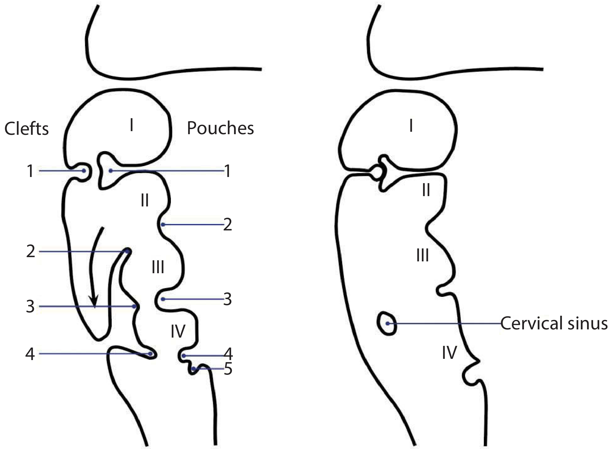
Figure: Left - early stage showing clefts (external) and pouches (internal). Right - 2nd arch overgrows clefts 2, 3, 4 forming the cervical sinus.
2. Components of Each Arch
Each branchial arch contains 4 core elements: Nerve + Muscle + Cartilage/Bone + Artery. Remember this with the mnemonic NMCA.
3. Derivatives of Each Arch (the High-Yield Table)
Arch I - Mandibular Arch
| Component | Derivative |
|---|---|
| Nerve | Trigeminal (V) |
| Muscles | Muscles of mastication, tensor veli palatini, mylohyoid, anterior belly of digastric, tensor tympani |
| Skeletal | Mandible, malleus, incus, sphenomandibular ligament, anterior malleal ligament |
| Artery | Maxillary artery |
| Pouch (1st) | Eustachian tube, middle ear cavity, mastoid antrum |
| Cleft (1st) | External auditory canal (EAC) |
Arch II - Hyoid Arch (Reichert's Cartilage)
| Component | Derivative |
|---|---|
| Nerve | Facial nerve (VII) |
| Muscles | Stapedius, stylohyoid, posterior belly of digastric, muscles of facial expression |
| Skeletal | Stapes superstructure, styloid process, stylohyoid ligament, lesser cornu + upper body of hyoid |
| Artery | Stapedial artery |
| Pouch (2nd) | Palatine tonsil |
Arch III
| Component | Derivative |
|---|---|
| Nerve | Glossopharyngeal (IX) |
| Muscles | Stylopharyngeus |
| Skeletal | Greater cornu + lower body of hyoid |
| Artery | Common carotid, proximal internal carotid |
| Pouch (3rd) | Thymus, inferior parathyroid gland |
Arch IV
| Component | Derivative |
|---|---|
| Nerve | Superior laryngeal nerve (branch of X) |
| Muscles | Pharyngeal constrictors, cricothyroid |
| Skeletal | Laryngeal cartilages (thyroid, arytenoid, cricoid) |
| Artery | Right: subclavian; Left: arch of aorta |
| Pouch (4th) | Superior parathyroid, parafollicular C cells of thyroid |
Arch VI (5th obliterates, so this is effectively the "6th")
| Component | Derivative |
|---|---|
| Nerve | Recurrent laryngeal nerve (X) |
| Muscles | Intrinsic muscles of the larynx (except cricothyroid) |
| Skeletal | Laryngeal cartilages |
| Artery | Right: pulmonary artery; Left: ductus arteriosus |
4. Summary Table - Pouches vs. Clefts
| # | Pouch (Endoderm - internal) | Cleft (Ectoderm - external) |
|---|---|---|
| 1st | Eustachian tube, middle ear, mastoid antrum | External auditory canal |
| 2nd | Palatine tonsil | (Obliterated into cervical sinus) |
| 3rd | Thymus, inferior parathyroid | (Obliterated) |
| 4th | Superior parathyroid, ultimobranchial body | (Obliterated) |
Key Mechanism - Cervical Sinus Formation:
During the 5th week, the 2nd arch grows downward and caudally to cover over clefts 2, 3, and 4. This creates a temporary enclosed space called the cervical sinus of His. Normally this obliterates completely. Failure to do so = branchial cleft anomalies.
5. Hillocks of His - External Ear Development
At week 5 of gestation, six small mesenchymal buds (hillocks) appear around the first branchial cleft - 3 from the first arch and 3 from the second arch. They fuse by week 12.
| Hillock | Arch | Forms |
|---|---|---|
| 1, 2, 3 | 1st arch | Tragus, helical crus, helix |
| 4, 5, 6 | 2nd arch | Antihelix, antitragus, lobule |
6. Branchial Anomalies - Three Types
When the branchial apparatus fails to develop normally, three types of remnants can result:
| Type | Description |
|---|---|
| Cyst | Closed sac with no skin opening |
| Sinus | Blind-ended tract with one opening (skin or pharynx) |
| Fistula | Complete tract with two openings (external skin + internal pharyngeal) |
Bailey's Classification of 2nd branchial cysts (4 types, by depth/position relative to carotid):
- Type I: Superficial to SCM, anterior to carotid
- Type II: Under SCM, adjacent to carotid sheath (most common)
- Type III: Between internal + external carotid arteries
- Type IV: Adjacent to pharyngeal wall (medial to carotid)
7. Specific Branchial Cleft Anomalies
First Branchial Cleft Anomalies (5% of all branchial anomalies)
Classified by Work (1972):
Type I:
- Ectodermal origin only (duplication of EAC)
- External opening: anterior and inferior to the tragus
- Tract runs parallel to EAC
- Relation to facial nerve: superficial
Type II:
- Ectodermal + mesodermal (full arch + cleft duplication)
- External opening: near angle of mandible
- Internal opening: EAC at osseocartilaginous junction or conchal bowl
- Tract passes through/near parotid gland - facial nerve relationship is variable and unpredictable (can be deep or superficial)
- Higher surgical risk
Clinical features: Recurrent discharge from ear or neck, preauricular/neck swelling, can mimic parotid abscess if infected
Treatment: Complete surgical excision; facial nerve monitoring essential
Second Branchial Cleft Anomalies (90% of all branchial anomalies)
The most common branchial anomaly. The classic presentation is:
- Cyst: Spherical swelling at angle of mandible, anterior to SCM - typically presents in young adults (3rd decade)
- Sinus: Opening on anterior border of SCM (lower third) - usually noted at birth
- Fistula: External on anterior SCM + internal on anterior tonsillar fossa
Tract course: Passes between internal and external carotid arteries, crossing over the hypoglossal and glossopharyngeal nerves, to open in the region of the tonsillar fossa.
Cyst contents: Lined by stratified squamous epithelium (90%) or respiratory epithelium (10%); 80% have lymphoid tissue in the wall; straw-coloured fluid with cholesterol crystals
Associated syndrome: Bilateral second branchial cleft sinuses = think Branchio-Oto-Renal (BOR) syndrome
Treatment:
- Surgical excision via transverse cervical incision
- Stepladder incisions if sinus extends high up
- Nerves at risk: hypoglossal, glossopharyngeal, vagus, accessory, marginal mandibular branch of facial
Important clinical point (Scott-Brown's key point): In patients over 35 years, a cystic neck swelling should be treated as metastatic squamous cell carcinoma (especially HPV-related oropharyngeal cancer) until proven otherwise.
Third and Fourth Branchial/Pharyngeal Pouch Anomalies (Rare)
- Arise from incomplete closure of the pyriform fossa sinus (thymopharyngeal duct of 3rd pouch)
- Almost always left-sided (due to asymmetric descent of the 4th arch artery on the right)
- Closely related to the left thyroid lobe
- Present as: recurrent left-sided thyroid abscess or neck infection (often misdiagnosed and just drained)
Investigations: MRI + rigid endoscopy under anaesthesia to identify opening in pyriform fossa
Treatment:
- Modern first-line: Endoscopic cautery of the pyriform fossa opening (minimally invasive, described by Bailey 2004)
- Open surgery (wide excision + ipsilateral thyroid lobectomy) for neonates or recurrences
8. Preauricular Sinus
Embryology: Incomplete fusion of the auricular hillocks at week 6 of gestation leads to a blind-ended, skin-lined pit anterior to the ear.
- A sinus above the tragus = isolated preauricular sinus
- A sinus below the tragus = consider first branchial cleft sinus
Clinical features: Usually asymptomatic pit; may discharge, get infected, form abscess
Treatment: Surgical excision, including the entire tract; recurrence common if incomplete excision
9. Theories of Origin of Branchial Cysts
Scott-Brown's lists four theories:
- Branchial apparatus theory - remnants of pharyngeal pouches or clefts (most widely accepted)
- Cervical sinus theory - remnant of the cervical sinus of His (formed when 2nd arch overgrows 3rd/4th clefts)
- Thymopharyngeal duct theory - remnant of the thymus descent tract
- Inclusion theory - epithelial elements trapped in cervical lymph nodes
10. Summary - Quick Recall
| Arch | Nerve | Key Structures |
|---|---|---|
| I | V (Trigeminal) | Mandible, malleus, incus, muscles of mastication, EAC (cleft) |
| II | VII (Facial) | Stapes, styloid, hyoid (lesser), facial muscles, palatine tonsil (pouch) |
| III | IX (Glossopharyngeal) | Hyoid (greater), stylopharyngeus, thymus + inf. parathyroid (pouch) |
| IV | X superior laryngeal | Laryngeal cartilages, constrictors, sup. parathyroid (pouch) |
| VI | X recurrent laryngeal | Intrinsic laryngeal muscles, pulmonary A / ductus arteriosus |
| Cleft | External Anomaly | Location |
|---|---|---|
| 1st | Type I/II sinus | Preauricular/angle of mandible to EAC |
| 2nd | Cyst/Sinus/Fistula (90%) | Anterior SCM, between carotids, to tonsil |
| 3rd/4th | Pyriform fossa sinus | Left-sided, near thyroid |
Sources: Scott-Brown's Otorhinolaryngology Head & Neck Surgery (Vols 1 & 2); KJ Lee's Essential Otolaryngology, Chapter 60.
This is a shared conversation. Sign in to Orris to start your own chat.