
Congenital pseudarthrosis of tibia
Reading File
Finding Sources
Finding Sources
Searching PubMed
"congenital pseudarthrosis tibia"
Searching the Web
congenital pseudarthrosis of tibia clinical management NF1 review
Searching Images
congenital pseudarthrosis tibia clinical radiograph anterolateral bowing
Reading File
Reading File
Reading Article
PMID: 39764600
Reading Web Page
https://pmc.ncbi.nlm.nih.gov/articles/PMC9840411
Congenital pseudarthrosis of the tibia (CPT)
A rare pediatric disorder in which the tibia is dysplastic from birth and may progressively bow, fracture with little or no trauma, and fail to unite, creating a persistent false joint (pseudarthrosis).
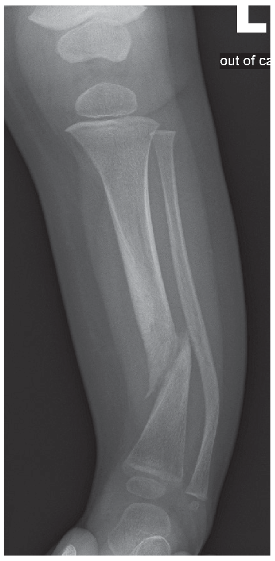
Congenital pseudarthrosis with associated fibular abnormality. Bailey and Love's Short Practice of Surgery, p. 646.
Key features
- Usually affects the distal half of the tibia, often with ipsilateral fibular involvement.
- Typical early sign: progressive anterolateral bowing of the lower leg.
- May present with congenital bowing or a pathological fracture in infancy or childhood.
- Radiographs may show cortical narrowing, tapering or cystic dysplasia, medullary canal narrowing, bowing, fracture, and established nonunion.
- The pseudarthrosis site contains poorly vascularized hamartomatous fibrous tissue, which helps explain the poor healing potential. Campbell's Operative Orthopaedics, 15th ed., Figure 31.55 and accompanying text.
Association
CPT is strongly associated with neurofibromatosis type 1 (NF1). Therefore, assess for café-au-lait macules, axillary/inguinal freckling, neurofibromas, family history, and other skeletal manifestations. Reported association rates vary by cohort and diagnostic criteria. Bailey and Love's Short Practice of Surgery, p. 646. A recent NF1-CPT review describes the genetic and clinical association.
Management principles
-
Before fracture or surgery
- Protect the limb with a custom brace or orthosis.
- Serial clinical and radiographic follow-up.
- Refer early to a pediatric orthopedic limb-reconstruction center.
-
After fracture or established pseudarthrosis
- Excision of abnormal pseudarthrosis tissue.
- Restoration of alignment and stable fixation, often intramedullary fixation with or without circular external fixation.
- Autologous bone grafting, commonly iliac crest graft.
- Address associated fibular pseudarthrosis, limb-length discrepancy, ankle valgus, and deformity as needed.
- Long-term protection is important because refracture can occur even after union.
The 2025 systematic review and consensus guideline found that commonly used combined procedures include pseudarthrosis excision, intramedullary fixation, external fixation, and autogenous grafting; vascularized fibular grafting or tibiofibular cross-union are alternatives in selected cases. Evidence for recombinant BMP remains inconclusive in this condition (2025 consensus review).
Prognosis
This is a difficult condition with risks of persistent nonunion, refracture, deformity, ankle valgus, limb shortening, and repeated surgery. The main endpoint is durable tibial union with a functional, well-aligned limb, not simply initial fracture healing.
Exam point: Anterolateral bowing of the tibia in a child should raise concern for congenital pseudarthrosis, particularly in the setting of NF1.
Please give all answers
Reading File
Finding Sources
Searching PubMed
"congenital pseudarthrosis tibia"
Searching the Web
congenital pseudarthrosis tibia review diagnosis treatment NF1
Congenital Pseudarthrosis of Tibia (CPT)
Definition
Congenital pseudarthrosis of the tibia is a rare congenital dysplasia of the tibia in which there is progressive anterolateral bowing, pathological fracture, and failure of union, producing a false joint. It may be evident at birth or develop during early childhood.
It is not a true fracture nonunion from trauma. The bone is intrinsically abnormal and has poor osteogenic and healing capacity.
Epidemiology
- Rare: estimated incidence about 1 in 140,000 to 250,000 live births.
- Usually presents in infancy or early childhood.
- Predominantly affects the distal or middle-distal tibia.
- The ipsilateral fibula is often affected.
- It is strongly associated with neurofibromatosis type 1 (NF1).
Etiology and pathogenesis
The precise cause is not completely established.
Association with NF1
CPT is closely related to NF1. A child with anterolateral tibial bowing or tibial pseudarthrosis should be evaluated for:
- Café-au-lait macules
- Axillary or inguinal freckling
- Neurofibromas
- Optic pathway glioma or Lisch nodules
- Scoliosis
- Family history of NF1
Local pathology
The pseudarthrosis site contains:
- Fibrous hamartomatous tissue
- Poor vascular ingrowth
- Weak osteogenic potential
- Increased osteoclastic resorption
- Abnormal periosteum and cortex
This produces fragile dysplastic bone that does not heal normally. Campbell's Operative Orthopaedics, 15th ed., section “Congenital Pseudarthrosis of the Fibula and Tibia.”
Clinical presentation
Typical presentation
- Congenital or early childhood anterolateral bowing of the leg
- Shortened lower limb
- Progressive deformity
- Pathological fracture after trivial trauma or spontaneously
- Painless abnormal mobility after fracture
- Established nonunion or pseudarthrosis
The key exam clue is: anterolateral bowing of the tibia in a young child, especially with features of NF1.
Fibular involvement
The fibula may show:
- Bowing
- Dysplasia
- Pseudarthrosis
- Shortening
- Ankle valgus due to loss of lateral support
Radiological features
Plain radiographs of the whole tibia and fibula, including ankle and knee, are the main investigation.
Findings may include:
- Anterolateral bowing of tibia
- Narrowed, sclerotic, tapered bone ends
- Medullary canal narrowing or obliteration
- Cortical thinning
- Cystic lesion at the dysplastic segment
- Pathological fracture
- Atrophic or hypertrophic pseudarthrosis
- Associated fibular dysplasia or pseudarthrosis
- Limb shortening and ankle valgus
The classic tibial bow is anterolateral, in contrast to posteromedial tibial bowing, which often improves with growth. Bailey and Love's Short Practice of Surgery, p. 646.
Classification
Several systems exist, including Anderson, Boyd, Crawford, and Paley classifications. The Crawford classification is commonly quoted:
| Type | Radiographic appearance |
|---|---|
| I | Anterior bowing with cortical thickening and narrowed medullary canal |
| II | Anterior bowing with cortical thinning and a narrowed medullary canal |
| III | Cystic lesion in the tibia, often with bowing |
| IV | Established pseudarthrosis with fracture and tapered bone ends |
Crawford type IV is the most severe form, with established nonunion.
Differential diagnosis
- Physiological bowing
- Posteromedial tibial bowing
- Fibular hemimelia
- Blount disease
- Osteogenesis imperfecta
- Rickets
- Fibrous dysplasia
- Chronic osteomyelitis
- Post-traumatic nonunion
- Other skeletal dysplasias
Investigations
-
Radiographs
- AP and lateral views of the tibia and fibula
- Evaluate deformity, pseudarthrosis, fibular involvement, ankle valgus, and limb length.
-
Assessment for NF1
- Full skin examination
- Eye examination when appropriate
- Family history
- Genetic consultation/testing if clinically indicated.
-
CT or MRI
- Not always required.
- May help assess anatomy, extent of dysplastic tissue, operative planning, and associated soft tissue abnormalities.
-
Limb-length assessment
- Clinical measurement and standing long-leg radiographs when feasible.
Management
Goals
- Obtain solid, durable union.
- Correct tibial angulation.
- Prevent refracture.
- Maintain limb length.
- Correct ankle valgus and associated fibular deformity.
- Preserve foot and ankle function.
Conservative treatment
Appropriate mainly before fracture or in selected young children:
- Protective custom brace or orthosis
- Avoidance of high-risk impact activity
- Regular clinical and radiological follow-up
- Monitoring of tibial bowing and fracture risk
Bracing can delay fracture but does not correct the underlying dysplasia.
Surgical treatment
Surgery is usually required after fracture or once pseudarthrosis is established. No single procedure is ideal for every child. Management should be in a specialized pediatric orthopedic limb-reconstruction center.
Core surgical principles:
- Radical excision of hamartomatous pseudarthrosis tissue.
- Freshening of bone ends until viable bleeding bone is reached.
- Correction of angulation.
- Rigid stabilization.
- Biological augmentation with autologous bone graft.
- Long-term protection against refracture.
1. Intramedullary fixation
- Rod or telescopic rod through the tibia, sometimes extending into the talus.
- Gives internal support during healing and growth.
- Often combined with bone grafting and/or external fixation.
- Telescopic rods can be advantageous in younger children because they accommodate growth.
2. Circular external fixation, Ilizarov or hexapod frame
Useful for:
- Compression at the pseudarthrosis site
- Correction of angulation
- Limb-lengthening
- Managing complex deformity
- Stabilization when internal fixation alone is insufficient
Complications include pin-tract infection, stiffness, axial deviation, and refracture.
3. Combined intramedullary rod plus external fixation
Often used in severe or recurrent CPT because it combines mechanical stability with deformity correction and compression.
4. Autologous bone graft
- Usually iliac crest corticocancellous graft.
- May be wrapped around the pseudarthrosis after excision.
- Aims to increase biological healing potential.
5. Vascularized fibular graft
- Used for large defects, recurrent nonunion, or failed previous procedures.
- Provides living vascularized bone.
- Technically demanding and associated with donor-site issues.
6. Tibiofibular cross-union
- Tibia and fibula are surgically united with grafting to create a broad, strong cross-sectional healing mass.
- Increasingly used in difficult and recurrent cases.
- Can reduce refracture risk, but ankle alignment and limb length still require surveillance.
7. Bone morphogenetic proteins
- BMPs have been used as adjuncts in selected cases.
- Their benefit remains uncertain, particularly in NF1-associated CPT.
- They should not replace thorough excision, stable fixation, and grafting.
A 2025 systematic review and consensus guideline reported that common combined treatment includes excision of the pseudarthrosis, intramedullary fixation, external fixation, and autogenous grafting. It identified vascularized fibular grafting and cross-union as options in selected patients, while evidence for rhBMP remains inconclusive (2025 CPT surgical consensus).
Postoperative care
- Immobilization and protective bracing for a prolonged period.
- Serial X-rays to confirm union.
- Monitoring for refracture, especially after removal or migration of implants.
- Physiotherapy to preserve knee and ankle motion.
- Long-term follow-up until skeletal maturity.
- Ongoing assessment and treatment of:
- Limb-length discrepancy
- Ankle valgus
- Tibial deformity
- Knee and ankle stiffness
- Refracture
- Implant migration or breakage
Complications
- Persistent nonunion
- Refracture after apparent union
- Limb shortening
- Tibial angulation
- Ankle valgus
- Fibular pseudarthrosis
- Knee and ankle stiffness
- Pin-tract infection with external fixation
- Neurovascular injury
- Implant failure
- Need for repeated surgery
Prognosis
CPT has a guarded prognosis because union is difficult and refracture remains a major long-term risk. Successful management requires not only initial union but also maintenance of a straight, stable tibia throughout growth.
The 2025 review found substantial variability among studies. Even after union, refracture rates remained high with all main methods, which supports prolonged protection and follow-up. The review reported pooled primary-union rates of 69% with intramedullary rodding alone, 89% with Ilizarov fixation, and 85% with combined Ilizarov plus intramedullary fixation. These results are derived mainly from observational studies, so procedure choice must be individualized (systematic review abstract).
Short exam answer
Congenital pseudarthrosis of the tibia is a rare congenital dysplasia characterized by anterolateral bowing of the tibia, pathological fracture, and failure of union. It commonly affects the distal tibia and may involve the fibula. It is strongly associated with NF1. X-ray shows anterior or anterolateral bowing, narrowing or tapering of the bone ends, cystic dysplasia, fracture, and established nonunion. Initial management is protective bracing before fracture. Established pseudarthrosis requires excision of abnormal fibrous tissue, correction of deformity, stable fixation with intramedullary rod and/or circular external fixator, and autologous bone grafting. Long-term bracing and follow-up are needed because refracture, limb shortening, ankle valgus, and recurrent nonunion are common.
One-line viva answer:
A child with anterolateral tibial bowing should be considered to have congenital pseudarthrosis of the tibia until proved otherwise, and should be assessed for neurofibromatosis type 1.
A child with anterolateral tibial bowing should be considered to have congenital pseudarthrosis of the tibia until proved otherwise, and should be assessed for neurofibromatosis type 1.
This is a shared conversation. Sign in to Orris to start your own chat.