
Meaning of consolidation of lungs
Reading File
Lung consolidation is when the air-filled spaces in the lung (alveoli) become filled with fluid, pus, blood, or cells - making that area of the lung "solid" instead of air-filled. It shows up on chest X-ray or CT as a dense, white opacity.
Searching sources...
Finding Sources
Searching the Web
lung consolidation meaning pathology radiology
Finding Sources
Reading File
Reading File
Here is a thorough explanation of lung consolidation:
Consolidation of the Lungs
Definition
Consolidation means "solidification" of the lung tissue. Normally, the alveoli (tiny air sacs) are filled with air. In consolidation, that air is replaced by an abnormal substance - such as inflammatory exudate (pus/fluid), blood, mucus, or cells - making the lung tissue dense and solid instead of airy.
As stated directly in Robbins & Kumar Basic Pathology:
"The term 'consolidation,' used frequently, refers to 'solidification' of the lung due to replacement of the air by exudate in the alveoli."
- Robbins & Kumar Basic Pathology, p. 470
What Fills the Alveoli?
The air space can be replaced by:
| Substance | Cause |
|---|---|
| Inflammatory exudate (pus, fibrin, neutrophils) | Bacterial pneumonia |
| Fluid/edema | Pulmonary edema, ARDS |
| Blood (red cells) | Pulmonary hemorrhage |
| Mucus/cells | Obstructive causes |
| Tumor cells | Bronchoalveolar carcinoma |
Patterns of Consolidation
There are two main anatomical patterns:
-
Lobar consolidation - an entire lobe or large portion of a lobe becomes solidified. Classic in pneumococcal (Streptococcus pneumoniae) pneumonia.
-
Bronchopneumonia (patchy consolidation) - multiple focal areas of consolidation scattered through the lung, often bilateral and basal. More common with other bacteria.
Stages in Lobar Pneumonia (Classic Teaching)
In lobar pneumonia, consolidation progresses through 4 classic stages:
- Congestion - lung is heavy, wet, red; vascular engorgement + fluid in alveoli
- Red hepatization - alveoli packed with neutrophils, red cells, and fibrin; lung becomes red, firm, and liver-like in texture
- Gray hepatization - red cells disintegrate; fibrinopurulent exudate remains; lung turns grayish-brown
- Resolution - exudate is enzymatically digested and cleared by macrophages, or organized by fibroblasts
(Robbins, Cotran & Kumar Pathologic Basis of Disease)
On Imaging (Radiology)
- Chest X-ray: Appears as a white/opaque area where the lung should be dark (air-filled). A classic sign is the air bronchogram - air-filled bronchi appearing as dark tubes against the white consolidated background. This confirms the opacity is in the alveoli (not a collapse or pleural effusion).
- CT scan: Consolidation is defined as increased lung density sufficient to obscure the margins of vessels and airway walls, per Radiopaedia's reference article.
Common Causes
- Infections: Pneumonia (bacterial, viral, fungal) - most common cause
- ARDS (Acute Respiratory Distress Syndrome): heterogeneous bilateral consolidation
- Pulmonary edema: fluid from heart failure fills alveoli
- Lung cancer: especially mucinous adenocarcinoma
- Pulmonary infarction: blood fills alveoli after embolism
- Cryptogenic Organizing Pneumonia (COP): inflammatory process with characteristic peripheral consolidation
- Aspiration
Clinical Significance
Consolidation causes:
- Impaired gas exchange - O2 cannot reach blood through fluid-filled alveoli
- Reduced breath sounds or bronchial breathing on auscultation
- Dullness to percussion over the affected area
- Hypoxia in severe cases
The key distinction from lung collapse (atelectasis) is that in consolidation the lung volume is preserved (sometimes even increased), whereas in collapse the lung shrinks due to loss of air without replacement. Both produce opacity on X-ray, but the air bronchogram sign is typically present only in consolidation.
Image of air bronchogram
Searching sources...
Finding Sources
Searching the Web
air bronchogram sign chest X-ray CT image radiology
Loading Image
Loading Image
Reading File
Finding Sources
Loading Image
Loading Image
Loading Image
Here are the images of the air bronchogram sign:
Chest X-ray - Air Bronchogram (Labeled)
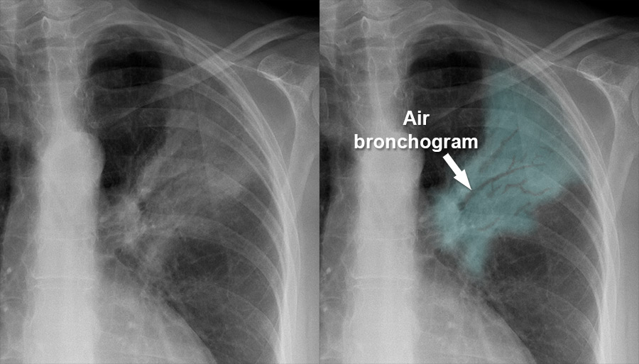
Left panel: Plain X-ray showing a white (consolidated) area in the left upper/mid zone.
Right panel (highlighted): The same X-ray with the consolidated area highlighted in teal, and an arrow pointing to the air bronchogram - the dark, branching tree-like structures (air-filled bronchi) visible within the white consolidated lung tissue.
(Source: Radiology Masterclass)
Chest X-ray vs. Close-up Comparison
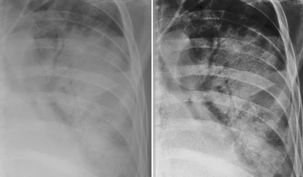
The right panel is a magnified view clearly showing the dark branching airways (air bronchograms) outlined against the surrounding white consolidated lung tissue.
(Source: St. Vincent's University Hospital Radiology)
What You Are Seeing
| Feature | Appearance |
|---|---|
| Consolidated lung | White/opaque (dense) area |
| Air bronchogram | Dark branching lines within the white area |
| Why visible? | Surrounding alveoli are fluid-filled (opaque), but the bronchi still contain air (dark) |
Clinical Significance of the Air Bronchogram
- Confirms alveolar (airspace) disease - the opacity is in the lung parenchyma, not from a pleural effusion or mass
- Indicates patent (open) proximal airways - the bronchi leading to the affected area are not blocked
- Rules against obstructing tumor as the cause - if airways were blocked, no air bronchogram would be visible
- Seen in: bacterial pneumonia, pulmonary edema, ARDS, organizing pneumonia, pulmonary hemorrhage
As stated in Grainger & Allison's Diagnostic Radiology:
"Consolidation refers to the increase in lung density on CXR or CT in which the margins of vessels and airways are obscured. An air bronchogram may or may not be seen. This radiological pattern indicates that air in the air spaces has been replaced (e.g. by inflammatory cells, blood or tumour)."
This is a shared conversation. Sign in to Orris to start your own chat.