
Femur
Reading File
The femur is the longest, strongest bone in the body, forming the thigh. It articulates with the acetabulum of the hip proximally and the tibia/patella distally at the knee.
Searching sources...
Finding Sources
Finding Sources
Reading File
Reading File
Reading File
Finding Sources
Finding Sources
Finding Sources
Reading File
Reading File
Now let me fetch the key anatomical images and get more detailed anatomy content.
Loading Image
Loading Image
Finding Sources
Finding Sources
Finding Sources
Reading File
Reading File
Loading Image
Loading Image
Here is a complete anatomy review of the femur, drawn from authoritative orthopaedic and anatomy textbooks.
The Femur
The femur is the longest and strongest bone in the human body. It forms the skeleton of the thigh, articulating proximally with the acetabulum of the pelvis (hip joint) and distally with the tibia and patella (knee joint).
Gross Anatomy
The femur is divided into proximal, shaft, and distal parts.
Proximal Femur
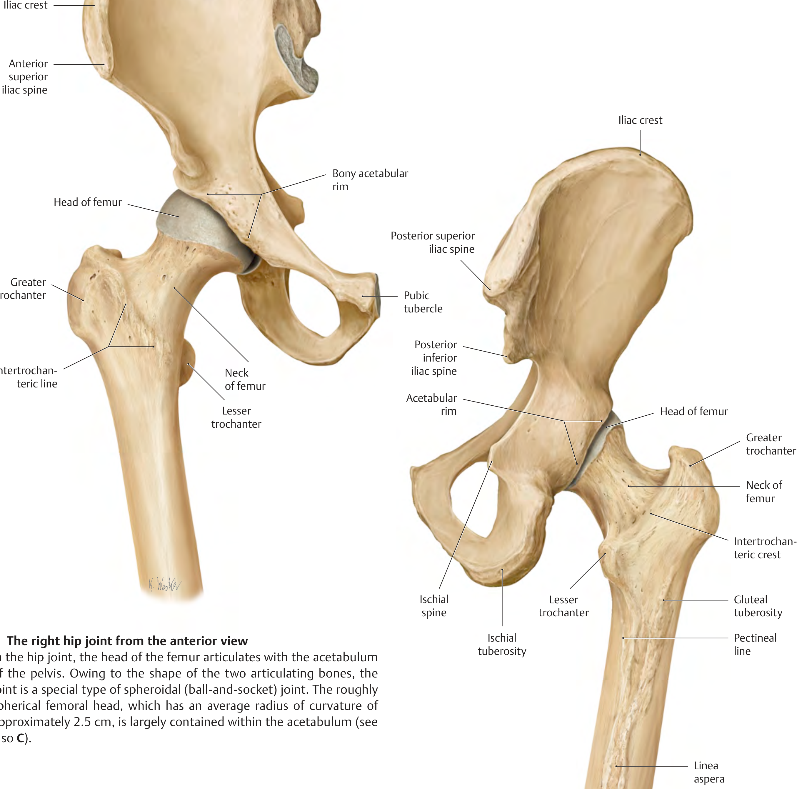
The right hip joint: anterior (left) and posterior (right) views, showing the head, neck, trochanters, and posterior surface of the proximal femur. - THIEME Atlas of General Anatomy
Head of femur
- Roughly spherical (~2.5 cm radius of curvature)
- Largely covered by hyaline articular cartilage except at the fovea capitis (a pit on the medial surface for attachment of the ligamentum teres / ligament of the head)
- Articulates with the acetabulum to form a ball-and-socket (spheroidal) joint
Neck of femur
- Connects the head to the shaft
- Neck-shaft angle averages 127° in adults (begins at ~141° in the fetus; decreases with age)
- Femoral neck is anteverted approximately 14° relative to the femoral condyles (range 1-40°)
- Largely intracapsular; the posterior neck is only partially covered by the joint capsule (basicervical and intertrochanteric regions are extracapsular)
Trochanters
| Feature | Greater Trochanter | Lesser Trochanter |
|---|---|---|
| Position | Lateral, proximal | Medial, posteromedial |
| Muscle attachments | Gluteus medius & minimus, obturators, piriformis, gemelli | Iliopsoas |
| Connecting ridge | Intertrochanteric line (anterior) | Intertrochanteric crest (posterior) |
The gluteal tuberosity is a roughened area on the posterior proximal shaft lateral to the lesser trochanter where gluteus maximus inserts.
Shaft (Diaphysis)
- Cylindrical; directed obliquely downward and medially, so the knee lies near the body's midline
- The posterior surface bears a prominent longitudinal ridge, the linea aspera, which serves as attachment for adductor and other thigh muscles
- The linea aspera splits distally into the medial and lateral supracondylar lines, which form the floor and sides of the popliteal fossa
- The pectineal line runs from the lesser trochanter to the linea aspera (adductor longus and brevis, pectineus insert here)
Distal Femur
- Expands into two large articular masses: the medial condyle (larger) and lateral condyle (smaller)
- The condyles articulate with the tibial plateaus and with the patella (via the patellar/trochlear surface anteriorly)
- Between the condyles posteriorly lies the intercondylar (intercondyloid) fossa:
- Lateral wall = attachment of the anterior cruciate ligament (ACL)
- Medial wall = attachment of the posterior cruciate ligament (PCL)
- The roof is demarcated by the intercondylar line
- Non-articular bony prominences above the condyles = medial and lateral epicondyles (attachment sites of collateral ligaments and muscles)
- The adductor tubercle sits above the medial epicondyle; it is the termination of the medial supracondylar line and receives the adductor magnus tendon
Blood Supply
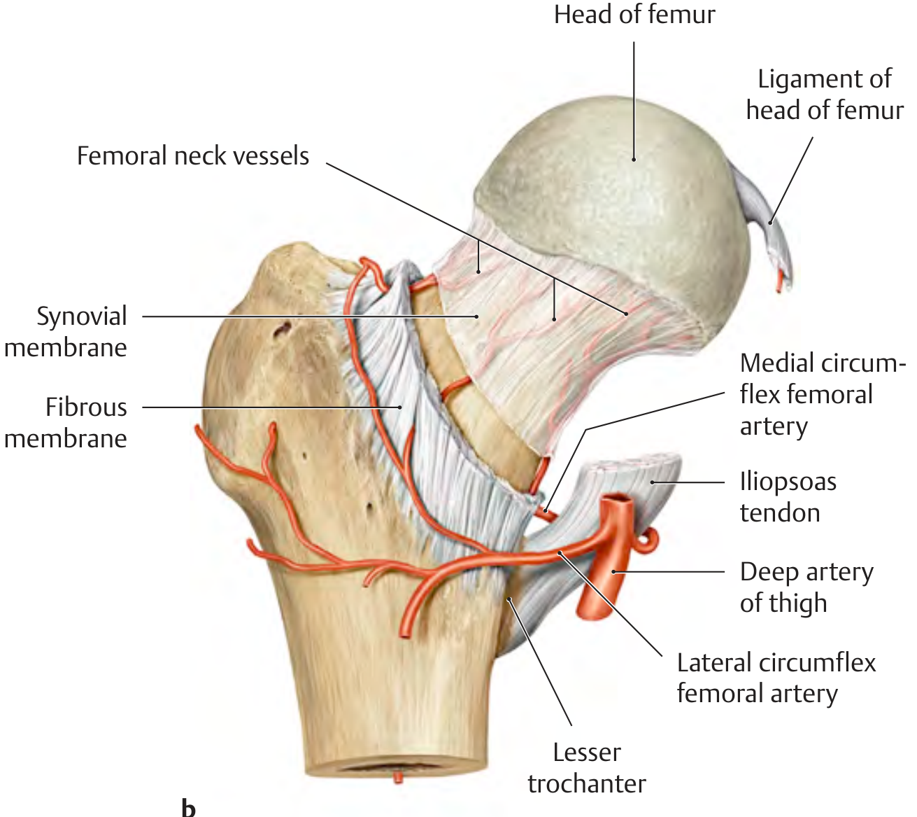
Course of femoral neck vessels and blood supply to the femoral head via medial and lateral circumflex femoral arteries. - THIEME Atlas of General Anatomy
The femoral head's blood supply is tenuous and clinically important:
- Medial circumflex femoral artery (MFCA) - the dominant supply; a branch of the profunda femoris (deep artery of thigh). It runs posteriorly around the neck and its deep branch penetrates retinacular vessels under the joint capsule to supply the femoral head
- Lateral circumflex femoral artery (LCFA) - contributes mainly to the anterior neck via ascending branch
- Artery of the ligamentum teres - a branch of the obturator artery; relatively minor in adults but contributes to the foveal area
Clinical point: Femoral neck fractures and hip dislocations can rupture the MFCA retinacular vessels, interrupting the main blood supply and causing avascular necrosis (AVN) of the femoral head. In children, the piriformis fossa nail entry point for intramedullary nailing risks damage to the posterosuperior retinacular vessels off the MFCA - this is why lateral trochanteric entry is preferred.
Ossification
| Epiphysis | Appearance | Fusion |
|---|---|---|
| Distal femoral epiphysis | Present at birth (important in forensic/gestational age assessment) | ~19 years |
| Femoral head epiphysis | ~11 months (one large physis including both trochanters) | ~18 years |
- The distal femur is responsible for the highest percentage of longitudinal growth in the lower extremity (~70% of femoral length; ~37% of total lower limb length)
- Slipped capital femoral epiphysis (SCFE) occurs through the femoral head physis at the zone of hypertrophy
Key Angles and Measurements
| Parameter | Normal Value |
|---|---|
| Neck-shaft angle | ~127° (coxa vara < 120°, coxa valga > 135°) |
| Anteversion of femoral neck | ~14° (range 1-40°) relative to condyles |
| Acetabular transverse angle | ~40° in adults (51° at birth) |
| Femoral head radius | ~2.5 cm |
Proximal Femoral Trabecular Architecture
The proximal femur has two main trabecular systems that resist loading:
- Compression trabeculae - arching from calcar to superolateral femoral head cortex
- Tension trabeculae - from the lateral cortex, arching medially toward the femoral head
- The Ward triangle is a relatively weak area between the two systems where trabecular density is low - a common site for stress fractures and osteoporotic fractures
Muscle Attachments Summary
| Region | Muscles Attaching |
|---|---|
| Greater trochanter | Gluteus medius, gluteus minimus, piriformis, obturator internus & externus, gemelli |
| Lesser trochanter | Iliopsoas |
| Gluteal tuberosity | Gluteus maximus |
| Linea aspera | Adductors (longus, brevis, magnus), vastus lateralis, vastus medialis, short head of biceps femoris |
| Adductor tubercle | Adductor magnus (lower/hamstring part) |
| Lateral epicondyle | Lateral collateral ligament, popliteus, gastrocnemius (lateral head) |
| Medial epicondyle | Medial collateral ligament, gastrocnemius (medial head) |
Clinical Correlations
- Femoral neck fractures: Commonest in elderly osteoporotic women (minimal trauma); risk of AVN and non-union due to tenuous blood supply. Classification: subcapital, transcervical, basicervical
- Intertrochanteric fractures: More vascular region, lower AVN risk; treated with intramedullary nail or dynamic hip screw
- Subtrochanteric fractures: High biomechanical stress; treated with intramedullary nail
- Femoral shaft fractures: High energy in young (MVA), lower energy in elderly; intramedullary nail is gold standard treatment
- Distal femur fractures: Up to 6% of all femur fractures; bimodal distribution (high energy in young, low energy in elderly); thin cortices and wide medullary canal make fixation challenging
Sources: THIEME Atlas of General Anatomy and Musculoskeletal System; Imaging Anatomy Vol. 3 Bones, Joints, Vessels and Nerves; Miller's Review of Orthopaedics 9th Ed; Rockwood and Green's Fractures in Adults 10th Ed 2025; Campbell's Operative Orthopaedics 15th Ed 2026
Femur
I have detailed notes on the femur from yesterday's session. Let me pull fresh content from the medical library for a thorough response.
Searching sources...
Finding Sources
Finding Sources
Reading File
Reading File
Reading File
Finding Sources
Reading File
Finding Sources
Reading File
Reading File
Reading File
Reading File
Loading Image
Loading Image
Loading Image
Loading Image
Loading Image
Now I have rich content from multiple authoritative sources. Here is the complete, well-illustrated anatomy of the femur.
The Femur
The femur is the longest and strongest bone in the human body. It forms the entire bony skeleton of the thigh, articulating proximally at the hip joint (with the acetabulum) and distally at the knee joint (with the tibia and patella). The shaft descends obliquely from lateral to medial at approximately 7 degrees from the vertical, bringing the knee toward the body's midline under the center of gravity.
Overview: Anatomical Position in the Body
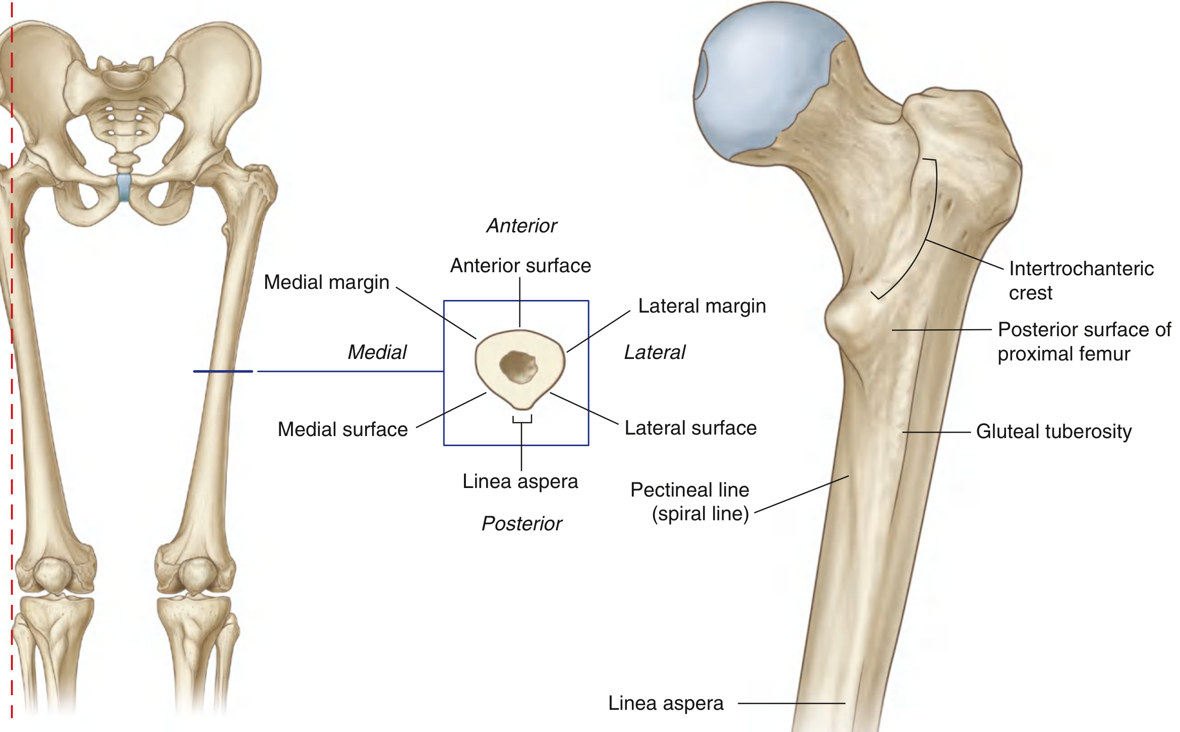
1. Proximal Femur
Head of Femur
- Smooth, roughly spherical (~2.5 cm radius)
- Covered by hyaline articular cartilage over most of its surface
- Articulates with the acetabulum to form a ball-and-socket (spheroidal) joint
- The fovea capitis (fovea) is a small pit on the superomedial surface - it is non-articular and is the attachment for the ligamentum teres (ligament of the head of the femur)
Neck of Femur
- Approximately 5 cm long; connects the head to the shaft
- Projects in a superomedial direction
- Neck-shaft angle: averages 127° in adults (starts at ~141° in the fetus; decreases with age)
- Coxa vara = angle < 120°; Coxa valga = angle > 135°
- Anteversion: the neck is rotated anteriorly 10-15° (range 1-40°) relative to the plane of the femoral condyles
- Capsular relationship: the anterior surface of the neck is fully intracapsular; the posterior neck is only partially covered by the capsule (the basicervical and intertrochanteric regions are extracapsular)
Greater Trochanter
- Large, lateral bony prominence at the neck-shaft junction
- Muscle attachments: gluteus medius (lateral facet), gluteus minimus (anterior facet), piriformis (superior border), obturator internus and gemelli (medial surface - trochanteric fossa), obturator externus
Lesser Trochanter
- Smaller, posteromedial prominence at the base of the neck
- Sole attachment: iliopsoas (psoas major + iliacus)
Intertrochanteric Line and Crest
| Feature | Surface | Course | Attachment |
|---|---|---|---|
| Intertrochanteric line | Anterior | Greater → lesser trochanter | Iliofemoral ligament (strongest ligament in the body) |
| Intertrochanteric crest | Posterior | Greater → lesser trochanter | - |
| Quadrate tubercle | On crest (upper half) | Prominent tubercle | Quadratus femoris |
2. Shaft (Diaphysis)
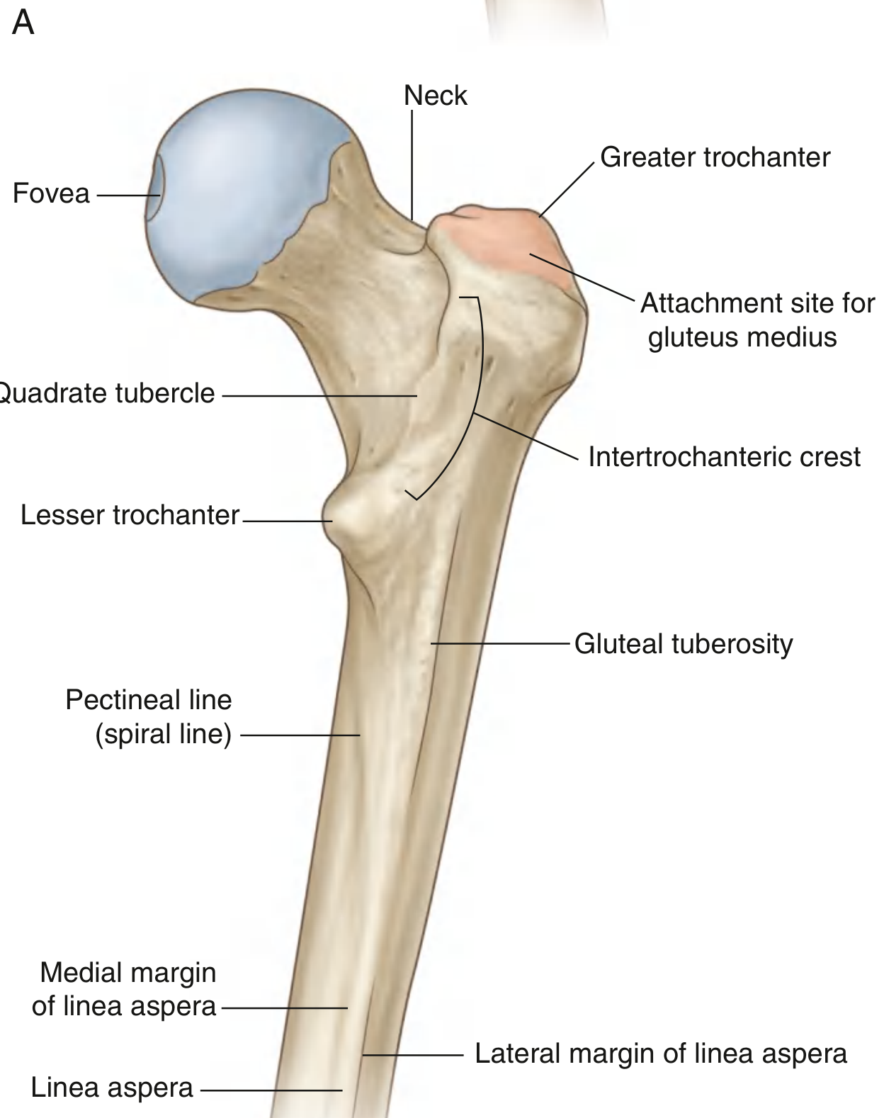
- The shaft is bowed anteriorly (slight anterior convexity)
- Mid-shaft cross-section is triangular: anterior surface, posteromedial surface, and posterolateral surface, with the posterior border forming the linea aspera
Linea Aspera
The linea aspera is a broad, roughened posterior crest running most of the femoral shaft length. It is the principal muscle attachment site in the thigh.
Proximally, the linea aspera diverges into two lines:
- Pectineal line (spiral line) - medial divergence, curves anteriorly under the lesser trochanter to join the intertrochanteric line. Attachment: pectineus
- Gluteal tuberosity - lateral divergence, broad roughening up to the greater trochanter. Attachment: gluteus maximus
Distally, the linea aspera again diverges into:
- Medial supracondylar line - ends at the adductor tubercle on the medial condyle. The adductor magnus inserts here
- Lateral supracondylar line - forms the lateral edge of the popliteal surface
- The area between these two lines forms the popliteal surface (floor of the popliteal fossa)
Key Muscle Attachments to Shaft
| Landmark | Muscles |
|---|---|
| Gluteal tuberosity | Gluteus maximus |
| Pectineal line | Pectineus |
| Linea aspera (medial lip) | Adductor longus, adductor brevis, adductor magnus, vastus medialis |
| Linea aspera (lateral lip) | Vastus lateralis, short head of biceps femoris |
| Adductor tubercle | Adductor magnus (adductor/hamstring part) |
3. Distal Femur
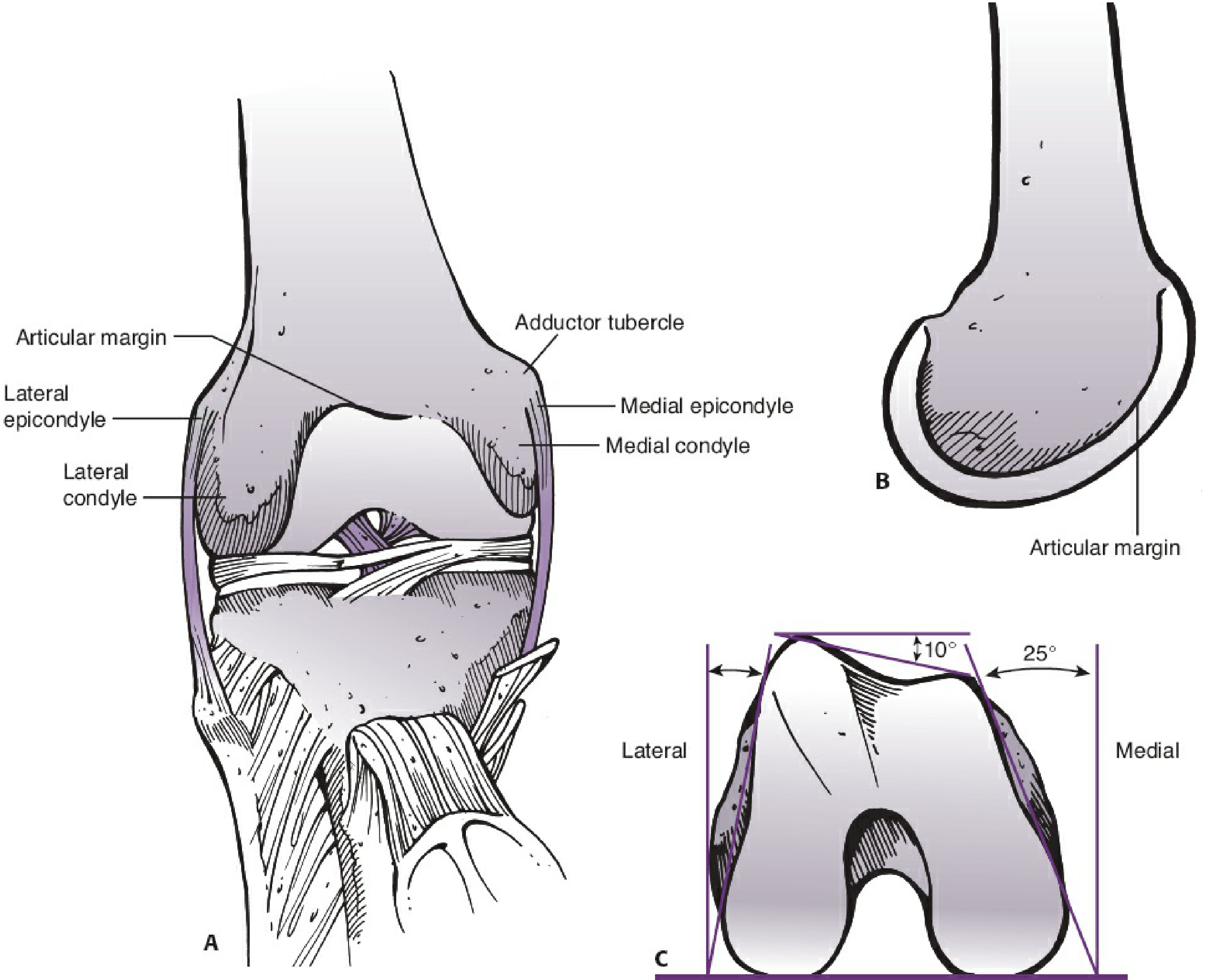
Condyles
- Two large articular masses: medial condyle (longer, extends farther distally) and lateral condyle (shorter, broader, extends farther proximally)
- Anteriorly, the two condyles unite to form the patellar (trochlear) surface - a groove for the patella
- Intercondylar (intercondyloid) fossa separates them posteriorly
- The distal femur, viewed on end, is trapezoidal in shape: the posterior part of the condyles is wider than the anterior part (creating ~25° inclination medially, ~15° laterally)
Intercondylar Fossa
| Wall | Cruciate Ligament Attachment |
|---|---|
| Lateral wall | Anterior cruciate ligament (ACL) |
| Medial wall | Posterior cruciate ligament (PCL) |
| Roof | Intercondylar line |
Epicondyles
- Medial epicondyle - bony non-articular prominence on the medial condyle; attachment for the medial (tibial) collateral ligament; medial head of gastrocnemius attaches just above it
- Lateral epicondyle - attachment for the lateral (fibular) collateral ligament and popliteus tendon (in an oblique groove just below it)
- Adductor tubercle - at the uppermost part of the medial condyle; where the medial supracondylar line terminates; adductor magnus inserts here
Femoral-Tibial Axis
- The anatomic axis of the femoral shaft makes ~6-7° of valgus relative to the knee joint line (range 2-10°)
- This valgus offset is factored into knee arthroplasty instrumentation
4. Blood Supply to the Femoral Head
The blood supply is precarious and of great clinical significance:
- Medial circumflex femoral artery (MFCA) - dominant supply. A branch of the profunda femoris (deep artery of thigh). Its deep branch passes posterior to the neck, then gives off superior retinacular arteries that run along the neck under the capsule → the main perfusion to the femoral head
- Lateral circumflex femoral artery (LCFA) - contributes via ascending branch; mainly supplies the anterior neck and greater trochanter
- Artery of the ligamentum teres - branch of the obturator artery; enters through the fovea; minor contribution in adults but important in children
Clinical: AVN risk - Femoral neck fractures can sever the retinacular vessels of the MFCA, cutting off the dominant blood supply and causing avascular necrosis (AVN) of the femoral head. The risk is highest with subcapital fractures (highest displacement, most proximal), lower with basicervical fractures (extracapsular, more vascular zone). This is why displaced femoral neck fractures in young adults are treated urgently with reduction and fixation (or hemiarthroplasty in elderly patients).
5. Ossification
| Epiphysis | Appearance | Fusion |
|---|---|---|
| Distal femoral epiphysis | Present at birth (important forensic/gestational marker) | ~19 years |
| Femoral head epiphysis | ~11 months (one physis including both trochanters) | ~18 years |
| Greater trochanter apophysis | ~4 years | ~18 years |
| Lesser trochanter apophysis | ~9 years | ~17 years |
- The distal femoral epiphysis contributes the highest proportion of longitudinal growth in the lower limb
- Slipped Capital Femoral Epiphysis (SCFE) occurs through the zone of hypertrophy of the femoral head physis
- In children, intramedullary nail insertion at the piriformis fossa risks damage to the posterosuperior retinacular vessels of the MFCA → AVN risk; trochanteric entry is preferred
6. Key Angles and Measurements
| Parameter | Normal | Notes |
|---|---|---|
| Neck-shaft angle | 127° | 141° at birth; decreases with age |
| Femoral anteversion | 10-15° (range 1-40°) | Relative to condylar plane |
| Femoral valgus (anatomic axis) | 6-7° | Range 2-10° |
| Femoral head radius | ~2.5 cm | Used in arthroplasty sizing |
| Supracondylar region length | Distal ~15 cm | Metaphysis-diaphysis junction |
7. Thigh Compartments Related to Femur
The femur serves as the central axis around which the thigh is divided by intermuscular septa (arising from the femur to the fascia lata) into three compartments:
| Compartment | Contents | Nerve |
|---|---|---|
| Anterior | Quadriceps, sartorius, iliopsoas | Femoral nerve |
| Medial | Adductors, gracilis, obturator externus | Obturator nerve |
| Posterior | Hamstrings (biceps femoris, semitendinosus, semimembranosus) | Sciatic nerve |
8. Clinical Fracture Classification Summary
Femoral Neck Fractures
By location:
- Subcapital (across head-neck junction) - highest AVN risk
- Transcervical (through mid-neck)
- Basicervical (at base of neck) - lowest AVN risk; extracapsular
Garden Classification (displacement):
| Grade | Description |
|---|---|
| I | Incomplete (valgus impacted) |
| II | Complete, non-displaced |
| III | Complete, partially displaced (trabeculae misaligned) |
| IV | Complete, fully displaced (trabeculae realigned with acetabulum) |
Pauwels Classification (fracture line angle - predicts shear stress):
- Type I: < 30° from horizontal (compressive forces - more stable)
- Type II: 30-50°
- Type III: > 50° (shear forces dominant - highest non-union risk)
Intertrochanteric Fractures
- Run between greater and lesser trochanters
- Extracapsular - blood supply to femoral head preserved, AVN risk is low
- Treated with intramedullary nail or dynamic hip screw (DHS)
Femoral Shaft Fractures
- High energy in young (MVAs); lower energy in elderly
- Intramedullary nail is gold standard
- "Damage control" approach for polytrauma patients
Distal Femur / Supracondylar Fractures
- The supracondylar zone = distal ~15 cm of femur
- Bimodal: high energy in young, ground-level falls in elderly osteoporotic patients
- Thin cortices and wide medullary canal make stable fixation difficult
- Goals: anatomic articular reduction, restore limb alignment, early mobilization
Sources: Gray's Anatomy for Students 4th Ed; THIEME Atlas of General Anatomy and Musculoskeletal System; Imaging Anatomy Vol. 3 - Bones, Joints, Vessels and Nerves; Miller's Review of Orthopaedics 9th Ed; Campbell's Operative Orthopaedics 15th Ed 2026; Rockwood and Green's Fractures in Adults 10th Ed 2025
This is a shared conversation. Sign in to Orris to start your own chat.