

Patient had this condition for 2 yrs and has a history of thyroid what ointment should be given
Diagnosis: Acanthosis Nigricans (AN) - Posterior Neck
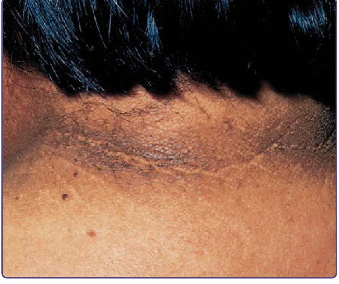
Why This Patient Has AN
Topical Treatment Options
| Agent | How to Use | Notes |
|---|---|---|
| Tretinoin cream (0.025-0.05%) | Apply thinly to affected neck at night | Topical retinoid; reduces hyperkeratosis; first-line topical choice |
| Calcipotriol (calcipotriene) 0.005% cream/ointment | Apply twice daily | Vitamin D analogue; improves skin texture |
| Salicylic acid 5-10% ointment/lotion | Apply once or twice daily | Keratolytic; softens and lightens the thickened skin |
| Glycolic acid 10-15% cream | Apply once daily | Chemical exfoliant; improves pigmentation |
| Urea 20-40% cream | Apply twice daily | Moisturizing keratolytic; helps with thickening |
- Tretinoin 0.025% cream at night (most evidence-supported topical)
- Urea 20% or salicylic acid cream in the morning for keratolysis
Key Management Points - Thyroid History
- Check thyroid function (TSH, T3, T4) - if hypothyroidism is undertreated, AN will not improve with topicals alone. Optimizing thyroid hormone levels can directly improve AN.
- Screen for insulin resistance - check fasting glucose and HbA1c, as hypothyroidism often co-exists with insulin resistance.
- If insulin resistance confirmed: Metformin (oral) improves insulin sensitivity and has a theoretic benefit in reducing AN.
- Topicals address appearance only - they will not resolve AN unless the underlying hormonal cause is managed.
What NOT to Use
- Avoid potent topical corticosteroids - they can worsen skin atrophy and may worsen insulin resistance.
- Avoid bleaching agents (like hydroquinone) as monotherapy - they do not address the texture/thickening, only pigment.
Important note: This patient should have a systemic evaluation done alongside topical treatment. Resolution will be slow (months) and partial with topicals alone if the thyroid/metabolic cause is not corrected.
- Fitzpatrick's Dermatology, Vol 1-2, p. 2525
- Andrews' Diseases of the Skin, Clinical Dermatology, p. 584
All about pitted keratolysis
pitted keratolysis sole of foot multiple pits
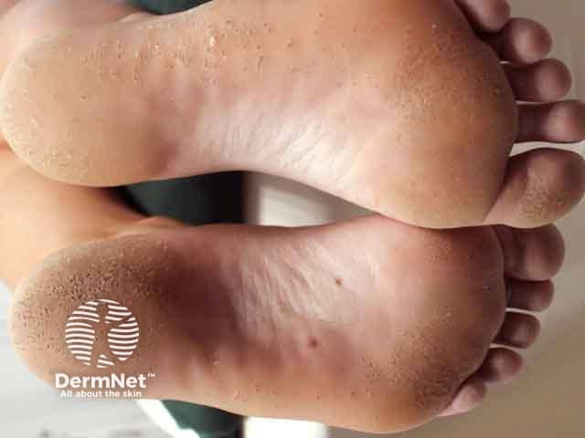
Clinical photography of bilateral plantar feet demonstrating characteristic pits of pitted keratolysis. This in vivo image captures the sole surfaces, primarily the weight-bearing plantar aspect from midfoot to toes, with a close-up view of the skin. Visual features include multiple shallow, punched-out depressions (1–3 mm) scattered across the plantar epidermis, well-defined circular to oval pits with smooth rims, and surrounding hyperkeratosis or desquamation. The intervening skin shows minimal erythema and subtle fissuring in flexural areas; overall texture is dry-to-moccasin-like. The pattern is bilateral and symmetrical, more pronounced on pressure-bearing regions. These pits result from superficial cornified layer loss and bacterial activity, classically due to Corynebacterium spp., and are often associated with hyperhidrosis or occlusive footwear in humid environments. Clinically, this finding is highly suggestive of pitted keratolysis and helps distinguish it from tinea pedis, plantar warts, and irritant dermatitis; differential diagnoses include keratolytic plaque and moccasin foot. Diagnostic significance lies in targeted therapy: topical antibiotics (erythromycin, clindamycin, or tetracycline) or antiseptics, improved foot hygiene, drying strategies, and breathable footwear. Potential clinical use cases include dermatology teaching, disease documentation, and electrodermal research on bacterial colonization of hyperhidrotic plantar skin. Clinical utility.
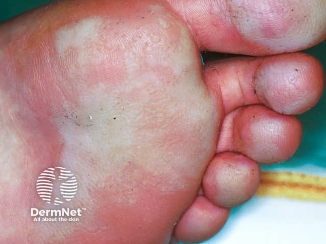
Clinical photograph of the plantar surface of the foot demonstrating classic pitted keratolysis. The image captures white, macerated, moist keratoderma with shallow, punched-out pits distributed across the weight-bearing sole, most prominently in the central metatarsal area. The surrounding skin shows mild erythema and desquamation; there is minimal scale at the periphery. This condition results from superficial bacterial decomposition of keratin in occluded, sweaty skin, commonly Corynebacterium species, and is favored by warm, moist environments and occlusive footwear. The pits appear as small depressions that may drain or secrete moisture; the lesion lacks true vesicles, purulence, or significant inflammatory infiltration. Clinically, pitted keratolysis is differentiated from tinea pedis by its characteristic pits rather than annular scaling; interdigital involvement is common; interdigital maceration may be present. Diagnostic significance lies in recognizing moisture-associated superficial infection, which responds to improved drying, antiseptic washes, and topical antibiotics. Potential clinical use cases include diagnostic aid in dermatology education, teledermatology triage, and differential diagnosis for plantar pits and maceration. This image provides educational reference to correlate appearance with microbiologic etiology and to guide management, including hygiene modification, barrier protection, and targeted topical therapy. These features reinforce a moisture-based bacterial etiology.
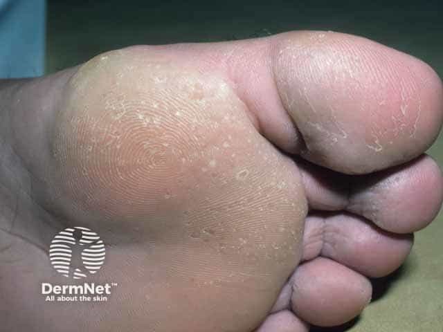
Plantarly focused clinical photograph illustrating pitted keratolysis on the ball of the foot. The image is a close‑up plantar view with natural color balance, designed for dermatology documentation and teaching. Multiple shallow, crater‑like pits (1–3 mm) are present in the weight‑bearing stratum corneum, often with surrounding mild hyperkeratosis and intermittent maceration between pits. The pits have well‑defined edges and show minimal surrounding erythema, indicating superficial epidermal involvement without deep ulceration. No nail or interdigital involvement is evident in this view. The distribution is concentrated to the plantar ball region and may be unilateral or bilateral depending on shoe wear and moisture. Pathognomonic features include cratering pits in a moist, occluded environment, frequently accompanied by odor and mild pruritus. Pathogenic mechanism involves superficial bacterial keratolysis caused by Corynebacterium species that secrete proteolytic enzymes in humid conditions. Clinically, these findings differentiate PK from tinea pedis, plantar warts, callosities, and interdigital dermatitis. Diagnostic significance lies in rapid recognition and referral for topical antibiotics and drying measures. This image provides an educational reference for diagnosis, differential diagnosis, and management planning, including topical keratolytics (salicylic acid, urea) and antibiotics, plus moisture control. This photograph supports dermatology education, teledermatology review, and research on plantar dermatologic infections. Correlation with symptoms guides treatment and prognosis in affected individuals.
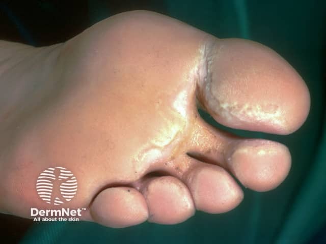
Clinical photograph of the plantar aspect of a foot highlighting interdigital spaces demonstrates classic pitted keratolysis manifesting as multiple shallow, craterlike pits in the stratum corneum between the toes and along the toe margins. The surrounding epidermis shows focal mild hyperkeratosis and subtle maceration with a waxy, pale appearance. The lesions are primarily superficial, non-erythematous, and do not form frank ulcers; the pits vary in size and are often arranged in clusters within web spaces. The appearance is characteristic for interdigital pitted keratolysis, a superficial bacterial infection caused by Corynebacterium spp. that thrives in moist environments. Clinically, the condition may be accompanied by mild odor and discharge in advanced cases, though odor is not a visual finding in the image. Pathogenesis involves keratolytic enzyme production leading to focal degradation of corneocytes, with epidermal involvement limited to the stratum corneum and granulosum. Diagnostic significance lies in distinguishing this entity from tinea pedis and eczematous interdigital dermatitis; management typically includes topical antibiotics or antiseptics and measures to reduce moisture and occlusion. This image is suitable for dermatology education, medical student training, and clinical reference libraries; it supports differential diagnoses, patient counseling, and treatment planning for interdigital foot skin infections and hyperhidrosis-associated dermatoses.
Pitted Keratolysis - Complete Overview
Clinical Appearance
Definition & Synonyms
Epidemiology
- Occurs worldwide, more common in tropical and humid climates
- Affects adults and children of both sexes, but adult males with sweaty feet are most susceptible (~90% of cases; male-to-female ratio ~8:1)
- Common in soldiers, athletes, and people wearing occlusive footwear for prolonged periods
- First described in people who went barefoot during the rainy season
Causative Organisms
| Organism | Role |
|---|---|
| Kytococcus sedentarius (formerly Micrococcus sedentarius) | Primary pathogen in most cases |
| Corynebacterium spp. | Common co-pathogen |
| Dermatophilus congolensis | Implicated in some cases |
| Actinomyces spp. | Occasionally implicated |
Predisposing Factors
- Hyperhidrosis (excessive sweating) - most important factor
- Prolonged skin occlusion (tight shoes, boots)
- Increased skin surface pH (alkaline moisture environment)
- Hot, humid weather
- Poor foot hygiene
Clinical Features
- Plantar (sole) surface - pressure-bearing areas primarily: ball of foot, heel, ventral aspect of toes
- Web spaces between toes (may be the only manifestation)
- Rarely: palmar surface
- Multiple shallow, crateriform pits, 1-7 mm in diameter
- Pits have elongated configurations along plantar furrows
- Pits may coalesce to form large craters, furrows, or rings with serpiginous borders
- No associated erythema (non-inflammatory)
- Skin may appear white and macerated in moist conditions
- Often asymptomatic and unnoticed initially
- Malodor - in two-thirds of patients (hallmark complaint)
- Sliminess - foot sticking to socks (~70% of cases)
- Pain, burning, or irritation - in one-third of patients
- Rarely pruritic
Pathology (Histology)
- Well-defined pits extend approximately two-thirds of the way into the stratum corneum (superficial; no deeper involvement)
- No inflammatory infiltrate
- Gram, PAS, and Gomori methenamine silver stains reveal bacteria in the walls and bases of the craters
- Coccoid forms superficially; filamentous forms with branches and septa in deeper locations
Diagnosis
- Clinical diagnosis based on characteristic appearance
- Gram stain of scrapings can detect organisms
- KOH exam is less helpful
- Wood's lamp - NO fluorescence (distinguishes from erythrasma, which shows coral-red fluorescence)
- Bacterial cultures often grow multiple species and are not diagnostically helpful
- Biopsy is generally unnecessary
Differential Diagnosis
| Condition | Distinguishing Feature |
|---|---|
| Tinea pedis | Annular scaling, positive KOH, no pits; interdigital tinea can mimic PK |
| Erythrasma (interdigital) | Hyperkeratotic, coral-red fluorescence on Wood's lamp |
| Plantar warts | Interrupted skin lines, black dots (thrombosed capillaries), painful on lateral squeeze |
| Palmoplantar keratoderma (punctate) | Genetic, no bacterial cause, pits are harder/keratotic |
| Darier disease | Pits with keratotic plugs, other skin/nail findings |
| Basal cell nevus syndrome | Pits on palms + jaw cysts + calcified falx |
Management
1. General / Preventive Measures (Address Hyperhidrosis)
- Keep feet as dry as possible at all times
- Wear well-fitted, non-occlusive shoes
- Change socks and shoes frequently
- Wash socks at ≥60°C (140°F) to kill bacteria
- Use inert antiseptic foot powders
2. Topical Antibiotics (First-Line)
| Drug | Form | Notes |
|---|---|---|
| Erythromycin 2% | Solution / gel | First-line; well-established |
| Clindamycin 1% | Solution / gel | Equally effective; well tolerated |
| Mupirocin 2% | Ointment/cream | Effective alternative |
| Fusidic acid | Cream | Useful alternative |
| Tetracycline | Topical solution | Reported effective |
3. Antiseptic / Keratolytic Agents
| Agent | Use |
|---|---|
| Benzoyl peroxide 5% gel/cream or cleanser | Highly effective; antibacterial + keratolytic; often first-line |
| Aluminum chloride 20% solution | Targets hyperhidrosis; reduces moisture = less bacterial growth |
| Whitfield's ointment (benzoic acid + salicylic acid) | Keratolytic; effective alternative |
| Chlorhexidine wash | Antiseptic; adjunctive |
| Miconazole / clotrimazole cream | Azole antifungals with some antibacterial effect |
| Salicylic acid / urea | Keratolytics to reduce hyperkeratosis and enhance drug penetration |
4. For Recalcitrant Hyperhidrosis
| Treatment | Notes |
|---|---|
| Topical glycopyrrolate | Anticholinergic; reduces focal sweating |
| Botulinum toxin injections | Plantar injections for severe hyperhidrosis associated with PK |
| Systemic clindamycin or erythromycin | Reserved for severe / refractory cases |
Key Teaching Points
- PK is a bacterial infection, NOT a fungal one - antifungals are used only as adjuncts
- The odor is caused by sulfur compounds from bacterial keratin degradation, not from hygiene alone
- Benzoyl peroxide is often the most practical first-line agent (antibacterial + keratolytic + cheap)
- Treating the underlying hyperhidrosis is as important as the antibiotic
- Wood's lamp is negative in PK (useful to rule out erythrasma which is positive)
- No inflammation = no need for corticosteroids
- Fitzpatrick's Dermatology, Vol 1-2, p. 2770
- Andrews' Diseases of the Skin, Clinical Dermatology, p. 14
- Dermatology 2-Volume Set 5e, p. 1532
Create a visual comparison poster: PK vs tinea pedis vs plantar warts
Medical education comparison poster titled "Plantar Foot Conditions: Differential Diagnosis" with three columns side by side comparing Pitted Keratolysis vs Tinea Pedis vs Plantar Warts. Clean clinical poster style with a white background and color-coded columns (teal/green for PK, orange for Tinea Pedis, purple for Plantar Warts). Each column has a header with the condition name and includes the following labeled rows: Cause, Appearance, Location, Symptoms, Wood's Lamp, Key Test, and Treatment. Column 1 - Pitted Keratolysis (teal): Cause: Kytococcus sedentarius bacteria; Appearance: Multiple 1-7mm crateriform pits, may coalesce; Location: Pressure-bearing sole, heels, toe webs; Symptoms: Foul odor, slimy feet, ± pain; Wood's Lamp: Negative (no fluorescence); Key Test: Gram stain of scrapings; Treatment: Topical erythromycin/clindamycin, benzoyl peroxide, aluminum chloride. Column 2 - Tinea Pedis (orange): Cause: Dermatophyte fungi (Trichophyton rubrum); Appearance: Annular scaling, maceration, vesicles; Location: Interdigital spaces, sole (moccasin type); Symptoms: Pruritus, burning, scaling; Wood's Lamp: Negative; Key Test: KOH prep (hyphae positive); Treatment: Topical azoles (clotrimazole, miconazole), terbinafine. Column 3 - Plantar Warts (purple): Cause: Human Papillomavirus (HPV types 1, 2, 4); Appearance: Hyperkeratotic papule, black dots (thrombosed capillaries), interrupted skin lines; Location: Pressure points on sole; Symptoms: Painful on lateral squeeze, not squeeze; Wood's Lamp: Negative; Key Test: Dermoscopy - black dots, interrupted ridges; Treatment: Salicylic acid, cryotherapy, laser. At the bottom a comparison strip: "Key Differentiator" row - PK: pits + odor + no KOH; Tinea: KOH positive hyphae; Warts: black dots + lateral squeeze pain. Professional medical illustration style, bold headings, clean icons or small illustrative diagrams next to each condition name showing characteristic lesion shape.
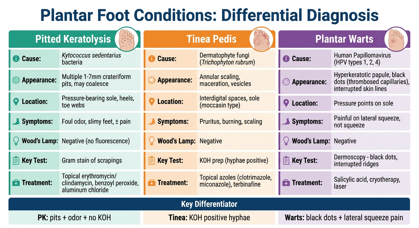
| Feature | Pitted Keratolysis | Tinea Pedis | Plantar Warts |
|---|---|---|---|
| Cause | Bacteria (Kytococcus) | Fungi (Trichophyton) | HPV (types 1, 2, 4) |
| Hallmark | Pits + foul odor | Scaling + itch | Black dots + lateral squeeze pain |
| KOH prep | Negative | Positive (hyphae) | Negative |
| Wood's lamp | Negative | Negative | Negative |
| 1st-line Rx | Topical erythromycin / BPO | Topical terbinafine / azole | Salicylic acid / cryotherapy |