
PITUITARY APOPLEXY
"pituitary apoplexy"[MeSH Terms] AND management
pituitary apoplexy MRI hemorrhage infarction
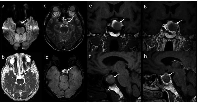
This figure presents multi-sequence cranial MRI images (axial, coronal, and sagittal planes) of a sellar mass, illustrating the diagnostic features of pituitary apoplexy. Axial Diffusion-Weighted Imaging (DWI) (a, b) demonstrates a focal area of central hyperintensity within the pituitary gland, indicating restricted diffusion consistent with acute infarction. On axial T2-weighted imaging (c), the lesion is relatively hyperintense, while the axial Susceptibility-Weighted Imaging (SWI) (d) reveals a central hypointense signal, or susceptibility artifact, indicative of intralesional hemorrhage. The coronal and sagittal T1-weighted pre-contrast images (e, f) show incomplete peripheral hyperintensity within the mass. Post-contrast T1-weighted images (g, h) display a characteristic peripheral ring enhancement pattern, which appears slightly less pronounced on follow-up compared to initial imaging. The combined findings of infarction (DWI restriction) and focal hemorrhage (SWI artifact) in a pituitary macroadenoma are hallmark radiologic features of pituitary apoplexy, a clinical emergency requiring neurosurgical and endocrinological evaluation.

This composite figure presents a clinical case of pituitary apoplexy through multimodality imaging and histopathology. Panel (a) is an axial non-contrast CT scan showing a hyperdense mass in the sellar/suprasellar region, characteristic of acute intratumoral hemorrhage. Panels (b) and (c) are coronal T1-weighted MRI scans; (b) shows a large, pre-contrast hyperintense pituitary tumor extending into the suprasellar cistern, while (c) demonstrates a lack of internal enhancement following gadolinium administration, indicative of tumor infarction or necrosis. Panel (d) is a coronal T2-weighted MRI revealing a heterogeneous tumor with a prominent low-signal-intensity (hypointense) peripheral rim, a classic sign of hemosiderin deposition or acute-on-chronic bleeding in pituitary apoplexy. Panel (e) provides a histopathological correlation with hematoxylin and eosin (H&E) staining, showing a clear demarcation between viable papillary-patterned pituitary adenoma tissue (left) and areas of extensive hemorrhage and necrosis (right). The sequence illustrates the diagnostic hallmark of sudden hemorrhagic transformation of a macroadenoma.
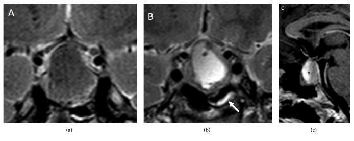
This set of diagnostic MRI scans illustrates the neuroradiological hallmarks of pituitary apoplexy across three views. Panels (a) and (b) are coronal T2-weighted images demonstrating a heterogeneous sellar and suprasellar mass. Panel (a) shows areas of low signal intensity corresponding to intralesional hemorrhage, while panel (b) reveals high-signal intensity cystic or necrotic regions and focal mucosal thickening of the sphenoid sinus (indicated by the white arrow), a secondary finding in acute apoplexy. Panel (c) provides a sagittal T1-weighted view showing a fluid-fluid level (marked by an asterisk) within the pituitary mass. This specific finding is pathognomonic for pituitary apoplexy, representing the layering of settled cellular blood components inferiorly and free extracellular methemoglobin superiorly. The images collectively demonstrate the typical mass effect of a pituitary adenoma complicated by acute infarction or hemorrhage, essential for medical students and clinicians to recognize in cases of acute pituitary failure or visual compromise.
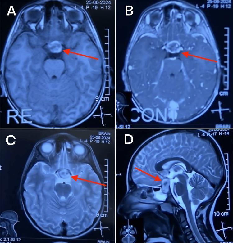
Diagnostic Magnetic Resonance Imaging (MRI) of the brain in axial and sagittal planes illustrating a sellar and suprasellar mass consistent with pituitary apoplexy. Panel A (Axial T1-weighted without contrast) reveals a mixed-intensity lesion (red arrow) indicating acute to subacute hemorrhage within the pituitary gland. Panel B (Axial T1-weighted with contrast) demonstrates peripheral enhancement around the non-enhancing core, suggesting peripheral vascularization or infarction. Panel C (Axial T2-weighted) shows a hyperintense signal within the lesion, indicative of edema or blood products. Panel D (Sagittal T2-weighted) provides anatomical orientation, showing the lesion's expansion within the sella turcica and its significant suprasellar extension, causing compression of the optic chiasm. Key landmarks include the sphenoid sinus, sella turcica, brainstem, and optic chiasm. This radiological series demonstrates the diagnostic features of pituitary macroadenoma complicated by hemorrhage (apoplexy), highlighting its relationship to surrounding neuro-anatomical structures for surgical planning.