
Explain malrotation and intestinal obstructions in surgery
Reading File
Reading File
Finding Sources
Finding Sources
Searching PubMed
intestinal malrotation AND obstruction
Searching the Web
intestinal malrotation volvulus diagnosis management pediatric surgery guideline
Reading File
Finding Sources
Reading File
Searching Images
upper gastrointestinal contrast study intestinal malrotation midgut volvulus corkscrew duodenum
Reading Web Page
https://pmc.ncbi.nlm.nih.gov/articles/PMC11271324/
Reading File
Reading File
Reading File
Intestinal malrotation
Intestinal malrotation is a congenital failure of normal fixation of the midgut. Normally, the bowel returns to the abdomen and becomes fixed with a broad mesenteric base. In malrotation, the duodenojejunal junction and caecum lie abnormally close, leaving the midgut suspended on a narrow mesenteric pedicle around the superior mesenteric artery (SMA).
This narrow base predisposes to twisting of the bowel, called midgut volvulus, causing acute obstruction, vascular compromise, intestinal gangrene, short-bowel syndrome, or death.
Embryological basis
Classically, normal midgut development involves:
- Physiological herniation of midgut
- Approximately 270° counterclockwise rotation around the SMA
- Return to abdomen and fixation
Failure of fixation results in:
- Right-sided small bowel
- Abnormally placed, often medial caecum
- Narrow mesenteric root
- Ladd bands: abnormal peritoneal bands extending from the malpositioned caecum across the duodenum to the right lateral abdominal wall. These can compress the duodenum and cause obstruction.
Sabiston Textbook of Surgery, p. 2683-2684.
Clinical presentation
About 90% of symptomatic cases present before 1 year of age, but malrotation may present in older children or adults.
Acute midgut volvulus
- Sudden bilious vomiting, especially in a neonate
- Colicky abdominal pain
- Lethargy, shock, hematochezia or melena in advanced ischemia
- Distension may be absent initially and is a late ominous finding
- Metabolic acidosis suggests bowel ischemia
Chronic or intermittent presentation
- Recurrent bilious or non-bilious vomiting
- Intermittent crampy pain
- Poor feeding, failure to thrive
- Early satiety, malabsorption, protein-losing enteropathy
- Symptoms may mimic reflux or functional abdominal pain
Bilious vomiting in a newborn should be assumed to be intestinal obstruction, including malrotation with volvulus, until proved otherwise.
Diagnosis
-
Plain abdominal radiograph
- May be normal, show proximal obstruction, or a gasless abdomen
- It is nonspecific and must not delay surgery if volvulus is strongly suspected.
-
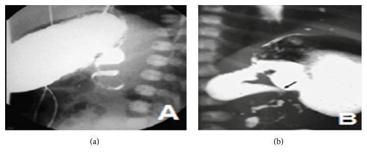
Upper GI contrast series
- Best confirmatory study in a stable child
- Findings:
- Duodenum fails to cross the midline
- Duodenojejunal junction lies abnormally to the right
- “Corkscrew” appearance of twisted proximal jejunum or duodenum in volvulus
- Tapered “bird-beak” obstruction in the third part of duodenum
-
Ultrasound with Doppler
- Abnormal SMA-SMV relationship
- SMV may lie left of or anterior to SMA
- “Whirlpool sign” indicates twisting of mesentery and vessels
Management
Suspected midgut volvulus is an emergency. Do not delay operative treatment in an unstable child for contrast imaging.
Initial management:
- Nil by mouth
- Nasogastric decompression
- IV fluids and correction of electrolyte and acid-base abnormalities
- Broad-spectrum antibiotics if ischemia, perforation, or sepsis is suspected
- Urgent paediatric surgical referral
Ladd procedure
The definitive operation includes:
- Counterclockwise detorsion of volvulus, if present
- Division of Ladd bands compressing the duodenum
- Broadening of the narrow mesenteric base
- Placement of small bowel on the right and colon on the left
- Appendicectomy, because the appendix will remain in an atypical position and could confuse later diagnosis
- Resection of nonviable bowel, if necessary
A questionable bowel segment may be preserved and reassessed at planned second-look laparotomy after 24-36 hours. A recent clinical series also describes the Ladd procedure as standard treatment, with emergency surgery for volvulus and resection when necrosis is present (2024 clinical review).
Sabiston Textbook of Surgery, p. 2684.
Sabiston Textbook of Surgery, p. 2684.
Intestinal obstruction
Intestinal obstruction is failure of normal onward passage of intestinal contents due to either a mechanical block or failure of intestinal motility.
Classification
1. By mechanism
| Type | Meaning | Examples |
|---|---|---|
| Mechanical obstruction | Physical blockage of the lumen or extrinsic compression | Adhesions, obstructed hernia, tumour, volvulus, intussusception, stricture |
| Dynamic obstruction | Failure of propulsion without a mechanical block | Paralytic ileus, acute colonic pseudo-obstruction, electrolyte disturbances |
2. By level
- Small-bowel obstruction (SBO): high or low
- Large-bowel obstruction (LBO)
3. By severity
- Partial or incomplete
- Complete
4. By vascular status
- Simple obstruction: blood supply intact
- Strangulating obstruction: venous then arterial compromise
- Closed-loop obstruction: bowel occluded at two points, high risk of rapid strangulation and perforation
Causes
Small-bowel obstruction in adults
Common causes:
- Postoperative adhesions: most common
- Incarcerated or strangulated hernia
- Crohn disease stricture
- Small-bowel tumour
- Intussusception
- Gallstone ileus
- Volvulus
- Foreign body or bezoar
Large-bowel obstruction
Common causes:
- Colorectal carcinoma
- Volvulus, especially sigmoid volvulus
- Diverticular stricture
- Faecal impaction
- Inflammatory stricture
Neonatal intestinal obstruction
Important causes include:
- Malrotation with midgut volvulus
- Duodenal atresia, web, or stenosis
- Jejunoileal atresia
- Meconium ileus
- Hirschsprung disease
- Anorectal malformation
- Necrotizing enterocolitis
Clinical features
The classical quartet is:
- Colicky abdominal pain
- Vomiting
- Abdominal distension
- Absolute constipation, meaning no stool or flatus
Bailey & Love’s Short Practice of Surgery, p. 1402.
Clinical pattern by site
| Feature | High small bowel | Low small bowel | Large bowel |
|---|---|---|---|
| Vomiting | Early, profuse | Later | Late, may become faeculent |
| Distension | Minimal | Central and marked | Early and pronounced |
| Pain | Colicky, central | Colicky, central | Less severe initially, lower abdominal |
| Dehydration | Early and severe | Moderate | Late |
| X-ray | Few proximal dilated loops | Multiple central small-bowel loops | Peripheral colonic dilatation |
Bailey & Love’s Short Practice of Surgery, p. 1402.
Features suggesting strangulation or ischemia
These require urgent surgery:
- Continuous severe pain, rather than intermittent colic
- Tachycardia, fever, hypotension
- Localized tenderness, guarding, rigidity, rebound tenderness
- Leukocytosis, rising lactate, metabolic acidosis
- Bloody nasogastric aspirate or rectal bleeding
- CT evidence of closed-loop obstruction, pneumatosis, portal venous gas, reduced bowel-wall enhancement, or free fluid
Investigations
Initial tests
- FBC, CRP
- Urea, creatinine, electrolytes
- Serum lactate and arterial/venous blood gas if ischemia suspected
- Group and save or crossmatch if surgery likely
Imaging
-
Abdominal radiograph
- Useful initial test
- Small bowel: central dilated loops, valvulae conniventes
- Large bowel: peripheral dilated colon with haustral markings
- May show free gas if perforation
-
CT abdomen and pelvis with IV contrast
- Preferred imaging in most stable adults
- Identifies level, cause, closed loop, ischemia, perforation, and complications.
-
Water-soluble contrast study
- Sometimes used in adhesive SBO
- Can help predict resolution and may have therapeutic value in selected uncomplicated cases.
-
Contrast enema
- Used selectively for colonic obstruction, volvulus, or neonatal distal obstruction.
Management principles
Immediate measures for all suspected obstructions
- Admit and assess severity
- Nil by mouth
- Two wide-bore IV cannulas
- IV crystalloid resuscitation
- Correct potassium and other electrolyte abnormalities
- Nasogastric tube for vomiting or significant distension
- Urinary catheter and monitor urine output
- Analgesia and antiemetics
- Broad-spectrum antibiotics if strangulation, perforation, sepsis, or operation is likely
- Early surgical assessment
The core treatment framework is nasogastric drainage, fluid and electrolyte replacement, and relief of the obstruction. Bailey & Love’s Short Practice of Surgery, p. 1407.
When conservative treatment may be appropriate
A carefully observed nonoperative approach can be used in selected patients with:
- Suspected uncomplicated adhesive partial SBO
- No peritonitis
- No ischemia or strangulation
- No closed-loop obstruction
- Stable vital signs and improving clinical status
Indications for urgent operation
- Peritonitis or perforation
- Suspected strangulation or bowel ischemia
- Closed-loop obstruction
- Obstructed or strangulated hernia
- Volvulus with ischemia
- Complete obstruction with deterioration
- Failure of nonoperative management
- Malrotation with suspected midgut volvulus
Operative goals
- Identify and relieve the obstruction
- Divide adhesions, reduce hernia, untwist volvulus, resect tumour, or remove obstructing lesion as appropriate
- Assess bowel viability
- Resect nonviable bowel
- Restore continuity or form a stoma when required
Exam-focused comparison: malrotation versus general obstruction
| Point | Malrotation with volvulus | General adult intestinal obstruction |
|---|---|---|
| Usual age | Neonates and infants, but any age possible | Usually adults |
| Main danger | Rapid total midgut ischemia | Strangulation, perforation, sepsis |
| Key symptom | Bilious vomiting | Pain, vomiting, distension, constipation |
| Best diagnostic test if stable | Upper GI contrast series | CT abdomen with IV contrast |
| Definitive surgery | Ladd procedure | Cause-specific operation |
| Surgical urgency | Immediate if volvulus suspected | Immediate if strangulation, closed loop, or perforation suspected |
Key takeaway: In a neonate with bilious vomiting, always exclude malrotation with midgut volvulus first because delay can cause catastrophic loss of bowel.
This is a shared conversation. Sign in to Orris to start your own chat.