
Paralytic ileus
"paralytic ileus" OR "postoperative ileus" management
paralytic ileus abdominal X-ray dilated bowel loops
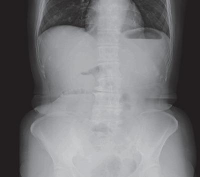
This abdominal plain radiograph (supine X-ray) demonstrates marked gaseous distension of multiple small bowel loops, primarily located in the central abdomen. The loops are dilated significantly beyond the normal 3cm threshold, suggesting a small bowel obstruction or paralytic ileus. A notable clinical feature is the 'gasless' appearance or lack of significant dilatation in the large bowel (colon), which is characteristic of an obstructive process occurring proximal to the ileocecal valve. Key radiological landmarks include visible valvulae conniventes within the distended loops and a gastric bubble in the left upper quadrant. The absence of gas in the rectum or distal colon supports a diagnosis of mechanical small bowel obstruction. This imaging modality is a primary diagnostic tool for assessing acute abdominal pain and distension, providing evidence of intestinal transition zones and potential complications such as perforation (though no pneumoperitoneum is visible here).
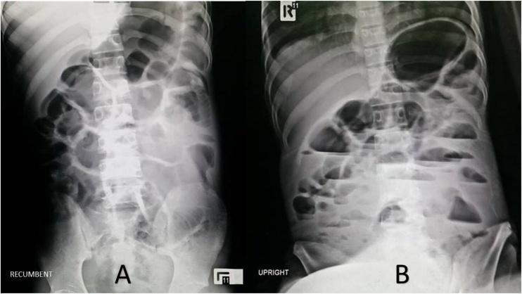
Diagnostic Image: This abdominal radiograph (X-ray) panel displays a comparison between recumbent (A) and upright (B) views of a 19-year-old patient. The images demonstrate significant pathological dilatation of both small and large bowel loops, characteristic of generalized ileus. In the recumbent view (A), gas-filled intestinal segments are dispersed across the abdominal cavity, revealing prominent valvulae conniventes and haustral markings indicative of gaseous distension. In the upright view (B), multiple air-fluid levels are clearly visible within the dilated loops, manifesting as horizontal lines where gas interfaces with fluid due to gravity. These findings suggest a functional bowel obstruction or paralytic ileus. The imaging is critical for differentiating between mechanical obstruction and generalized dysmotility. Educational concepts illustrated include the radiographic signs of bowel distension, the diagnostic utility of positional X-rays in emergency medicine, and the identification of air-fluid levels as markers of impaired intestinal transit.
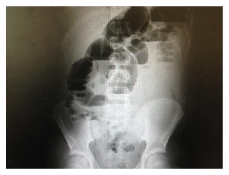
This abdominal radiograph (X-ray) shows a diagnostic image of the abdomen in a patient presenting with postoperative symptoms. The primary finding is the presence of several markedly dilated small bowel loops, primarily located in the upper and central abdominal regions. These loops exhibit significant gaseous distension and multiple air-fluid levels, characteristic of an intestinal obstruction or paralytic ileus. The degree of small bowel dilatation is disproportionate to the sparse amount of gas visible within the colon. Anatomical landmarks including the lumbar spine, sacrum, and pelvic bones (iliac crests and pubic symphysis) are clearly visible, providing spatial orientation. The radiographic presentation is highly suggestive of a mechanical small bowel obstruction, potentially related to postoperative adhesions or complications. This visual evidence serves as an educational tool for identifying typical radiological signs of bowel distension and air-fluid levels in acute surgical abdominal pathologies.
Paralytic Ileus
Definition
- Bailey and Love's Short Practice of Surgery, 28th Ed.
Pathophysiology
- Neurogenic factors: Disruption of the enteric nervous system (ENS) and central nervous system input
- Inflammatory mediators: Surgical trauma triggers an inflammatory cascade that inhibits smooth muscle contractility
- Enteric hormones and neuropeptides: Released in response to stress, they inhibit coordinated propulsion
- Electrolyte disturbances: Hypokalemia, hypomagnesaemia, and hyponatraemia reduce smooth muscle excitability
- Opioids: Bind to mu-receptors in the gut wall, directly suppressing motility
- Hypoxia and bowel oedema: Relative splanchnic ischaemia (e.g., in pancreatitis) contributes via reflex vasoconstriction
- Sabiston Textbook of Surgery, 21st Ed.
Causes / Varieties
| Category | Examples |
|---|---|
| Postoperative | Most abdominal procedures (self-limiting, 24-72 hours); prolonged if hypoproteinaemia or metabolic derangement present |
| Intra-abdominal sepsis | Peritonitis, anastomotic leak, abscesses - causes localised or generalised ileus |
| Reflex ileus | Spinal/rib fractures, retroperitoneal haemorrhage, plaster jacket application |
| Metabolic | Hypokalemia, uraemia, hypomagnesaemia, diabetic ketoacidosis |
| Drugs | Opioids, anticholinergics, tricyclic antidepressants |
| Pancreatitis | Regional ileus affecting the duodenum, proximal jejunum, or transverse colon due to proximity of inflammation |
| Systemic illness | Sepsis, myocardial infarction, pneumonia, trauma |
| Pseudo-obstruction (Ogilvie's syndrome) | A variant - massive colonic dilatation (especially the caecum) in patients with major non-abdominal illness; risk of perforation |
- Bailey and Love's, Pye's Surgical Handicraft, Maingot's Abdominal Operations
Return of Bowel Function After Surgery
- Small bowel - within hours (some contractile activity within hours)
- Stomach - 24-48 hours
- Colon - 3-5 days
- Bailey and Love's; Sabiston Textbook of Surgery
Clinical Features
- Nausea and vomiting
- Abdominal distension (tympanic on percussion)
- Loss of appetite
- Absent or markedly reduced bowel sounds
- Absolute constipation - failure to pass flatus or stool
- Diffuse, non-colicky abdominal discomfort (no cramping, which would suggest mechanical obstruction)
Key distinguishing point: mechanical obstruction features colicky pain and high-pitched, tinkling bowel sounds early; paralytic ileus features silent abdomen and no colic.
Radiology
- Dilatation of both small and large bowel (unlike mechanical obstruction, where dilatation is proximal to the block only)
- Multiple air-fluid levels on erect views
- In pancreatitis: "sentinel loop" (localised dilated loop near inflamed pancreas) and "colon cut-off sign" (abrupt loss of gas in transverse colon)
- Sensitivity of AXR approximately 66% for small bowel obstruction vs ileus
-
No transition point (the hallmark of mechanical obstruction - an abrupt calibre change)
-
Generalised dilatation without a defined obstruction site
-
Can exclude mechanical cause and identify intra-abdominal complications
-
Grainger & Allison's Diagnostic Radiology
Management
1. Treat the Underlying Cause
- Correct electrolyte imbalances (especially K+, Mg2+, Na+)
- Treat intra-abdominal sepsis (drainage, antibiotics)
- Reduce/discontinue opioids where possible
- Address pancreatitis, retroperitoneal pathology
2. Supportive Measures
- IV fluid replacement and maintenance of adequate hydration
- Nasogastric (NG) tube for gastric decompression (drainage purposes; relieves nausea and vomiting)
- Nil by mouth initially; advance diet as tolerated
- Early ambulation - promotes return of bowel motility
3. Nutritional Support
- Nutrient absorption from the gut is impaired in paralytic ileus
- Parenteral nutrition (TPN) should be considered in prolonged ileus to prevent malnutrition
4. Pharmacological Options
- Neostigmine (a cholinesterase inhibitor): used in Ogilvie's syndrome/adynamic ileus to stimulate intestinal motility; also useful for urinary bladder atony
- Prokinetics (e.g., metoclopramide) have limited evidence
- Alvimopan (mu-opioid receptor antagonist): used to accelerate recovery of postoperative GI function after opioid-containing anaesthesia (not universally available)
5. Ogilvie's Syndrome (Pseudo-obstruction)
-
Enormous caecal dilatation (>12 cm) carries risk of perforation
-
Colonoscopic decompression is the treatment of choice
-
Neostigmine may avoid colonoscopy in some cases
-
Bailey and Love's; Sabiston Textbook of Surgery; Goodman & Gilman's Pharmacology; Pye's Surgical Handicraft
Paralytic Ileus vs. Mechanical Obstruction
| Feature | Paralytic Ileus | Mechanical Obstruction |
|---|---|---|
| Pain | Dull, continuous | Colicky, intermittent |
| Bowel sounds | Absent/very reduced | High-pitched, tinkling |
| AXR | Generalised dilation (small + large) | Dilation proximal to obstruction |
| CT | No transition point | Clear transition point |
| Onset post-op | Expected, usually resolves 3-5 days | Unusual if bowel function had returned |
| Cause | Functional/neurogenic | Adhesions, hernia, volvulus, tumour |
Key Points Summary
- Paralytic ileus = failure of neuromuscular transmission (Auerbach's and Meissner's plexuses)
- Most common cause is post-laparotomy (self-limiting, 24-72 h for small bowel; 3-5 days for colon)
- Hypokalemia and opioids are major reversible contributing factors
- Bowel sounds are absent; no colic (distinguishes from mechanical obstruction)
- AXR shows generalised bowel dilatation; CT excludes mechanical cause
- Management is largely supportive: fluids, electrolyte correction, NG tube, early ambulation, treat underlying cause
- TPN for prolonged cases; neostigmine/colonoscopy for Ogilvie's syndrome
Relevant anatomy for understanding headaches.
anatomy pain sensitive structures headache cranial nerves meninges
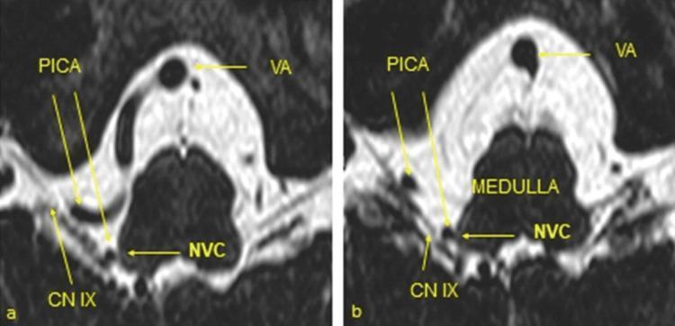
Two high-resolution axial MRI images (a and b) utilizing a heavily T2-weighted CISS (Constructive Interference in Steady State) sequence to demonstrate the neurovascular anatomy of the posterior cranial fossa. The images focus on the ventrolateral medulla oblongata and its associated cranial nerves. Labeled structures include the vertebral artery (VA), the posterior inferior cerebellar artery (PICA), and the glossopharyngeal nerve (CN IX). On the right side, a vascular loop of the PICA is shown in direct contact with the glossopharyngeal nerve at its root entry zone (REZ). This point of contact is labeled as neurovascular compression (NVC), which is the primary pathological finding associated with glossopharyngeal neuralgia. The CISS sequence provides high contrast between the dark (hypointense) vascular and neural structures and the bright (hyperintense) cerebrospinal fluid (CSF), allowing for precise localization of the compression. These images serve as a diagnostic tool for identifying microvascular conflict in patients presenting with cranial nerve syndromes.
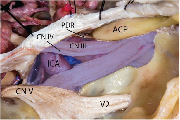
This clinical photograph displays a high-magnification cadaveric dissection of the human middle cranial fossa, specifically focused on the neurovascular anatomy of the cavernous sinus region. The image utilizes a blue color overlay to represent the boundaries of the cavernous sinus venous space. Key anatomical structures labeled include the internal carotid artery (ICA), the anterior clinoid process (ACP), and the proximal dural ring (PDR). The spatial relationships of several cranial nerves (CN) are demonstrated: the oculomotor nerve (CN III) and trochlear nerve (CN IV) are seen coursing superiorly and anteriorly to the ICA. The trigeminal nerve (CN V) and its second division, the maxillary nerve (V2), are situated inferiorly. Notably, the V2 is shown passing outside the demarcated blue venous space of the cavernous sinus, illustrating its anatomical position relative to the dural envelop. This educational visual is intended for neurosurgical training and advanced anatomical study of the skull base, highlighting the complex arrangement of structures relevant to transsphenoidal or cavernous sinus surgical approaches.
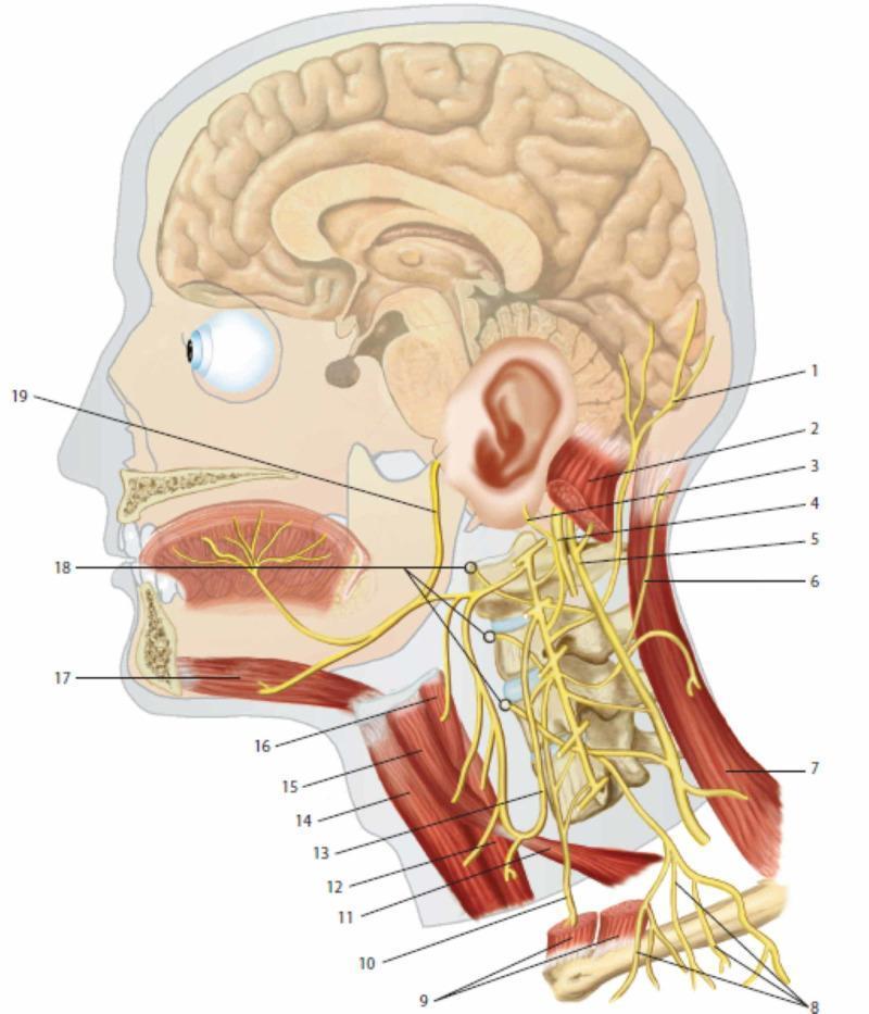
This medical anatomical diagram provides a lateral view of the human head and neck, focusing on the cervical plexus and its integration with cranial nerves. Key neural structures include the lesser occipital nerve (1), greater auricular nerve (3), and transverse cutaneous nerve of the neck (6), demonstrating the sensory distribution of the cervical plexus. The phrenic nerve (10) is shown descending inferiorly anterior to the prevertebral muscles (18). Cranial nerves X (vagus, 4), XI (accessory, 5), and XII (hypoglossal, 19) are illustrated to show their spatial relationships with cervical spinal branches. Muscular landmarks include the sternocleidomastoid (2, 9), trapezius (7), and infrahyoid muscles such as the omohyoid (11, 15) and thyrohyoid (16). The ansa cervicalis (13) is depicted innervating the sub-hyoid musculature. The diagram also illustrates the sagittal section of the brain and oral cavity, highlighting the innervation of the tongue and the relationship between the cervical spine vertebrae and emerging nerve roots. This resource is intended for medical education regarding the peripheral nervous system and regional anatomy of the neck.
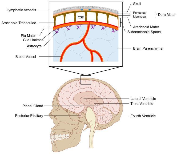
This medical anatomical diagram illustrates the layers of the human meninges and the organization of the subarachnoid space. The lower panel displays a sagittal view of the head, identifying internal structures such as the lateral, third, and fourth ventricles, alongside circumventricular organs including the pineal gland and posterior pituitary. The upper panel provides a high-magnification cross-sectional view of the cranial protective layers. From superficial to deep, it depicts the skull, the dura mater (consisting of periosteal and meningeal layers containing lymphatic vessels), the arachnoid mater, and the subarachnoid space filled with cerebrospinal fluid (CSF). The arachnoid trabeculae are shown spanning the subarachnoid space to connect with the pia mater. Below the pia mater, the glia limitans is illustrated, formed by astrocytic foot processes that interface with the underlying brain parenchyma and cortical blood vessels. This diagram is designed to explain leptomeningeal anatomy and the fluid dynamics between the ventricular system, subarachnoid space, and the blood-brain barrier interface.
trigeminovascular system trigeminal nucleus caudalis headache pathway diagram
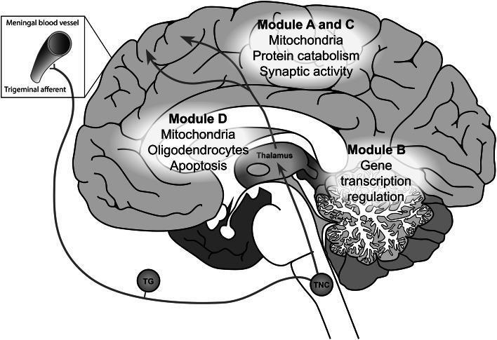
A medical schematic illustrating the pathophysiology of migraine, specifically the trigeminovascular pathway and associated gene co-expression modules in the brain. The diagram depicts a sagittal view of the human brain with key anatomical structures labeled, including the cortex, cerebellum, thalamus, and trigeminal nucleus caudalis (TNC). The trigeminovascular pathway is traced from meningeal blood vessels via trigeminal afferents through the trigeminal ganglion (TG) and TNC, ascending to the thalamus and ultimately the cerebral cortex. Overlaid on the anatomy are localized gene expression modules (A-D) highlighting specific cellular functions and components: Modules A and C in the cortex are associated with mitochondria, protein catabolism, and synaptic activity; Module B in the cerebellum is linked to gene transcription regulation; and Module D in the subcortical white matter and thalamic region is associated with mitochondria, oligodendrocytes, and apoptosis. The illustration serves as an educational tool for understanding the integration of genetic expression patterns with neuroanatomical pain pathways in migraine headache disorders.
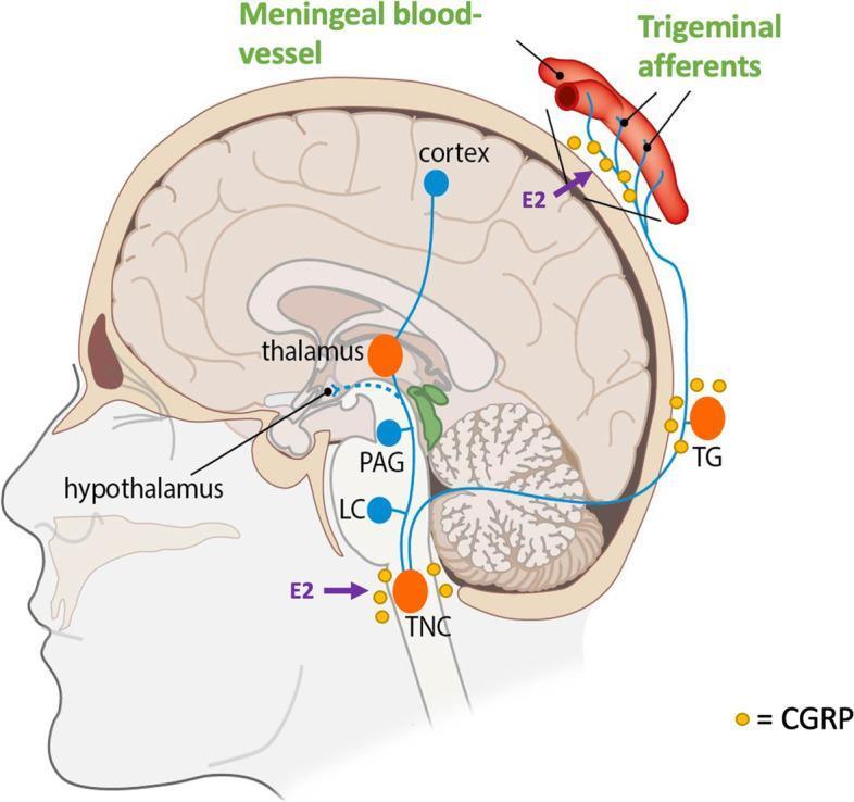
This pathophysiology diagram illustrates the human trigeminovascular system and its activation during a migraine attack. Presented in a sagittal cross-section of the head, the diagram highlights key anatomical structures including the meningeal blood vessels, cerebral cortex, thalamus, hypothalamus, and brainstem components such as the periaqueductal gray (PAG), locus coeruleus (LC), and trigeminal nucleus caudalis (TNC). The trigeminal ganglion (TG) is shown as a peripheral relay point. The diagram emphasizes the role of Calcitonin Gene-Related Peptide (CGRP), represented by yellow icons, localized at the meningeal vessels, TG, and TNC, indicating its role in neurotransmission and vasodilation. Purple arrows denote the influence of estradiol (E2) on CGRP release at the level of the TNC and the trigeminal afferents. Blue lines trace the ascending and descending pathways between these centers, demonstrating the functional connectivity involved in headache phase signaling. This visual is designed for medical education regarding migraine pathophysiology and the neuro-hormonal modulation of the trigeminal system.
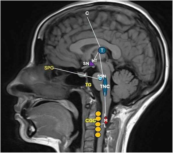
This diagnostic image consists of a midsagittal T1-weighted MRI scan of the human head and neck, overlaid with a schematic pathophysiology diagram illustrating the neuroanatomical pathways of cluster headache (CH). The diagram maps three primary systems: the trigeminovascular system, the autonomic reflex, and the hypothalamic system. Key labeled structures include the Primary Sensory Cortex (C), Thalamus (T), Hypothalamus (HT), and Suprachiasmatic Nucleus (SN). In the brainstem and peripheral regions, the diagram highlights the Superior Salivatory Nucleus (SSN), Trigeminal Caudal Nucleus (TNC), Trigeminal Ganglion (TG), and Sphenopalatine Ganglion (SPG). Descending pathways are shown reaching the Intermediolateral Nucleus (IML) of the spinal cord and the Cervical Ganglionic Complex (CGC). Functional connections illustrate nociceptive afferents from the TG to the TNC, ascending projections to the thalamus and cortex for pain perception, and hypothalamic activation of autonomic centers. This educational visual explains the clinical manifestations of CH, including trigeminal distribution pain, cranial autonomic symptoms like lacrimation via the SPG, and sympathetic dysfunction leading to Horner's syndrome via the IML-CGC pathway.
Relevant Anatomy for Understanding Headaches
The Core Principle: The Brain Itself Does Not Hurt
- Neuroanatomy Through Clinical Cases, 3rd Ed.; Harrison's Principles of Internal Medicine, 22nd Ed.
1. Pain-Sensitive Structures of the Head
Intracranial Structures (Pain-Sensitive)
| Structure | Notes |
|---|---|
| Meningeal (dural) arteries | Highly pain-sensitive; middle meningeal artery most important |
| Large pial arteries (proximal segments) | Circle of Willis and first few centimetres of their branches |
| Dural venous sinuses (e.g., superior sagittal, transverse) | Distension or thrombosis causes headache |
| Falx cerebri and tentorium cerebelli | Dural folds; traction on these causes referred pain |
| Dura mater (near blood vessels) | Most sensitive; base of skull dura especially |
| Pia mater and small cerebral vessels | Recent evidence (awake craniotomy studies) suggests these are also pain-sensitive |
Intracranial Structures (NOT Pain-Sensitive)
- Brain parenchyma
- Most of the brain's white and grey matter
- Ventricular ependyma
- Choroid plexus
Extracranial Structures (Pain-Sensitive)
| Structure | Notes |
|---|---|
| Scalp | Skin, subcutaneous tissue, muscles (temporalis, occipitalis) |
| Scalp arteries: superficial temporal, occipital | Inflamed in giant cell arteritis |
| External carotid artery and branches | |
| Neck muscles (attached to occiput) | Muscle spasm = tension-type headache mechanism |
| Cervical nerve roots (C1-C3) | Refer pain to the back of head and neck |
| Nasal and paranasal sinus mucosa | Infection/congestion causes frontal or facial headache |
| Teeth and dental pulp | Dental pain can be referred to the head |
| Eyes | Raised intraocular pressure (glaucoma) causes orbital headache |
- Bradley & Daroff's Neurology in Clinical Practice; Guyton & Hall Textbook of Medical Physiology
2. Innervation of Pain-Sensitive Structures
Trigeminal Nerve (CN V) - the dominant headache nerve
- Innervates all supratentorial dura (the largest part of the intracranial cavity)
- Specifically the ophthalmic division (V1) carries most intracranial nociception - this is why frontal and periorbital pain is common even with posterior lesions
- V1 fibres also innervate the cornea, forehead, nose, and upper eyelid
- V2 (maxillary) innervates the mid-face, upper teeth, and maxillary sinuses
- V3 (mandibular) innervates the lower teeth, jaw muscles, and temporal skin
Posterior Fossa Innervation
- The infratentorial dura (posterior fossa) is innervated mainly by CN X (vagus), with contributions from CN IX (glossopharyngeal) and the upper cervical nerves (C1-C3)
- This explains why posterior fossa lesions tend to refer pain to the occiput and back of the neck
Cervical Nerve Roots (C1-C3)
- Supply the back of the scalp and upper neck
- Converge on the same nucleus as the trigeminal nerve (see trigeminocervical complex below)
- Cervical spine pathology commonly causes occipital and sub-occipital headache
Other Cranial Nerves
-
CN VII (facial), CN IX, CN X also carry pain from extracranial structures
-
CN IX - pharynx and posterior tongue (glossopharyngeal neuralgia)
-
CN X - ear, pharynx, larynx
-
Neuroanatomy Through Clinical Cases; Bradley & Daroff's Neurology
3. The Trigeminovascular System
Pathway:
- Peripheral afferents: Trigeminal (and C2/C3) sensory fibres innervate the dura mater, meningeal blood vessels, and large cerebral arteries
- Trigeminal ganglion (Gasserian ganglion): Located in Meckel's cave at the skull base - fibres from all three divisions coalesce here
- Trigeminal nucleus caudalis (TNC): The first central synapse - located in the dorsomedial medulla and extending into the dorsal horn of C1-C2 (see trigeminocervical complex)
- Thalamus (ventroposteromedial nucleus): Second-order relay
- Somatosensory cortex and other cortical areas: Final pain perception
- Bradley & Daroff's Neurology in Clinical Practice; Goldman-Cecil Medicine
4. The Trigeminocervical Complex
- The caudal trigeminal nucleus extends from the medulla down to the dorsal horn at C2-C3
- Cervical nerve inputs from C2 and C3 converge onto the same neurons as trigeminal fibres
- This convergence means:
- An occipital lobe tumour can cause frontal head pain (trigeminal referral)
- Cervical spine disease can cause frontal headache
- Posterior fossa lesions produce occipital pain (C2/C3 referral)
- Neck stiffness and headache often co-occur (meningeal irritation activates both systems)
- Harrison's; Bradley & Daroff's
5. Pain Modulation - Descending Control
| Structure | Role |
|---|---|
| Periaqueductal grey (PAG) | Major inhibitory centre; activated by opioids |
| Rostral ventromedial medulla | Inhibitory and facilitatory control |
| Locus coeruleus (noradrenergic) | Modulates trigeminal pain; may be dysfunctional in migraine |
| Dorsal raphe nuclei (serotonergic) | 5-HT pathways; target of triptans |
| Hypothalamus | Circadian regulation; strongly activated in cluster headache |
6. Supratentorial vs Infratentorial - Pain Referral Patterns
| Location of pathology | Referred pain area |
|---|---|
| Supratentorial (most of the brain, frontal to tentorium) | Frontal, temporal, parietal head (CN V distribution) |
| Infratentorial (posterior fossa, cerebellum, brainstem) | Occiput and upper neck (C2/C3 distribution) |
| Base of skull / posterior communicating artery aneurysm | Ipsilateral eye and brow |
| Carotid dissection | Ipsilateral eye, forehead, and face |
| Vertebral artery occlusion/dissection | Upper neck, postauricular area |
| Basilar artery thrombosis | Occiput and forehead |
| Temporal artery (giant cell arteritis) | Localized scalp tenderness, then diffuse |
| Raised ICP | Bifrontal and bioccipital, worse when supine |
| Low CSF pressure | Worse upright, relieved by lying flat |
- Adams & Victor's Principles of Neurology, 12th Ed.
7. Specific Extracranial Headache Sources
Muscle Tension
- Spasm of temporalis, frontalis, occipitalis, and neck muscles (sternocleidomastoid, trapezius, suboccipitals) refers pain to the scalp
- The mechanism of tension-type headache: sustained muscle contraction and/or central sensitization
Nasal Sinuses
- Maxillary sinusitis → pain over the cheeks and below the eyes, sometimes referred to the forehead
- Frontal sinusitis → pain over the forehead (not "behind the eyes" as often thought)
- Important: autonomic features of migraine (nasal congestion, rhinorrhoea, lacrimation) mimic sinus disease - migraine is vastly over-diagnosed as "sinus headache"
Eyes
- Raised intraocular pressure (acute angle-closure glaucoma) → severe orbital and periorbital headache, often with nausea
- Refractive error and eye strain → mild frontal headache
Neck and Cervical Spine
- C2 neuralgia → greater occipital nerve (runs from C2 dorsal ramus up to the scalp) causes occipital headache and scalp tenderness
- Cervicogenic headache: unilateral pain radiating from neck to forehead, with restricted neck movement
8. The Meninges in Detail
- Dura mater: Double-layered (periosteal + meningeal); contains the dural venous sinuses between the layers; innervated by CN V (supratentorial) and CN IX, X, upper cervicals (infratentorial)
- Arachnoid mater: Not pain-sensitive; contains the subarachnoid space (CSF)
- Pia mater: Closely adherent to the brain; some pain sensitivity in larger pial vessels
Summary Diagram of Headache Anatomy
PAIN-SENSITIVE STRUCTURES
         │
    ┌────┴─────────────┐
Intracranial           Extracranial
    │                      │
Dural arteries         Scalp arteries
Dural sinuses          Scalp muscles
Large pial vessels     Neck muscles (C1-C3)
Dura mater             Sinuses, teeth, eyes
    │                      │
    └────────┬─────────────┘
             │
    Trigeminal nerve (CN V)
    + CN IX, X (post. fossa)
    + C1-C3 nerve roots
             │
    Trigeminal Ganglion
             │
    Trigeminal Nucleus Caudalis
    (trigeminocervical complex - extends to C2/C3)
             │
    Thalamus (VPM nucleus)
             │
    Somatosensory Cortex
    ← Modulated by: PAG, raphe, locus coeruleus, hypothalamus
Key Points
- The brain parenchyma has no pain receptors - all headache arises from peripheral structures
- Supratentorial dura = CN V (V1 mainly) → frontal/periorbital pain referral
- Infratentorial dura = CN IX, X, C1-C3 → occipital/neck pain referral
- The trigeminocervical complex (TNC extending to C2/C3) explains why neck and head pain converge
- The trigeminovascular system - trigeminal innervation of dural/meningeal vessels - is the anatomical basis for migraine; CGRP is the key neuropeptide
- Descending modulation (PAG, raphe, hypothalamus) explains why psychological and hormonal factors powerfully affect headache
- Pain referral patterns follow the innervation territory - supratentorial lesions → frontal pain; posterior fossa lesions → occipital pain
- Raised ICP alone does not cause headache - it is the traction on pain-sensitive vessels and dura that matters
Primary and secondary headaches
migraine headache types comparison diagram
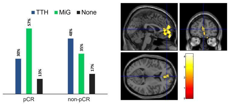
This composite educational graphic consists of a side-by-side comparison between a clinical bar chart and neuroimaging statistical maps. On the left, a grouped bar graph illustrates the distribution of headache types—Tension-Type Headache (TTH), Migraine (MiG), and No Headache (None)—in cancer patients categorized by their pathological complete response (pCR) to neoadjuvant chemotherapy (NAC). The pCR group shows a predominance of Migraine (57%), while the non-pCR group shows a higher prevalence of TTH (48%). On the right, a statistical map overlay on a T1-weighted MRI template displays regions of altered brain metabolism in sagittal, coronal, and axial views. Yellow-red clusters indicate a positive test statistic (intensity scale 0 to 4) localized predominantly in the bilateral anterior medial frontal cortex. The visual highlights a specific neuro-oncological correlation between chemotherapy response clinical outcomes and functional brain imaging biomarkers, specifically targeting metabolic variations in patients suffering from different primary headache disorders.
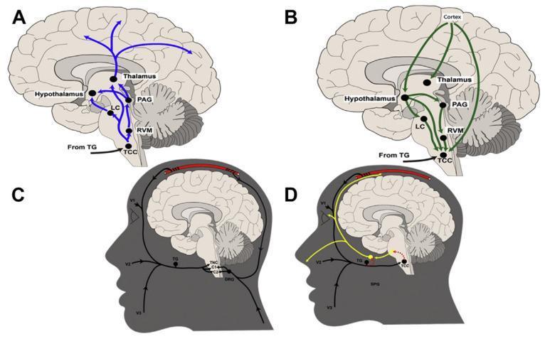
A multi-panel pathophysiology diagram illustrating the ascending and descending neuroanatomical pathways of migraine. Panel A depicts ascending pathways (blue) originating from the trigeminocervical complex (TCC) and projecting to the rostral ventromedial medulla (RVM), locus coeruleus (LC), periaqueductal gray (PAG), hypothalamus, and thalamus, eventually reaching the cortex. Panel B illustrates descending inhibitory and modulatory pathways (green) from the cortex and diencephalic structures (hypothalamus, thalamus, PAG) back to the TCC. Panels C and D provide sagittal head views demonstrating the integration of peripheral and central nervous systems. Panel C highlights the convergence of dural, cervical (C1, C2 via dorsal root ganglia), and trigeminal (V1, V2, V3) afferent inputs at the TCC via the trigeminal ganglion (TG). Panel D shows the trigeminal autonomic reflex interface, including connections between the TG and the sphenopalatine ganglion (SPG), illustrating the parasympathetic arm involved in headache pathophysiology. The schematic emphasizes the TCC as a critical relay center for head and neck nociception.
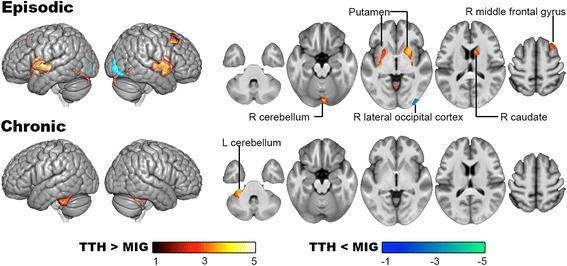
This diagnostic image displays voxel-based morphometry (VBM) results from a post-hoc 2x2 ANCOVA analysis comparing gray matter (GM) volume in patients with tension-type headache (TTH) and migraine (MIG). The visualization includes lateral cortical surface maps and sequential axial slices of the brain, divided into 'Episodic' and 'Chronic' rows. In the Episodic comparison, TTH shows significantly higher GM volume (indicated by red-yellow heat maps) in the bilateral putamen, right caudate, right middle frontal gyrus, and right cerebellum, while lower GM volume (indicated by blue-cyan) is visible in the right lateral occipital cortex compared to episodic migraine. In the Chronic comparison, significantly higher GM volume in TTH is localized only to the left cerebellum, with no significant regions of lower GM volume relative to chronic migraine. These findings illustrate neuroanatomical differences in pain processing and inhibitory regions between primary headache types, highlighting a reduction in structural divergence as the conditions transition from episodic to chronic forms.
Primary and Secondary Headaches
Overview
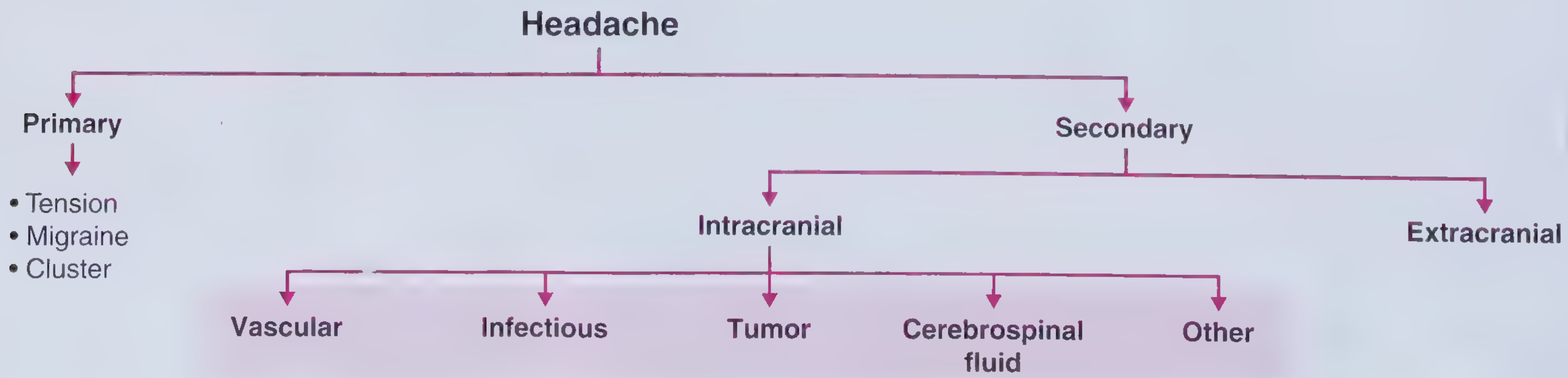
- Primary headaches - head pain is the disorder itself; no underlying structural or systemic disease
- Secondary headaches - head pain is a symptom of an identifiable underlying condition
PART 1: PRIMARY HEADACHES
1. Migraine
Definition and Phases
- Premonitory phase (prodrome): Hours to days before - mood change, food cravings, yawning, neck stiffness, fatigue
- Aura: Focal neurological symptoms lasting 20-60 minutes, fully reversible (present in ~30% of migraineurs)
- Headache phase: The pain itself, plus associated symptoms
- Postdrome: After the headache - fatigue, cognitive "fog", mood change
Epidemiology
- 1-year prevalence: 12% overall (18% in women, 6% in men)
- Female:male ratio ~3:1 (hormonal influence via estrogen)
- Peak prevalence: 4th decade (24% of women, 7% of men)
- Lifetime prevalence: ~33% women, ~13% men
- First-degree relatives of migraineurs are 2-4x more likely to develop migraine
- WHO ranks migraine among the most disabling medical conditions globally
ICHD-3 Diagnostic Criteria
| Criterion | Details |
|---|---|
| Duration | 4-72 hours (untreated or unsuccessfully treated) |
| Character (≥2 of 4) | Unilateral; Pulsating/throbbing; Moderate-severe intensity; Aggravated by routine physical activity (walking, stairs) |
| Associated symptoms (≥1) | Nausea ± vomiting; OR Photophobia AND phonophobia |
- Visual (most common): Zigzag fortification spectra, scintillating scotoma, photopsia - caused by cortical spreading depression (a slow wave of depolarization spreading at 3-5 mm/min)
- Sensory: Ipsilateral arm or periorbital numbness/tingling with a "marching" character
- Motor: Spreading motor deficit (e.g., in familial hemiplegic migraine - CACNA1A gene mutation)
- Speech: Mild dysphasia
Migraine Subtypes
| Subtype | Features |
|---|---|
| Basilar-type migraine | Aura referable to brainstem/bilateral hemispheres: dysarthria, vertigo, tinnitus, diplopia, bilateral paraesthesiae |
| Retinal migraine | Reversible monocular visual disturbances |
| Status migrainosus | Migraine attack lasting >72 hours |
| Migrainous infarction | Cerebral infarct on neuroimaging associated with migraine |
| Chronic migraine | ≥15 headache days/month for >3 months, of which ≥8 are migrainous |
Triggers
Pathophysiology Summary
- Cortical spreading depression → activates trigeminal afferents → meningeal nociceptors
- Trigeminovascular activation → release of CGRP, substance P, VIP, PACAP, nitric oxide → neurogenic inflammation of dural vessels
- Central sensitisation at trigeminal nucleus caudalis → allodynia (non-painful stimuli perceived as painful - scalp tenderness)
- Dysfunctional descending pain modulation (PAG, locus coeruleus, raphe) amplifies the experience
Treatment
| Drug Class | Examples | Notes |
|---|---|---|
| Triptans (5-HT1B/D agonists) | Sumatriptan 6 mg SC; Zolmitriptan 5 mg nasal; oral triptans | First-line for moderate-severe migraine; SC fastest onset |
| Ergotamines | DHE (dihydroergotamine) IV/IM/nasal | Effective; contraindicated in vascular disease |
| NSAIDs/analgesics | Ibuprofen, naproxen, ketorolac IM/IV | Mild-moderate attacks |
| Antiemetics | Metoclopramide, prochlorperazine | Also have direct antimigraine effect |
| CGRP antagonists (gepants) | Rimegepant, ubrogepant | Newer agents; no vasoconstriction |
- Beta-blockers (propranolol, metoprolol)
- Tricyclics (amitriptyline)
- Valproate, topiramate
- CGRP monoclonal antibodies (erenumab, fremanezumab) - newest, highly effective
- Verapamil (especially if with aura)
2. Tension-Type Headache (TTH)
ICHD-3 Criteria
| Feature | TTH | Migraine (contrast) |
|---|---|---|
| Location | Bilateral | Often unilateral |
| Quality | Pressing/tightening (band-like, non-pulsating) | Pulsating/throbbing |
| Intensity | Mild to moderate | Moderate to severe |
| Physical activity | Not aggravated | Worsened by activity |
| Nausea/vomiting | Absent | Present |
| Phono/photophobia | At most ONE of these | Both often present |
| Duration | 30 minutes to 7 days | 4-72 hours |
Treatment
- Acute: Simple analgesics (aspirin, ibuprofen, paracetamol), avoid opioids/overuse
- Prophylactic: Amitriptyline (most evidence), stress management, physiotherapy, biofeedback
3. Cluster Headache and Trigeminal Autonomic Cephalalgias (TACs)
Cluster Headache
- Severe unilateral orbital/supraorbital/temporal pain
- Duration: 15-180 minutes (shorter than migraine)
- Ipsilateral cranial autonomic features (≥1): Conjunctival injection, lacrimation, nasal congestion, rhinorrhoea, forehead/facial sweating, miosis, ptosis, eyelid oedema
- Restlessness/agitation during attack
- Attacks may awaken from sleep (circadian pattern - hypothalamic involvement)
| Setting | Drug | Dose |
|---|---|---|
| Acute (first-line) | High-flow O₂ | ≥15 L/min via non-rebreather mask until remission |
| Acute (first-line) | Sumatriptan | 6 mg SC |
| Acute (second-line) | Octreotide | 100 mcg SC |
| Preventive | Verapamil | 240-480 mg/day (drug of choice) |
| Bridge | Dexamethasone | 10 mg IM/IV |
| Preventive adjunct | Melatonin | 10 mg at night |
Other Trigeminal Autonomic Cephalalgias (TACs)
| TAC | Attack Duration | Frequency | Key feature |
|---|---|---|---|
| Cluster headache | 15-180 min | 1-8/day | Periods of remission; male dominant |
| Paroxysmal hemicrania (PH) | 2-30 min | >5/day | Absolutely responsive to indomethacin |
| SUNCT (Short-lasting Unilateral Neuralgiform with Conjunctival injection and Tearing) | 5-240 sec | Dozens/day | Very brief; refractory to most drugs |
| SUNA | 1-600 sec | Dozens/day | Single autonomic feature (vs SUNCT) |
| Hemicrania continua | Continuous | - | Constant unilateral + autonomic features; indomethacin-responsive |
4. Other Primary Headache Disorders
- Primary cough headache: Bilateral, precipitated by coughing/Valsalva; must exclude Chiari malformation
- Primary exercise headache: During or after exertion; must exclude SAH
- Primary headache associated with sexual activity: "Sex headache" - orgasmic headache; must exclude SAH (thunderclap pattern)
- Primary thunderclap headache: Severe headache reaching max intensity within 1 minute; diagnosis of exclusion after SAH ruled out
- Hypnic headache: Awakens from sleep, elderly patients; often bilateral; no autonomic features
PART 2: SECONDARY HEADACHES
Intracranial Causes
A. Vascular
| Condition | Headache Character | Key Features |
|---|---|---|
| Subarachnoid haemorrhage (SAH) | Thunderclap - "worst headache of life", maximal within seconds | ± neck stiffness, photophobia, syncope, CN III palsy (posterior communicating artery aneurysm); ~80% from berry aneurysms at circle of Willis |
| Intracerebral haemorrhage | Sudden severe; location-related focal deficits | Hypertension, coagulopathy |
| Cerebral venous sinus thrombosis | Progressive over days-weeks; may mimic raised ICP | Young women on OCP; papilloedema; seizures |
| Arterial dissection (carotid/vertebral) | Carotid: ipsilateral eye/neck/face + Horner syndrome; Vertebral: occipital/neck pain | Sudden onset; stroke risk |
| Giant cell arteritis | Temporal/scalp tenderness, jaw claudication; >50 years | ESR >50; risk of blindness; steroid-responsive emergency |
| Hypertensive crisis | Bilateral, bifrontal/occipital; pulsatile | BP >180/120; papilloedema |
B. Infectious
| Condition | Headache | Key Features |
|---|---|---|
| Bacterial meningitis | Severe, rapid onset; worst headache of life | Fever + neck stiffness + headache triad; photophobia; rash (meningococcus); LP (turbid CSF, ↑WBC, ↑protein, ↓glucose) |
| Viral meningitis | Severe; similar to bacterial but less acute | Lymphocytic pleocytosis; self-limiting |
| Encephalitis | Headache + altered consciousness | Herpes simplex = most important; fever, personality change, seizures |
| Brain abscess | Progressive, focal | Fever, focal deficit, raised ICP |
| Sinusitis | Facial/frontal pressure; worse bending | Percussion tenderness over sinus; purulent discharge |
C. Raised/Altered ICP
| Condition | Notes |
|---|---|
| Brain tumour | Progressive; worse in morning; worse supine; vomiting; papilloedema - rare cause overall (<0.1% of headaches) |
| Idiopathic intracranial hypertension (pseudotumor cerebri) | Obese women of childbearing age; pulsatile tinnitus, visual obscurations, papilloedema; LP shows raised opening pressure; treat with acetazolamide, weight loss |
| Low CSF pressure | Worse upright, relieved lying flat; typically post-LP or spontaneous; bilateral frontal and occipital |
| Hydrocephalus | Positional or progressive; papilloedema |
D. Tumours and Mass Lesions
- Headache caused by traction on pain-sensitive meninges and vessels, not by raised ICP itself
- Character depends on location: supratentorial → frontal pain; posterior fossa → occipital pain
- "Red flag" features: morning predominance, positional worsening, progressive course, new neurological deficits
Extracranial Causes
| Category | Examples |
|---|---|
| Cervicogenic | Cervical spine OA/disc disease; C2 neuralgia; greater occipital neuralgia - unilateral occipital radiation to vertex |
| Eye | Acute angle-closure glaucoma (orbital pain, halos, red eye, N&V); refractive error |
| Dental/TMJ | Temporomandibular joint dysfunction; dental abscess referred to temple |
| Sinusitis | Acute bacterial sinusitis - mucopurulent discharge, fever, sinus tenderness |
| Giant cell arteritis | Temporal artery tenderness/nodularity; visual loss risk; ESR elevated |
| Systemic illness | Viral infections, sepsis, CO poisoning (bilateral, in household contacts), anaemia, hypoxia |
| Drugs/substances | Medication overuse headache (MOH); nitrates; caffeine withdrawal; alcohol |
Medication Overuse Headache (MOH)
RED FLAGS: When to Suspect Secondary Headache (SNOOP mnemonic)
| Letter | Red Flag |
|---|---|
| S | Systemic symptoms (fever, weight loss, rash, night sweats) |
| N | Neurologic symptoms or signs (focal deficit, altered consciousness, papilloedema, meningism) |
| O | Onset sudden/thunderclap (maximal intensity <5 minutes) - until proven otherwise = SAH |
| O | Older age (new headache >50 years - consider giant cell arteritis, tumour) |
| P | Previous headache history - change in pattern; progressively worsening; "worst ever" |
- New headache in a patient with HIV or cancer
- Headache precipitated by cough, exertion, or Valsalva
- Headache in pregnancy or postpartum
- Headache waking from sleep
- New-onset seizures
- Optic disc oedema (papilloedema)
Primary vs Secondary: Comparison at a Glance
| Feature | Primary | Secondary |
|---|---|---|
| Underlying cause | None identifiable | Structural, vascular, infectious, metabolic |
| Neurological exam | Normal | May be abnormal |
| Onset | Recurrent, established pattern | New, recent-onset, or changed pattern |
| Red flags | Absent | Often present |
| Imaging | Usually normal | Often abnormal |
| Treatment focus | Headache itself | Underlying condition |