tricep surae
Reading File
Finding Sources
Finding Sources
Reading File
Reading File
Reading File
Loading Image
Loading Image
Here is a thorough overview of the triceps surae drawn from the THIEME Atlas of General Anatomy and Musculoskeletal System and the THIEME Imaging Anatomy atlas.
Triceps Surae
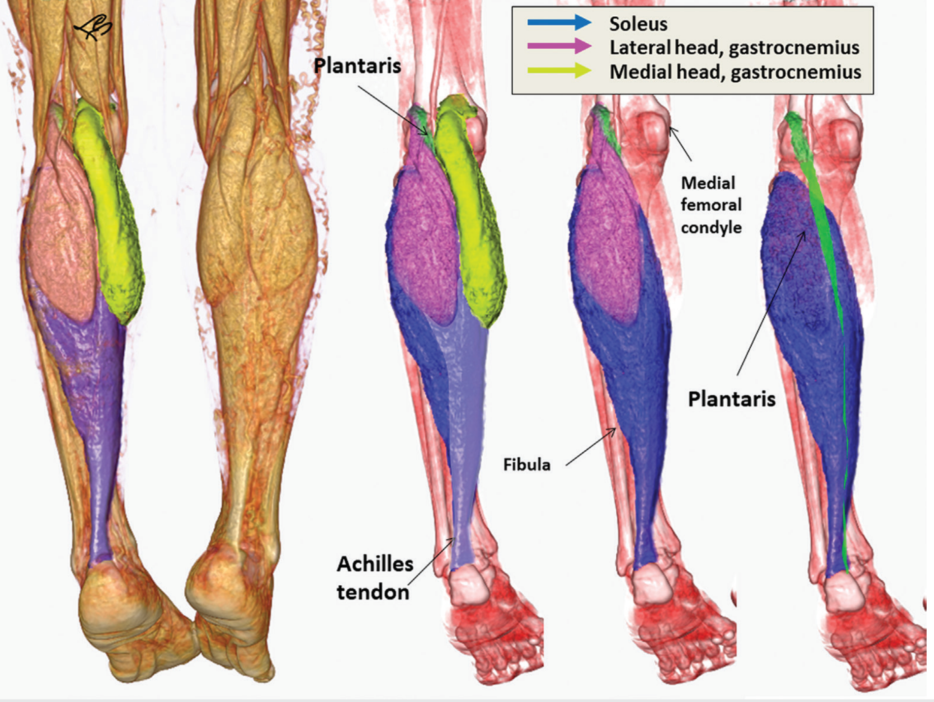
The triceps surae ("three-headed calf muscle") is the dominant muscle group of the superficial posterior compartment of the leg. It is also called the "superficial crural group." The name refers to its three muscular heads: the medial gastrocnemius, lateral gastrocnemius, and the soleus.
Constituent Muscles
1. Gastrocnemius (2 heads)
| Medial head | Lateral head | |
|---|---|---|
| Origin | Medial epicondyle of the femur | Lateral epicondyle of the femur |
| Note | Longer, inserts higher | Shorter |
Both heads originate from the posterior femoral condyles and the joint capsule. In the middle-to-distal third of the calf, the muscular fibers of each head converge through a myotendinous junction into a broad, flat tendon. This tendon then merges with the aponeurosis of the soleus.
2. Soleus
- Origin: Posterior surface of the head and neck of the fibula; attached to the soleal line of the tibia via a tendinous arch
- The soleus is a bipennate muscle and lies deep to the gastrocnemius
- Between its fibular and tibial origins there is a fibrous arch through which the posterior tibial neurovascular bundle passes
3. Plantaris (often considered the "4th head")
- Origin: Lateral epicondyle of the femur, proximal to the lateral gastrocnemius head
- Has a very small cross-section - its functional contribution is negligible
- May act to prevent compression of posterior leg musculature during knee flexion
Common Insertion
All three heads (plus plantaris) insert via the calcaneal (Achilles') tendon onto the calcaneal tuberosity.
The Achilles tendon is composed of twisted fascicles from all three heads:
- Soleus fibers occupy the central and medial portion
- Lateral gastrocnemius fibers are mainly lateral
- The tendon forms approximately 15 cm superior to the heel, where the broad aponeurosis narrows and thickens
Actions
| Joint | Action |
|---|---|
| Talocrural (ankle) joint | Plantar flexion (primary action) |
| Knee joint | Flexion (gastrocnemius only, as it crosses the knee) |
The triceps surae, together with tibialis anterior, stabilizes the upper ankle joint in the sagittal plane. It is the primary driver of push-off during the stance phase of gait.
Innervation
Tibial nerve (S1, S2) for all components.
The Achilles Tendon - Key Clinical Points
The calcaneal tendon is the strongest tendon in the body:
- Average length: 20-25 cm
- Mean cross-sectional area: ~70-80 mm²
- Breaking strength: 60-100 N/mm² (can bear nearly 1 ton)
Rupture is typically not a result of a single traumatic event on a healthy tendon. Instead:
- Repetitive microtrauma compromises blood supply, causing degeneration
- The watershed zone (~2-6 cm proximal to the calcaneal insertion) has the least blood flow and is the most common rupture site
- A trivial injury then precipitates the final rupture, accompanied by a characteristic whip-like snapping sound
- After rupture, active plantar flexion is lost - only residual flexion from the deep flexors remains
Clinical Relevance
- Calcaneus gait: Weakness of the triceps surae (gastrocnemius-soleus) causes increased ankle dorsiflexion during heel strike - a recognizable gait abnormality
- S1 nerve root lesion: Weakness particularly affects plantar flexion and the ankle jerk reflex (S1)
- Thompson test: Squeezing the calf with the patient prone - absence of plantar flexion indicates Achilles tendon rupture
Sources: General Anatomy and Musculoskeletal System, THIEME Atlas of Anatomy; Imaging Anatomy Volume 3, THIEME Atlas of Anatomy; Miller's Review of Orthopaedics 9th Ed.
This is a shared conversation. Sign in to Orris to start your own chat.