So now I'm starting CVS revision from first aid and then I'll solve all the questions from jumbled. So before I begin, I want to understand like in a few topics because that's important how the murmurs and all other things are tested, right? The Valsalva maneuver, explain me what exactly is happening, what is this straining, like sitting, standing positions can change the murmurs and explain me what Valsalva maneuver actually means, like squatting, what actually it does. So the conceptual things so that you use your brain to answer the question, I mean, conceptual wise rather than a memory wise. small concept notes to build blocks easily short notre

Reading File
Finding Sources
Finding Sources
Reading File
Reading File
Loading Image
Loading Image
I now have everything I need to build a thorough, concept-first answer. Here it is:

Murmur Dynamics: The Conceptual Framework

The core idea behind all positional and maneuver changes is just two variables:
  1. Preload (how much blood fills the ventricle before it contracts = end-diastolic volume)
  2. Afterload (the resistance the ventricle pushes against)
Once you understand what each maneuver does to these two, you can figure out any murmur question without memorizing.

The One Rule to Build Everything On

A bigger LV cavity = gradient decreases in dynamic obstruction (HOCM/MVP) A smaller LV cavity = gradient increases in dynamic obstruction (HOCM/MVP) All fixed obstruction murmurs (AS, MR, VSD, PDA) behave opposite - they are louder when more blood flows through them (increase with preload)

Valsalva Maneuver - What Actually Happens

You strain hard, like you're bearing down trying to open a really stuck jar, or like defecating against resistance. Here's the physiology step by step:
Phase I - Initial squeeze:
  • You forcefully close your glottis and contract your abdomen/thorax
  • This briefly squeezes blood out of the pulmonary veins into the left side - tiny transient BP rise
Phase II - The strain phase (most important for exams):
  • Sustained high intrathoracic pressure acts like squeezing the chest from outside
  • This compresses the vena cava - venous return to the right heart drops
  • Right heart fills less → less blood reaches the lungs → left heart filling drops
  • Preload decreases. The LV becomes small and underfilled.
  • Reflex tachycardia kicks in, pulse pressure narrows, BP drops
Phase III - Release:
  • Thoracic pressure drops suddenly
  • Blood rushes back into the now-decompressed pulmonary circulation
  • Brief further drop in aortic pressure
Phase IV - Overshoot:
  • Venous return suddenly normalizes
  • Preload restored, BP overshoots above baseline
  • Reflex bradycardia
For murmurs, the "strain phase" (Phase II) is the test-relevant phase.

Squatting vs. Standing - What Actually Happens

Squatting

When you squat down:
  • The leg muscles compress the leg veins like squeezing a tube of toothpaste
  • Venous return shoots up → preload increases
  • At the same time, squatting also kinks the femoral arteries and increases peripheral vascular resistance → afterload increases
  • Net effect: LV fills more, becomes bigger and fuller

Standing (rapidly from squat)

When you abruptly stand up:
  • Blood pools in the legs by gravity (venous pooling)
  • Venous return drops → preload decreases
  • Peripheral resistance drops too (afterload decreases)
  • Net effect: LV fills less, becomes smaller

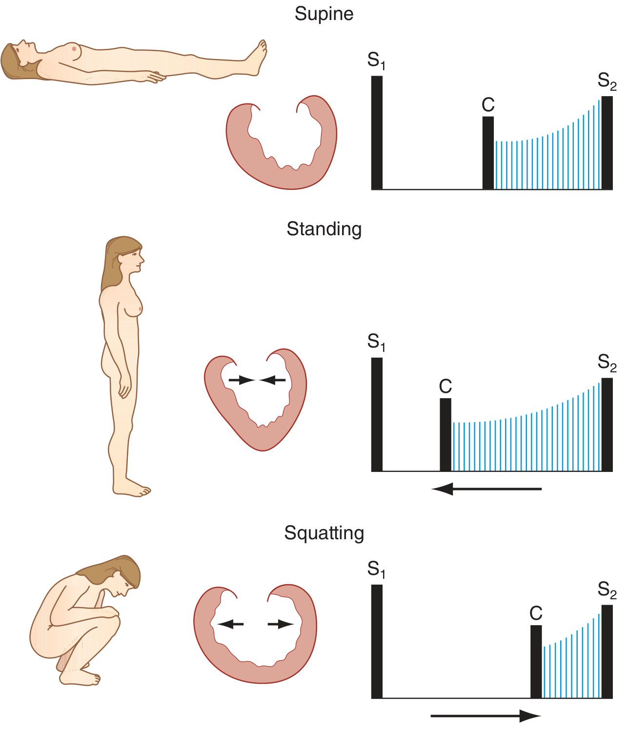
The Diagram Showing This (from Harrison's):

MVP click and murmur changes with position - Harrison's Fig 44-3
Look at the middle heart shape (standing) - the arrows are pointing inward - smaller heart, leaflets prolapse earlier, click moves toward S1. In squatting, arrows point outward - bigger heart, leaflets held tighter, click moves toward S2.

Applying This to the Two Key Murmurs

HOCM (Hypertrophic Obstructive Cardiomyopathy)

The obstruction in HOCM is dynamic - the hypertrophied septum + anterior mitral leaflet are sucked toward each other during systole (Venturi effect). This suction is worst when the LV cavity is small.
ManeuverPreloadLV sizeObstructionMurmur
Valsalva (strain)Small↑ MORELouder
StandingSmall↑ MORELouder
SquattingBig↓ LESSSofter
Leg raise (passive)Big↓ LESSSofter
Think of it this way: when the LV is nearly empty, the walls are close together, and the suction mechanism works better. When the LV is full and stretched, the walls are far apart and there's no suction.

MVP (Mitral Valve Prolapse)

The mitral leaflets are too long (or chordae too lax). During systole, as the LV empties and gets smaller, at some point the leaflets buckle backward into the left atrium - that's the click - and then the regurgitation starts (the murmur). The click timing tells you when the LV got small enough for prolapse to occur.
ManeuverLV sizeWhen does prolapse happenClick timingMurmur duration
StandingSmallHappens earlier in systoleClick → moves toward S1Longer murmur
SquattingBigHappens later (LV stays full longer)Click → moves toward S2Shorter murmur
Valsalva (strain)SmallHappens earlierClick → toward S1Longer murmur
Mnemonic to lock this in: HOCM and MVP are the "same team" - both get louder/longer with Valsalva/standing, and softer/shorter with squatting. They are the two exceptions to the general rule.

All Other Murmurs (AS, MR-organic, VSD, TR, PS, etc.)

These are fixed or flow-dependent. More blood flowing = louder murmur.
ManeuverVenous return / FlowMurmur
Squatting↑ preload + ↑ afterloadLouder
Valsalva (strain)↓ preloadSofter
Standing↓ preloadSofter
Handgrip (isometric)↑ afterloadLouder (especially MR, AR, VSD)
AS specifically: Softer with Valsalva (less flow), louder with squatting. This is the opposite of HOCM - that's how you distinguish them at the bedside.

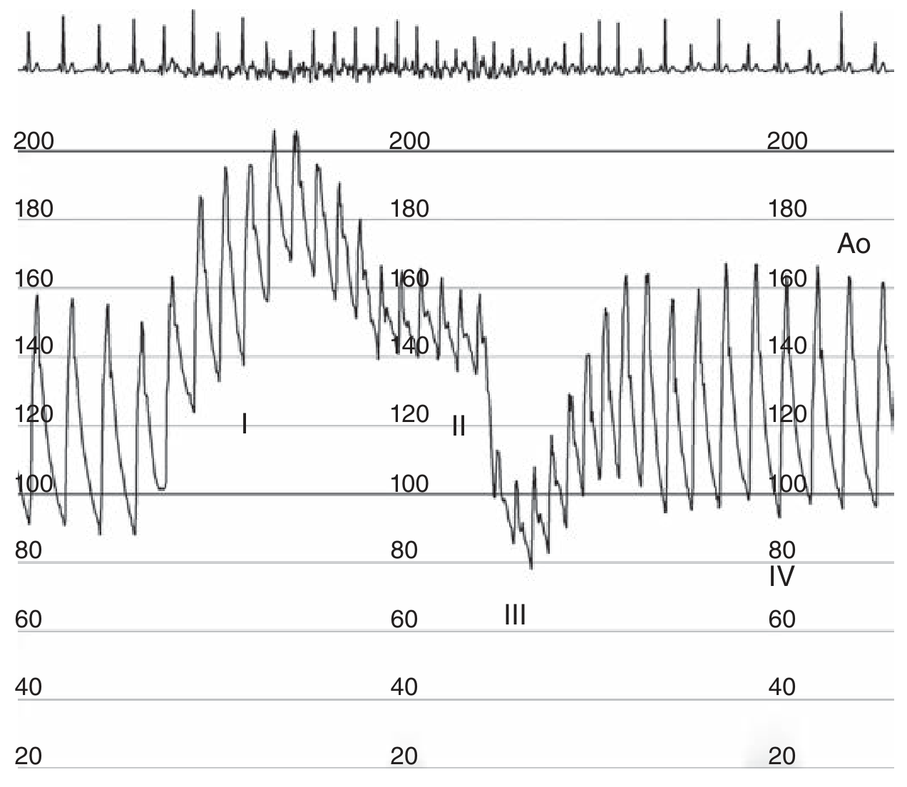
The Valsalva Aortic Pressure Trace (from Fuster & Hurst):

Valsalva maneuver 4 phases aortic pressure tracing
See the 4 phases labeled I, II, III, IV. Phase II is the sustained low-pressure phase where venous return is impaired. Phase IV is the overshoot. All murmurs increase in Phase IV (restored/increased flow) - except HOCM (LV is now full again so gradient decreases).

Summary Table for Quick Reference

ConditionValsalva (strain)StandingSquatting
HOCM↑ louder↑ louder↓ softer
MVP↑ longer + earlier click↑ longer + earlier click↓ shorter + later click
AS↓ softer↓ softer↑ louder
MR (organic)↓ softer↓ softer↑ louder
VSD↓ softer↓ softer↑ louder
AR↓ softer↓ softer↑ louder

One-Line Core Concept to Never Forget

Valsalva / Standing = Less blood in LV = HOCM & MVP louder/earlier Squatting = More blood in LV = HOCM & MVP softer/later; everyone else louder
This one concept lets you derive every answer conceptually rather than memorizing a table. Good luck with your First Aid CVS revision!

I told you very, very short note. I can't read. I'll get saturated if I have such long essays. Like exam-based NBME-ruled question-solving method, easy-to-remember and conceptual-based explanation, ultra-ultra-high-yield compact note.

Murmur Dynamics - Ultra Compact

One Core Concept

LV size = everything
Small LV → HOCM/MVP worse Big LV → HOCM/MVP better, everyone else worse

What Each Maneuver Does to LV Size

ManeuverWhat happensLV size
Valsalva (strain)Bearing down → ↓ venous returnSmall
StandingBlood pools in legs → ↓ venous returnSmall
SquattingLeg veins compressed + ↑ resistance → ↑ venous returnBig

The 2 Exceptions (HOCM + MVP)

Small LV → worse (louder/earlier) Big LV → better (softer/later)
Everything else is the opposite.

Master Table

Valsalva / StandingSquatting
HOCM↑ louder↓ softer
MVP↑ longer, click → S1↓ shorter, click → S2
AS / MR / VSD / AR↓ softer↑ louder

NBME Answer Trigger

  • "Louder with standing/Valsalva" → HOCM or MVP
  • "Softer with squatting" → HOCM or MVP
  • Distinguish HOCM vs AS? → Valsalva makes HOCM louder, AS softer

endocardial cushions mesns ELI5

Endocardial Cushions - ELI5

Imagine the early heart is just one big open room - no walls, no doors separating left from right, top from bottom.
Endocardial cushions = the construction crew that builds the dividing walls.
They are swellings of tissue that grow from the top and bottom of the middle of the heart and fuse in the center - like two people walking toward each other from opposite sides of a room and shaking hands in the middle.

What They Build

StructureWhat it becomes
Top + bottom cushions fuseAV septum (divides mitral from tricuspid)
Same fusionLower part of atrial septum (primum)
Same fusionUpper part of ventricular septum (membranous)
Cushion tissue itselfMitral + tricuspid valve leaflets

If They Fail to Fuse (Down Syndrome classically)

Endocardial cushion defect = AV septal defect (AVSD)
  • Hole between atria (primum ASD) + hole between ventricles (VSD) + abnormal AV valves
  • All because the "handshake never happened"

One-Line Memory

Endocardial cushions = the middle wall of the heart. No fusion = everything in the middle is missing.

Technically, if you're seeing from the top to the bottom, it's basically like splitting atria into two and ventricle into two. But if you're talking about atrioventricular defects, it means a horizontal line, right? Side to side, like two people from the side are what walking towards the middle and fusing there. Hence, the atrioventricular septum is formed where the mitral valve and also the tricuspid valve are formed. If it's coming from the middle, there are no septums or walls where we have mitral and tricuspid. When it's horizontal line, we have atrial and mitral. Sorry, we have mitral and tricuspid. If it's a vertical line from the middle, then we have no defects in the middle, right?

Yes, you've got it exactly right. Let me confirm your mental model cleanly.

Your Model is Correct

        LA  |  RA
        ----+----   ← endocardial cushions fuse HERE (horizontal)
        LV  |  RV
The cushions grow from left wall and right wall, meet in the horizontal middle - this is the AV junction level.

What That Horizontal Fusion Creates

  • The floor of the atria / roof of the ventricles = AV septum
  • Mitral valve (left side of fusion)
  • Tricuspid valve (right side of fusion)

The Other Walls are Different Structures

WallBuilt by
Vertical atrial wall (LA vs RA)Septum primum + septum secundum (separate story)
Vertical ventricular wall (LV vs RV)Muscular + membranous ventricular septum
Cushions only own the horizontal middle zone - the AV plane.

So Your Logic Holds

Cushion defect = hole at the AV plane = primum ASD + inlet VSD + bad valves Vertical walls = built by completely different embryological structures, cushions not involved
Your spatial reasoning is exactly how to think about it. Clean model.

So, your explanation of coming from top and coming from bottom and meeting in the middle is not endocardial cushion, right? Endocardial cushion means coming from the left wall, right wall and meeting in the centre. That is what forms the atrioventricular septum and this is where the left side forms the mitral valve and the right side forms the tricuspid valve. These are the cushion defects. Am I right? Am I clear?

You're right to call me out, but let me be precise - your model is mostly right but the anatomy is slightly different.

Actual Direction of Cushion Growth

The cushions grow from the superior wall and inferior wall of the AV canal - so top and bottom of the AV canal - and meet in the middle.
BUT - and this is key - "top and bottom" here means dorsal and ventral walls of the AV canal, NOT atrial wall vs ventricular wall.
Think of the AV canal as a horizontal tube connecting atria to ventricles. The cushions are bulges on the inner top and bottom surfaces of that tube, growing inward until they fuse.

What Your Model Gets Right

  • Fusion happens at the center of the AV plane
  • Left side → mitral valve ✓
  • Right side → tricuspid valve ✓
  • Defect = hole at that plane + bad valves ✓

One Correction

My original "top and bottom" was actually correct anatomically - it's the superior and inferior endocardial cushions (those are literally their names). Your "left and right wall" intuition gives the same functional result though - the key point is they meet in the center of the AV junction.
Your conceptual understanding of what the defect looks like and what it produces is 100% correct. The direction detail is minor.

So, is Eisenmenger syndrome reversible or irreversible? Irreversible. Because after the chronic left-to-right shunt, there will be fibrosis, hypertrophy of smooth muscles and hyperplasia of the intima and the pulmonary vasculature, and eventually it causes increasing pulmonary vascular resistance. So, and after some point, it will undergo a reversal of the shunt from right to left. This is how where cyanotic symptoms start appearing. So my next question, what NMBE always will attest it if Eisenmenger is reversible or irreversible? That is what the question in the asks, right?

Your pathophysiology is perfect. Now the NBME angle:

What NBME Actually Tests

Not "is it reversible" directly.
They test it as: "Can you now fix the underlying defect (VSD/ASD/PDA)?"

The One High-Yield Rule

Eisenmenger = surgical repair is CONTRAINDICATED
Because the pulmonary vascular disease is now fixed and irreversible. If you close the shunt now, the RV has nowhere to decompress → acute RV failure → death.
The shunt at this point is actually keeping the patient alive by acting as a pressure release valve.

NBME Trigger Phrase

"Late presentation of VSD/ASD/PDA + cyanosis + clubbing + polycythemia" → Question asks: next step? → Answer: NOT surgery = lung transplant (or heart-lung transplant) is the only fix

Why Polycythemia?

Right-to-left shunt → deoxygenated blood enters systemic circulation → chronic hypoxia → EPO ↑ → RBC ↑

Three-Word Summary

StageShuntSurgery?
Early L→RL→R✅ Fix it
EisenmengerR→L❌ Too late
That's the whole testable concept.

Now make a small note of Eisenegger syndrome, including my knowledge, concept, functions, and only what we're going to do, and late what we're going to do. Small high yield, MB and MB level, compact, high yield, short note. Very, very short.

Eisenmenger Syndrome

Trigger

Uncorrected L→R shunt (VSD > ASD > PDA)

Mechanism

Chronic ↑ pulmonary flow → smooth muscle hypertrophy + intimal fibrosis → irreversible ↑ PVR → shunt flips R→L

Symptoms (when R→L starts)

Cyanosis + clubbing + polycythemia (hypoxia → EPO↑)

NBME Rule

TimingAction
Early (L→R, no cyanosis)Fix the defect surgically ✅
Late (Eisenmenger, cyanosis)Surgery CONTRAINDICATED
Why no surgery? Shunt is now the RV's only escape - close it → RV failure → death.
Only option late: Lung transplant / heart-lung transplant

One-Line Trigger for MCQ

Late VSD/ASD/PDA + cyanosis + clubbing = don't touch the defect
This is a shared conversation. Sign in to Orris to start your own chat.