
COPD
"chronic obstructive pulmonary disease" AND management
COPD emphysema pathology lung diagram
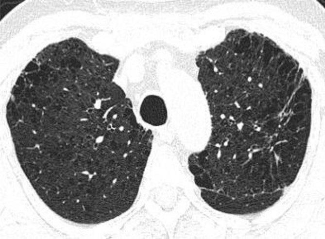
This diagnostic image is an axial non-contrast-enhanced CT scan of the chest at the level of the upper lobes, demonstrating significant chronic obstructive pulmonary disease (COPD) pathology. The lung parenchyma shows extensive centrilobular emphysema, characterized by multiple small, focal areas of low attenuation representing alveolar destruction within the secondary pulmonary lobules. Complementary paraseptal emphysematous changes are visible as subpleural lucencies along the peripheral lung margins. In the apicoposterior segment of the left upper lobe, there is a localized area of increased attenuation consistent with a fibrotic lesion or reticular opacity. The distribution is primarily bilateral and upper-lobe predominant, which is characteristic of smoking-related lung disease. Key educational concepts include the radiographic differentiation between centrilobular and paraseptal emphysema and the identification of concurrent interstitial lung changes in a patient with multi-faceted parenchymal disease.
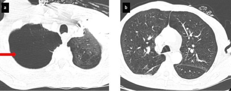
Two axial CT pulmonary angiography (CTPA) slices displayed in a lung window, illustrating chronic obstructive pulmonary disease (COPD) pathology. Image (a) shows a large, well-circumscribed, air-filled bulla in the right apical region, indicated by a red arrow. This bulla causes focal compression of the adjacent lung tissue and significant rarefaction of the vascular markings. Image (b) shows a lower thoracic section demonstrating diffuse, bilateral emphysematous changes. The lung parenchyma exhibits increased lucency, multiple small air-filled lucent spaces, and a reduction in the density and caliber of peripheral pulmonary vessels, creating a 'spongy' texture. These findings are characteristic of extensive centrilobular and paraseptal emphysema. The images provide clinical evidence of severe parenchymal destruction, contributing to decreased gas exchange and potential secondary pulmonary hypertension.
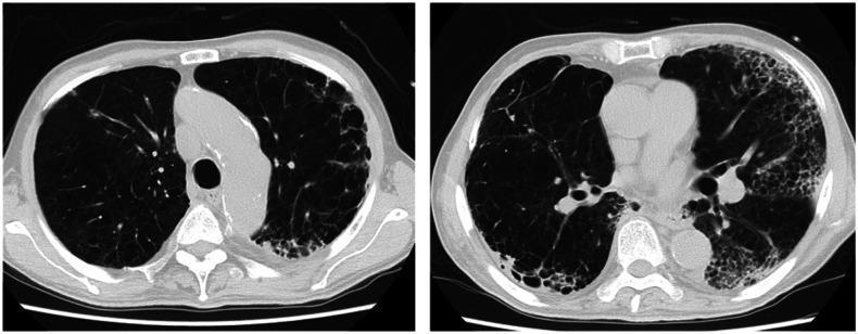
Two axial non-contrast computed tomography (CT) sections of the thorax demonstrate advanced pulmonary pathology. The images show significant bilateral emphysematous changes characterized by diffuse areas of low attenuation, representing lung parenchyma destruction and air trapping. There is a prominent peripheral and subpleural distribution of these lucent areas, particularly in the posterior lung zones. Notably, there are increased linear and reticular opacities along the margins of the emphysematous regions, suggesting a honeycomb-like pattern or fibrotic architectural distortion. These findings are consistent with severe chronic obstructive pulmonary disease (COPD) with underlying paraseptal or bullous emphysema, and potentially secondary interstitial changes. The visual highlights the importance of recognizing the distribution patterns of emphysema and associated peripheral opacities in geriatric patients with progressive dyspnea and obstructive lung disease.
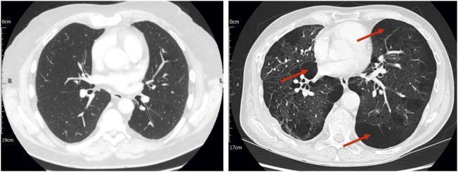
This Comparison Chart features two axial non-contrast Computed Tomography (CT) scans of the thorax at the level of the main pulmonary artery, illustrating different phenotypes of Stage 2 Chronic Obstructive Pulmonary Disease (COPD). The left panel displays a patient with relatively preserved lung parenchyma, characterized by normal attenuation and intact vascular markings without visible emphysematous destruction. The right panel demonstrates advanced structural pathology in a second patient, showcasing significant centrilobular and paraseptal emphysema. Red arrows highlight multiple pulmonary bullae, appearing as focal areas of low attenuation (increased lucency) with thin, well-defined walls, representing localized tissue destruction. A diffuse reduction in vascular density is also evident in the emphysematous lung compared to the normal scan. The educational focus is the radiological manifestation of COPD phenotypes, specifically the correlation between parenchymal destruction, anatomical deadspace volume, and lung inhomogeneity as visualized through diagnostic imaging.
Chronic Obstructive Pulmonary Disease (COPD)
Definition
Epidemiology & Risk Factors
- 3rd most common cause of death in the United States; costs exceed $40 billion/year in direct and indirect healthcare expenses - Katzung's Basic & Clinical Pharmacology, 16e
- Cigarette smoking is by far the dominant risk factor; 15-30% of habitual smokers were traditionally thought to develop COPD, but radiographic studies now show progressive bronchial wall thickening and lung tissue loss even in smokers with preserved spirometry
- Other causes: prolonged exposure to dusts, indoor/outdoor air pollution, recurrent childhood respiratory infections
- Alpha-1 antitrypsin (A1AT) deficiency: genetic cause of panacinar emphysema (see below)
Pathophysiology
Emphysema
- Loss of elastic recoil -> airflow obstruction
- Enlarged airspaces -> increased lung compliance and static hyperinflation
- Reduced surface area for gas exchange -> V/Q mismatch -> hypoxemia
| Type | Location | Cause |
|---|---|---|
| Centriacinar (centrilobular) | Upper lobe predominant, around respiratory bronchioles | Cigarette smoking (most common) |
| Panacinar | Entire acinus, lower lobe predominant | A1AT deficiency |
| Paraseptal | Adjacent to pleura and septa | Associated with spontaneous pneumothorax |
Chronic Bronchitis
Dynamic Hyperinflation & Exercise Limitation
Clinical Features
| Feature | Emphysema ("Pink Puffer") | Chronic Bronchitis ("Blue Bloater") |
|---|---|---|
| Build | Thin, barrel-chested | Overweight, cyanotic |
| Sputum | Scant | Copious, purulent |
| PaO2 | Near normal at rest | Low (hypoxemia) |
| PaCO2 | Low (hyperventilation) | High (hypercapnia) |
| Cor pulmonale | Late | Earlier |
| Polycythemia | Uncommon | Common |
- PaO2: 60 mmHg (↓), PaCO2: 30 mmHg (low - hyperventilating to compensate), pH 7.47 (mild respiratory alkalosis)
- A-a gradient widened due to V/Q mismatch
Diagnosis
- FEV1/FVC < 0.70 confirms airflow obstruction
- FVC normal or near-normal (unlike restrictive disease)
GOLD Spirometric Grading (post-bronchodilator FEV1 % predicted):
| GOLD Grade | FEV1 % predicted | Severity |
|---|---|---|
| 1 | ≥ 80% | Mild |
| 2 | 50-79% | Moderate |
| 3 | 30-49% | Severe |
| 4 | < 30% | Very Severe |
ABCD Assessment (beyond spirometry):
- Spirometry grade (GOLD 1-4)
- Symptoms - mMRC dyspnea scale or CAT score
- Exacerbation history (frequency and severity in prior year)
CT Imaging
Management
Non-Pharmacological (for all patients)
- Smoking cessation: Most important intervention at any disease stage; slows decline in FEV1
- Pulmonary rehabilitation
- Vaccinations (influenza, pneumococcal, COVID-19)
- Nutritional support
Pharmacological
| Indication | First-line agents |
|---|---|
| Acute symptom relief | SABA (albuterol/salbutamol) or SAMA (ipratropium) |
| Persistent exertional dyspnea (Group A/B) | LABA (salmeterol, formoterol, indacaterol) or LAMA (tiotropium, umeclidinium) |
| High symptom burden or exacerbation risk (Group B/E) | LABA + LAMA combination |
| Frequent exacerbations + eosinophils ≥300 cells/µL | Add ICS to LABA/LAMA (triple therapy) |
- LAMAs (e.g., tiotropium) are considered the best single agent for long-term maintenance in COPD - Swanson's Family Medicine Review
- ICS: Less central than in asthma; associated with increased pneumonia risk. Use only in severe obstruction, frequent exacerbations, or high eosinophil count. Guided by blood eosinophils (≥300 cells/µL = likely to benefit) - Katzung's
- Roflumilast (PDE-4 inhibitor): Improves pulmonary function and reduces exacerbation frequency; useful add-on for severe COPD with chronic bronchitis phenotype
- Theophylline: A large RCT showed low-dose theophylline did NOT reduce exacerbation frequency - its routine use is no longer recommended
Oxygen Therapy
- PaO2 ≤ 55 mmHg at rest, OR
- PaO2 ≤ 60 mmHg with evidence of tissue hypoxia (cor pulmonale or polycythemia)
- Increases survival in severe resting hypoxemia - Swanson's Family Medicine Review
Acute Exacerbations (AECOPD)
- Short-acting bronchodilators (SABA + SAMA)
- Systemic corticosteroids (oral prednisolone)
- Antibiotics if: (1) increased dyspnea + (2) increased sputum volume + (3) increased sputum purulence (Anthonisen criteria)
- Common antibiotics: amoxicillin, doxycycline, TMP-SMX, cefaclor; fluoroquinolones for severe exacerbations
- Non-invasive ventilation (BiPAP/NIV) for hypercapnic respiratory failure
Surgical Options
- Lung volume reduction surgery (LVRS): For severe emphysema with upper-lobe predominant disease
- Lung transplantation: Selected end-stage patients
Complications
- Cor pulmonale: Right heart failure from chronic pulmonary hypertension (hypoxic vasoconstriction)
- Polycythemia: Compensatory increase in RBC mass from chronic hypoxemia
- Spontaneous pneumothorax: From rupture of bullae (especially in paraseptal emphysema)
- Respiratory failure: Hypercapnic (type 2) failure in advanced disease
- Lung cancer: Markedly elevated risk in smokers with COPD
Key Differentiating Points from Asthma
| Feature | COPD | Asthma |
|---|---|---|
| Age of onset | >40 years (usually) | Often childhood/young adult |
| Cause | Smoking (predominantly) | Allergens, viral infections |
| Airflow obstruction | Irreversible (or minimally reversible) | Reversible |
| Inflammation | Neutrophilic | Eosinophilic |
| ICS response | Poor | Excellent |
| Progression | Relentless | Episodic, stable between attacks |