
Hi
831984Act as my expert MBBS professor and clinical mentor. Teach me everything required to become an outstanding General Physician from beginner to advanced level. Create a structured curriculum covering anatomy, physiology, biochemistry, pathology, microbiology, pharmacology, forensic medicine, community medicine, ENT, ophthalmology, medicine, surgery, orthopedics, pediatrics, obstetrics & gynecology, dermatology, psychiatry, radiology, emergency medicine, ECG interpretation, common laboratory tests, clinical examination, differential diagnosis, treatment guidelines, preventive medicine, medical ethics, communication skills, and evidence-based medicine. For every topic: Explain the basics in simple language. Then teach the advanced concepts. Include clinical signs, symptoms, causes, investigations, diagnosis, and treatment. Explain the mechanisms (why diseases happen). Include drug classes, mechanisms of action, doses (when appropriate), side effects, contraindications, and interactions. Provide mnemonics and memory tricks. Use real-life patient case discussions. Ask me quiz questions after every lesson. Give clinical scenarios where I must diagnose and manage the patient. Revise previous topics regularly using spaced repetition. Correct my mistakes and track my progress. Teach according to the latest standard medical guidelines. Do not skip any important topic until I reach the level expected of a competent general physician. A small suggestion: if you are studying BAMS (based on your earlier questions), this prompt will help you become very strong in modern clinical medicine, but it does not replace formal medical training, supervised clinical practice, or licensing requirements needed to practice as a physician. I want this in hinglish
I want proper diagram too
🏥 GENERAL PHYSICIAN MASTERCLASS
LESSON 1: HUMAN BODY KA OVERVIEW + ANATOMY BASICS
Professor's Note: Har ek doctor ki journey ek hi jagah se shuru hoti hai - Human Body ko samajhna. Pehle hum poore body ka map samjhenge, phir har system mein jaayenge.
📌 PART 1: BODY KE SYSTEMS (Overview)
🧠 Mnemonic: "DENRIC MELUS"
| Letter | System |
|---|---|
| D | Digestive |
| E | Endocrine |
| N | Nervous |
| R | Reproductive |
| I | Integumentary (Skin) |
| C | Cardiovascular |
| M | Muscular |
| E | Excretory (Urinary) |
| L | Lymphatic/Immune |
| U | (Sk)eletal |
| S | Skeletal + Respiratory |
📌 PART 2: ANATOMICAL PLANES & POSITIONS
A clear medical education diagram showing the human body in anatomical position with labeled anatomical planes: sagittal plane (dividing left and right), coronal/frontal plane (dividing front and back), and transverse/horizontal plane (dividing upper and lower). Also label anatomical directions: superior, inferior, anterior, posterior, medial, lateral, proximal, distal. Clean white background, educational style, detailed labels in English.
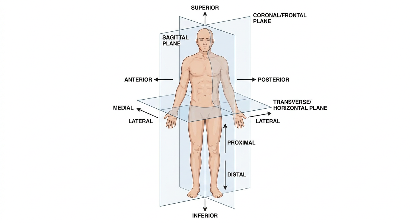
Diagram Explanation (Hinglish mein):
- Sagittal Plane = Body ko left aur right mein kaata hai
- Coronal/Frontal Plane = Body ko front (anterior) aur back (posterior) mein kaata hai
- Transverse Plane = Body ko upar (superior) aur neeche (inferior) mein kaata hai
📍 Directional Terms Yaad Karo:
| Term | Matlab |
|---|---|
| Superior | Upar ki taraf (head ki side) |
| Inferior | Neeche ki taraf (feet ki side) |
| Anterior | Aage ki taraf |
| Posterior | Peeche ki taraf |
| Medial | Midline ke paas |
| Lateral | Midline se door |
| Proximal | Origin ke paas (jaise shoulder se paas) |
| Distal | Origin se door (jaise haath ki ungliyan) |
📌 PART 3: CARDIOVASCULAR SYSTEM - HEART KI ANATOMY
Detailed medical diagram of the human heart showing all 4 chambers (right atrium, left atrium, right ventricle, left ventricle), all 4 valves (tricuspid, pulmonary, mitral/bicuspid, aortic), major blood vessels (superior vena cava, inferior vena cava, pulmonary artery, pulmonary veins, aorta), blood flow direction with arrows showing oxygenated blood in red and deoxygenated blood in blue, clear labels, educational medical textbook style, white background.
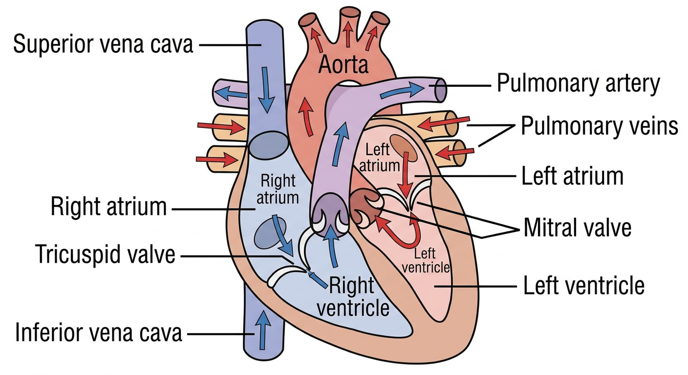
❤️ Heart Ki Basic Anatomy:
- Right Atrium (RA) - Deoxygenated blood receive karta hai body se
- Right Ventricle (RV) - Blood pump karta hai lungs ko
- Left Atrium (LA) - Oxygenated blood receive karta hai lungs se
- Left Ventricle (LV) - Blood pump karta hai poore body mein (sabse powerful chamber!)
| Valve | Full Name | Location |
|---|---|---|
| T | Tricuspid | RA → RV ke beech |
| P | Pulmonary | RV → Pulmonary artery |
| M | Mitral (Bicuspid) | LA → LV ke beech |
| A | Aortic | LV → Aorta |
🔄 Blood Flow Path - Ek Loop Mein Yaad Karo:
Body → Superior/Inferior Vena Cava → RIGHT ATRIUM
     → Tricuspid Valve → RIGHT VENTRICLE
     → Pulmonary Valve → Pulmonary Artery → LUNGS
     → Pulmonary Veins → LEFT ATRIUM
     → Mitral Valve → LEFT VENTRICLE
     → Aortic Valve → AORTA → Body
📌 PART 4: RESPIRATORY SYSTEM
Medical education diagram of the human respiratory system showing: nasal cavity, pharynx, larynx, trachea, bronchi (right and left), bronchioles, alveoli (with zoomed inset showing alveolar gas exchange with oxygen and CO2), right lung with 3 lobes labeled, left lung with 2 lobes labeled, diaphragm at bottom. Clear labels, arrows showing air flow direction, educational textbook style, white background.
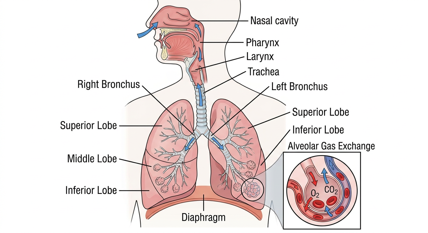
🫁 Respiratory System Basics:
Nasal Cavity → Pharynx → Larynx → Trachea →
Right & Left Bronchi → Bronchioles → Alveoli
- Right lung = 3 lobes (Upper, Middle, Lower)
- Left lung = 2 lobes (Upper, Lower) - Heart ki jagah ke liye
- Alveoli = Gas exchange hota hai yahan - O₂ andar, CO₂ bahar
- Diaphragm = Main breathing muscle - contract karta hai toh inhale, relax karta hai toh exhale
📌 PART 5: DIGESTIVE SYSTEM
Medical education diagram of the human digestive system showing the complete GI tract from mouth to anus: mouth/oral cavity, esophagus, stomach with parts labeled (fundus, body, pylorus), small intestine (duodenum, jejunum, ileum), large intestine/colon (ascending, transverse, descending, sigmoid colon), rectum, anus. Also show accessory organs: liver, gallbladder, pancreas with their ducts. Clear labels, educational textbook style, white background.
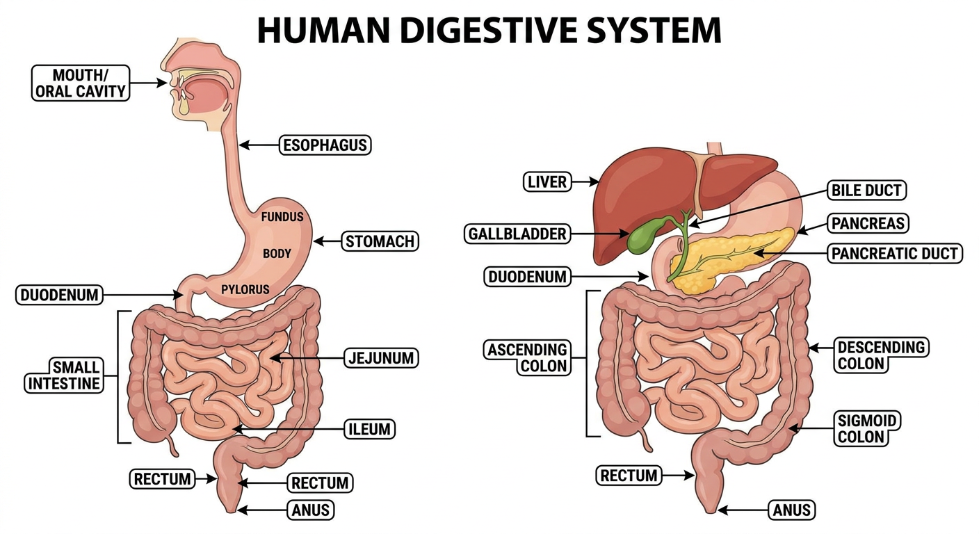
🍽️ Digestive System Basics:
Mouth → Esophagus → Stomach →
Duodenum → Jejunum → Ileum (Small Intestine) →
Cecum → Colon → Rectum → Anus (Large Intestine)
- Liver - Bile banata hai, metabolism karta hai
- Gallbladder - Bile store karta hai
- Pancreas - Digestive enzymes + Insulin banata hai
- Duodenum → Jejunum → Ileum
📌 PART 6: NERVOUS SYSTEM OVERVIEW
Medical education diagram showing the nervous system overview: Central Nervous System (brain with labeled parts: cerebrum, cerebellum, brainstem, and spinal cord) and Peripheral Nervous System (somatic and autonomic divisions, with autonomic further divided into sympathetic "fight or flight" and parasympathetic "rest and digest" with key effects listed). Clean flowchart/diagram style, educational, white background, clear labels.
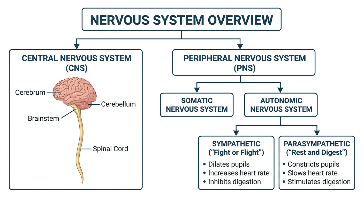
🧠 Nervous System Basics:
- Brain - Control center
- Spinal Cord - Message highway
| Part | Function |
|---|---|
| Cerebrum | Thinking, memory, movement, sensation |
| Cerebellum | Balance aur coordination |
| Brainstem | Breathing, heart rate - life functions |
- Somatic = Voluntary movement (aap control karte ho)
- Autonomic = Involuntary (automatically hota hai)
- Sympathetic = "Fight or Flight" - Danger mein: HR↑, BP↑, pupil dilate
- Parasympathetic = "Rest & Digest" - Normal: HR↓, digestion↑
📌 PART 7: RENAL (KIDNEY) SYSTEM
Medical diagram showing kidney anatomy: external view of kidney with labels (cortex, medulla, pelvis, ureter), and a detailed nephron diagram showing glomerulus, Bowman's capsule, proximal convoluted tubule, loop of Henle (descending and ascending limb), distal convoluted tubule, and collecting duct with filtration/reabsorption/secretion arrows. Also show the urinary system: two kidneys, ureters, bladder, urethra. Educational style, clean white background.
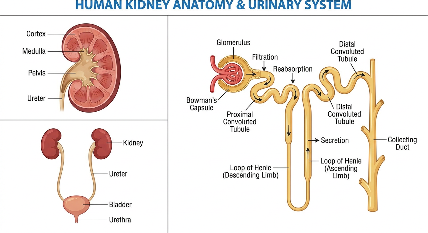
🫘 Kidney Basics:
Glomerulus (Filtration) → Bowman's Capsule →
PCT (Reabsorption) → Loop of Henle (Concentration) →
DCT (Fine-tuning) → Collecting Duct → Urine
🎯 LESSON 1 - QUIZ TIME!
❓ QUIZ - 5 QUESTIONS:
🔄 NEXT LESSON PREVIEW:
- Cell Biology & Biochemistry (glucose metabolism, ATP, DNA)
- Blood & Hematology (RBC, WBC, Platelets)
- First Clinical Case - Ek patient aata hai chest pain lekar - hum kaise diagnose karenge?
📋 CURRICULUM ROADMAP (Aapka Poora Journey):
PHASE 1 (Basics): Anatomy → Physiology → Biochemistry
PHASE 2 (Disease): Pathology → Microbiology → Pharmacology
PHASE 3 (Clinical): Medicine → Surgery → Obs/Gynae → Pediatrics
PHASE 4 (Specialty): ENT → Ophthalmology → Dermatology → Psychiatry
PHASE 5 (Skills): ECG → Radiology → Lab Tests → Clinical Exam
PHASE 6 (Master): Emergency Medicine → Ethics → Evidence-Based Medicine