
Explain causes of hyperkalemia and levels
Reading File
Hyperkalemia is defined as serum potassium above 5.0-5.5 mEq/L. Causes include reduced renal excretion (CKD, ACE inhibitors, aldosterone deficiency), transcellular shifts out of cells (acidosis, insulin deficiency, beta-blockers, cell lysis), and excessive potassium intake. Severity is graded as mild (5.1-5.9), moderate (6.0-6.4), and severe (≥6.5 mEq/L), with the latter carrying risk of fatal arrhythmias.
Searching sources...
Finding Sources
Searching the Web
hyperkalemia causes levels classification 2024
Reading File
Reading File
Searching Images
hyperkalemia ECG changes peaked T waves causes diagram
Finding Sources
Finding Sources
Reading File
Finding Sources
Reading File
Reading File
Finding Sources
Reading File
I now have comprehensive information from multiple authoritative textbooks. Let me compile the complete answer.
Hyperkalemia: Causes and Levels
Definition and Normal Range
Serum potassium is normally maintained between 3.5 and 5.0 mEq/L. Hyperkalemia is defined as a serum potassium > 5.0-5.5 mEq/L. The kidneys can normally excrete up to 500 mEq of K+ per day, so hyperkalemia in healthy individuals is rare. - Morgan and Mikhail's Clinical Anesthesiology, p. 2176
Severity Levels
| Grade | Serum K+ (mEq/L) | Clinical Significance |
|---|---|---|
| Mild | 5.1 - 5.9 | Often asymptomatic |
| Moderate | 6.0 - 6.4 | ECG changes may appear; muscle weakness |
| Severe | ≥ 6.5 | Life-threatening arrhythmias, paralysis |
| Critical | ≥ 7.0 | Risk of fatal cardiac arrest |
A K+ of 6 mEq/L should be considered potentially dangerous, even in patients who appear tolerant. The rate of rise matters as much as the absolute value - patients with chronic hyperkalemia tolerate higher levels better than those with acute rises. - Rosen's Emergency Medicine, p. 1367
Causes
Causes are best organized into three main mechanisms:
1. Pseudohyperkalemia (Artifactual)
This is a falsely elevated measurement with no true excess in the body:
- Hemolysis of the blood sample (most common) - mechanical trauma during venipuncture
- Excessive fist clenching during blood draw
- Leukocytosis (WBC > 70,000 × 10⁹/L) - K+ leaks from WBCs in vitro
- Thrombocytosis (platelets > 1,000,000 × 10⁹/L) - K+ released from platelets during clotting
- Erythrocytosis
- Hereditary pseudohyperkalemia - rare genetic increase in passive erythrocyte K+ permeability (e.g., SLC4A1 mutations)
- Cooling of blood after venipuncture (reduces cellular uptake)
Always exclude pseudohyperkalemia by repeating the sample before treatment if the patient is asymptomatic. - Harrison's Principles of Internal Medicine 22E (2025), Washington Manual of Medical Therapeutics
2. Transcellular Shift (K+ moves out of cells into ECF)
These causes increase extracellular K+ without total body excess:
| Cause | Mechanism |
|---|---|
| Metabolic acidosis (non-anion gap) | H+ enters cells, K+ exits to maintain electroneutrality. Note: lactic acidosis and ketoacidosis do NOT typically cause this shift |
| Insulin deficiency (e.g., DKA) | Insulin normally drives K+ into cells via Na+/K+-ATPase |
| Hyperosmolality / hypertonic states | "Solvent drag" - water exits cells osmotically, carrying K+ |
| Succinylcholine | Depolarizing agent causes K+ efflux from muscle (~0.5 mEq/L rise; can be 4-8 mEq/L in burns, denervation, trauma) |
| Beta-2 blockers (non-selective) | Block catecholamine-mediated K+ uptake into cells |
| Digitalis toxicity / overdose | Inhibits Na+/K+-ATPase, impairing cellular K+ uptake |
| Rhabdomyolysis | Crush injury, excessive exercise, seizures - releases intracellular K+ |
| Tumor lysis syndrome | Rapid cell death releases massive intracellular K+ |
| Hemolysis (in vivo) | Burns, hemolytic transfusion reactions, intravascular hemolysis |
| Cationic amino acids | Arginine, lysine, epsilon-aminocaproic acid - cause K+-cation exchange |
| Fluoride poisoning | Inhibits Na+/K+-ATPase |
| Hyperkalemic periodic paralysis | Hereditary channelopathy with pathological K+ efflux |
| Acute exercise | Transient shift (usually self-limited) |
- Harrison's Principles 22E (2025), Washington Manual, Morgan & Mikhail
3. Decreased Renal Excretion (Most Common True Cause)
The kidney is ultimately responsible for 90% of K+ excretion. Impairment here is the dominant cause of sustained hyperkalemia:
A. Reduced GFR / Renal Failure
- Chronic kidney disease (CKD) - hyperkalemia typically does not occur until GFR < 30 mL/min
- Acute kidney injury (AKI) with oliguria/anuria - oligo/anuria is a major cause
- Volume depletion (dehydration, hemorrhage, CHF, cirrhosis) - reduced distal tubular flow
B. Aldosterone Deficiency (Hypoaldosteronism)
Aldosterone drives K+ secretion in the collecting duct. Its deficiency is a major cause:
- Addison disease (primary adrenal insufficiency) - autoimmune, infectious (TB, HIV, CMV, fungal), infiltrative (amyloidosis, malignancy), hereditary, hemorrhagic
- Hyporeninemic hypoaldosteronism (Type 4 RTA) - common in diabetic nephropathy and elderly patients; low renin → low aldosterone despite intact adrenal glands
- Congenital enzyme defects: 21-hydroxylase deficiency, aldosterone synthase deficiency
C. Aldosterone Resistance (Pseudohypoaldosteronism)
- Type 1 PHA - hereditary end-organ resistance to aldosterone (salt-wasting, low BP)
- Type 2 PHA (Gordon syndrome) - familial hyperkalemic hypertension (salt-retaining, high BP)
D. Drugs that Impair Renal K+ Excretion (Major Cause)
| Drug Class | Mechanism |
|---|---|
| ACE inhibitors | Reduce angiotensin II → reduce aldosterone |
| Angiotensin receptor blockers (ARBs) | Same net effect as ACEi |
| Potassium-sparing diuretics (spironolactone, eplerenone, amiloride, triamterene) | Block aldosterone action or ENaC channel |
| NSAIDs / COX-2 inhibitors | Suppress renin release → reduce aldosterone; also reduce GFR |
| Heparin / LMWH | Inhibits aldosterone synthesis; antagonizes angiotensin II receptors |
| Trimethoprim | Blocks ENaC (like amiloride) in collecting duct |
| Pentamidine | Similar to trimethoprim |
| Cyclosporine / Tacrolimus (calcineurin inhibitors) | Reduce tubular K+ secretion |
| Ketoconazole | Decreases aldosterone production |
| Beta-blockers | Reduce renin secretion; also block cellular uptake |
- Washington Manual of Medical Therapeutics, Morgan & Mikhail, Brenner & Rector's The Kidney
4. Increased Potassium Intake
Rarely the sole cause unless there is concurrent renal impairment, but can tip the balance:
- Dietary excess: salt substitutes (KCl), dried fruits, nuts, bananas, potatoes, spinach, tomatoes
- Potassium supplements
- Massive blood transfusions (stored blood has elevated K+)
- Penicillin G (contains K+)
ECG Changes with Levels
ECG changes correlate roughly with rising K+ levels and are a clinical emergency when present:
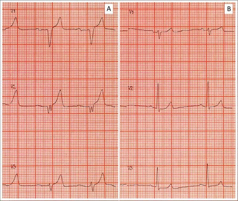
| K+ Level (mEq/L) | ECG Finding |
|---|---|
| 5.5 - 6.5 | Tall, symmetrically peaked T waves (narrow base); shortened QT |
| 6.5 - 7.5 | P wave flattening/loss; prolonged PR interval |
| 7.0 - 8.0 | QRS widening |
| > 8.0 | Sine-wave pattern, bundle branch block |
| > 9-10 | Ventricular fibrillation, asystole |
A normal ECG does NOT rule out hyperkalemia - ECG changes can be absent even at severely elevated levels. - Harrison's 22E, Rosen's Emergency Medicine, Barash Clinical Anesthesia
Key Clinical Points
- Critically ill patients often develop hyperkalemia from multiple simultaneous factors (e.g., AKI + ACE inhibitor + volume depletion + metabolic acidosis)
- In CKD patients, even drugs with minimal K+ effects in healthy people (beta-blockers, ACE inhibitors) can cause dangerous hyperkalemia
- Succinylcholine is contraindicated in burns, severe muscle trauma, and denervation injuries due to exaggerated K+ release
- The rate of rise is often more important than the absolute value - acute hyperkalemia at 6.5 mEq/L may be more dangerous than chronic hyperkalemia at 6.8 mEq/L
Sources: Harrison's Principles of Internal Medicine 22E (2025) | Washington Manual of Medical Therapeutics | Morgan & Mikhail's Clinical Anesthesiology | Brenner & Rector's The Kidney | Rosen's Emergency Medicine | StatPearls - Hyperkalemia
Explain transcellular shift more clearly
Transcellular shift means potassium moves from inside cells (intracellular fluid, ICF) to outside cells (extracellular fluid, ECF) without any change in total body potassium. Since 98% of body K+ is intracellular, even a small shift causes a large rise in serum K+.
Searching sources...
Finding Sources
Searching the Web
transcellular potassium shift hyperkalemia mechanism Na K ATPase physiology
Finding Sources
Finding Sources
Finding Sources
Finding Sources
Reading File
Reading File
Reading File
Loading Image
I have everything needed for a clear, thorough explanation. Here it is:
Transcellular Shift of Potassium - Explained Clearly
The Foundation: Why Shifts Matter So Much
The body's potassium is distributed very unevenly:
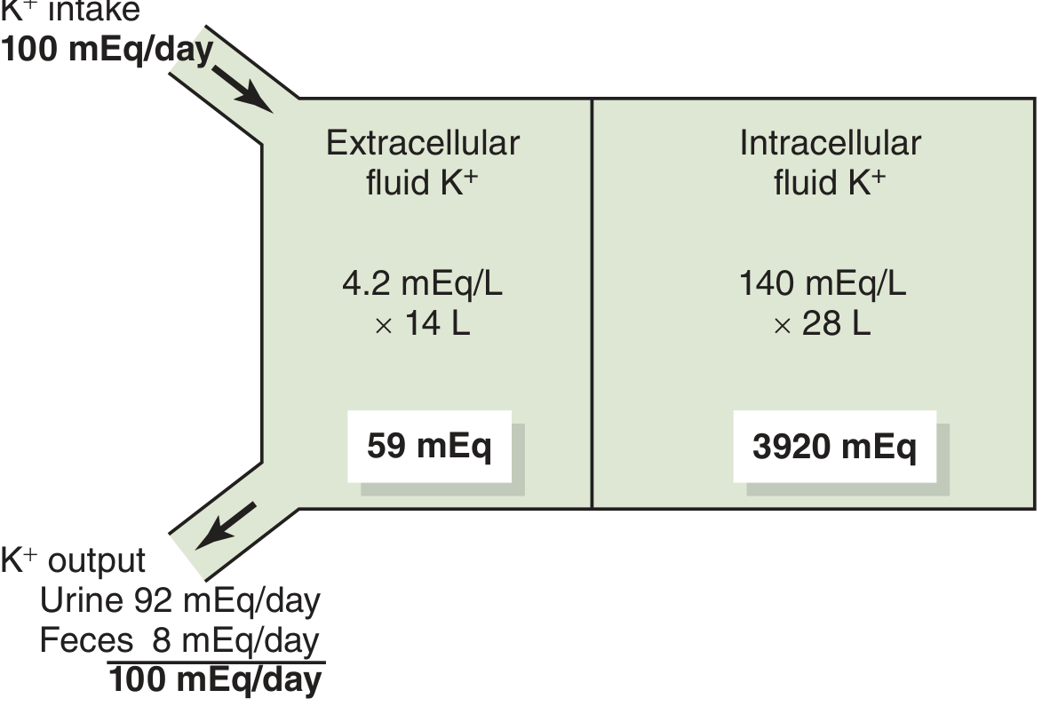
- Intracellular (ICF): ~140 mEq/L × 28 L = 3,920 mEq stored inside cells
- Extracellular (ECF): ~4.2 mEq/L × 14 L = only 59 mEq in blood/plasma
This 98:2 ratio means cells are a massive reservoir. Even a tiny fraction of intracellular K+ leaking out produces a large rise in serum K+. For example, moving just 42 mEq (about 1% of intracellular stores) into the ECF with no excretion would raise serum K+ by ~3 mEq/L - enough to be fatal. - Guyton & Hall Textbook of Medical Physiology
The Gatekeeper: Na+/K+-ATPase Pump
The pump responsible for keeping K+ inside cells is the Na+/K+-ATPase (sodium-potassium pump), located in every cell membrane. It:
- Pumps 3 Na+ OUT of the cell
- Pumps 2 K+ IN to the cell
- Runs continuously using ATP energy
- Maintains the steep K+ gradient (140 mEq/L inside vs. 4 mEq/L outside)
Anything that activates this pump drives K+ into cells (lowers serum K+). Anything that inhibits or bypasses this pump allows K+ to leak out (raises serum K+).
Mechanisms of Transcellular Shift (K+ OUT of cells → Hyperkalemia)
1. Acidosis
Rule: For every 0.1 unit drop in blood pH, serum K+ rises by ~0.6 mEq/L.
How it works: When the blood becomes acidic, excess H+ ions enter cells to be buffered. To maintain electrical neutrality across the membrane, K+ is pushed out of the cell in exchange - a direct H+/K+ swap. This also reduces Na+/K+-ATPase activity, further impairing K+ uptake. - Morgan & Mikhail's Clinical Anesthesiology
Important exception: Lactic acidosis and ketoacidosis (anion gap acidoses) do NOT cause significant K+ shift because the organic acid anions (lactate, beta-hydroxybutyrate) can cross cell membranes along with H+, so the K+ exchange is not needed for electroneutrality. - Harrison's Principles 22E (2025)
2. Insulin Deficiency
How it works: Insulin is the primary hormonal signal for K+ to enter cells. After a meal, insulin release drives dietary K+ rapidly into liver and skeletal muscle cells by stimulating Na+/K+-ATPase.
Without insulin (e.g., diabetic ketoacidosis), this pump activity drops, and K+ accumulates in the ECF. This is why DKA patients often present with hyperkalemia even though total body K+ is depleted - the serum K+ is falsely high due to shift, while the real deficit is intracellular. - Guyton & Hall
3. Beta-blockade (Non-selective Beta-blockers)
How it works: The sympathetic nervous system (epinephrine via beta-2 receptors) is a key driver of K+ into cells - it activates Na+/K+-ATPase in skeletal muscle and liver.
- Beta-2 stimulation → K+ enters cells → serum K+ falls
- Beta-blockade (propranolol, nadolol, carvedilol) → pump activity drops → K+ shifts OUT → hyperkalemia
This is why non-selective beta-blockers are more problematic than beta-1 selective ones (like metoprolol) for K+ balance. - Guyton & Hall, Morgan & Mikhail
4. Hyperosmolality (High Osmolarity States)
How it works: When the ECF becomes hypertonic (hypernatremia, uncontrolled hyperglycemia without insulin, mannitol), water is pulled out of cells osmotically. As water leaves, intracellular K+ concentration rises, creating a concentration gradient that drives K+ out of the cell - called "solvent drag."
Rise: approximately 0.6 mEq/L per 10 mOsm/L increase in osmolality. - Morgan & Mikhail
5. Cell Lysis (Release of Intracellular K+)
When cells die or rupture, all intracellular K+ (~140 mEq/L) floods into the ECF. The degree of hyperkalemia depends on the mass of tissue destroyed:
| Cause | Mechanism |
|---|---|
| Rhabdomyolysis | Crush injury, prolonged seizures, extreme exercise - muscle cells rupture |
| Tumor lysis syndrome | Chemotherapy kills large numbers of cancer cells simultaneously |
| Hemolysis (in vivo) | Transfusion reactions, hemolytic anemias - RBCs release K+ |
| Burns | Massive tissue destruction |
| Trauma / surgery | Large tissue mass injury |
6. Succinylcholine
How it works: Succinylcholine is a depolarizing neuromuscular blocker. It binds acetylcholine receptors and causes sustained depolarization of the muscle membrane, which opens K+ channels and causes a prolonged efflux of K+ from muscle cells.
- Normal, healthy patients: rise of ~0.5 mEq/L (safe)
- Burns, denervation, crush injury, prolonged immobility: rise of 4-8+ mEq/L (potentially fatal) because these conditions up-regulate acetylcholine receptors ("extra-junctional receptors") massively, multiplying the K+ release
- Succinylcholine is contraindicated in these situations
7. Digoxin Toxicity
How it works: Digoxin directly inhibits Na+/K+-ATPase. Without the pump actively pulling K+ in, K+ leaks out of cells down its concentration gradient. In overdose, this effect is pronounced enough to cause life-threatening hyperkalemia.
8. Aldosterone Deficiency
Beyond its renal effects, aldosterone also directly stimulates Na+/K+-ATPase in muscle and other tissues, driving K+ into cells. In Addison disease, loss of this effect contributes to the hyperkalemia alongside renal retention. - Guyton & Hall
9. Strenuous Exercise
During contraction, skeletal muscle cells cannot keep up with K+ entry fast enough, so K+ temporarily accumulates in the ECF (interstitial fluid around the exercising muscle). Usually mild and self-correcting at rest. More significant in patients with:
- Beta-blockade (blunted re-uptake)
- Insulin deficiency
- High-intensity or prolonged exercise
Summary Table
| Factor | Normal Role | When Absent/Disrupted |
|---|---|---|
| Insulin | Activates Na+/K+-ATPase → K+ into cells | Deficiency → K+ stays outside |
| Beta-2 adrenergic activity | Activates Na+/K+-ATPase | Blockade → K+ out of cells |
| Aldosterone | Stimulates Na+/K+-ATPase in muscle | Deficiency → K+ accumulates |
| Normal pH | Maintains pump activity | Acidosis → H+/K+ exchange outward |
| Normal osmolality | Water stays in cells | Hyperosmolality → solvent drag K+ out |
| Intact cell membranes | K+ stays inside | Lysis → massive K+ release |
| No Na+/K+-ATPase inhibitors | Pump runs freely | Digoxin → pump blocked, K+ leaks out |
The key concept: total body potassium has not changed in pure transcellular shift. Treatment therefore focuses on driving K+ back into cells (insulin + glucose, beta-2 agonists) or stabilizing cell membranes (calcium), rather than removing K+ from the body.
Sources: Guyton & Hall Textbook of Medical Physiology | Morgan & Mikhail's Clinical Anesthesiology | Harrison's Principles of Internal Medicine 22E (2025) | Washington Manual of Medical Therapeutics
Hyperkalemia diagnosis why ecg related to ecg
The ECG is used in hyperkalemia because high serum K+ directly alters the resting membrane potential of cardiac cells, disrupting normal depolarization and repolarization. This produces characteristic, progressive ECG changes - from peaked T waves at mild levels to a sine-wave pattern and cardiac arrest at severe levels.
Searching sources...
Finding Sources
Finding Sources
Searching the Web
hyperkalemia ECG changes mechanism physiology cardiac action potential 2024
Reading File
Reading File
Finding Sources
Reading File
Finding Sources
Finding Sources
Loading Image
Loading Image
Finding Sources
I now have everything needed for a clear, mechanistically complete explanation. Here it is:
Why Hyperkalemia Causes ECG Changes - The Complete Mechanism
Step 1: Understand the Normal Cardiac Action Potential
Before understanding what goes wrong, you need to know what's normal. A ventricular myocyte's action potential has 5 phases:
| Phase | Name | What Happens | Ions Involved |
|---|---|---|---|
| Phase 4 | Resting | Cell sits at -90 mV (very negative = polarized) | K+ leaking slowly out via background channels |
| Phase 0 | Rapid depolarization | Voltage-gated Na+ channels open → Na+ rushes IN → spike to +30 mV | Fast Na+ channels (INa) |
| Phase 1 | Early repolarization | Na+ channels close; brief K+ efflux | K+ out (Ito) |
| Phase 2 | Plateau | Ca2+ enters balancing K+ out - keeps cell depolarized | Ca2+ in, K+ out |
| Phase 3 | Repolarization | K+ channels fully open → K+ rushes OUT → back to -90 mV | K+ out (IKr, IKs) |
The ECG is simply the sum of all these action potentials happening across the heart, viewed from body surface electrodes:
- P wave = atrial depolarization (Phase 0 of atrial cells)
- QRS complex = ventricular depolarization (Phase 0 of ventricular cells)
- T wave = ventricular repolarization (Phase 3)
Step 2: What High K+ Does to the Resting Membrane Potential
The resting membrane potential (RMP) is determined by the ratio of intracellular to extracellular K+, described by the Nernst equation:
Normal: Inside cell = 140 mEq/L K+, Outside = 4 mEq/L → RMP = -90 mV
When serum K+ rises (e.g., to 7 mEq/L), the outside concentration increases, which reduces the gradient. The cell can no longer maintain -90 mV - it becomes less negative (less polarized), drifting toward -70 mV or -60 mV.
This single change - a less negative resting membrane potential - explains every ECG abnormality in hyperkalemia. - Rosen's Emergency Medicine, Medical Physiology
Step 3: How Each ECG Change is Produced
Early Hyperkalemia (K+ 5.5-6.5 mEq/L): Peaked T Waves + Short QT
Mechanism:
High extracellular K+ increases the conductance of IKr channels (the rapid delayed rectifier K+ channels responsible for Phase 3 repolarization). These channels open wider and faster, so K+ exits the cell more rapidly during repolarization.
- Faster Phase 3 → repolarization is quicker and more complete
- On the ECG: T wave becomes taller, narrower, and "peaked" (symmetric, narrow base)
- QT interval shortens (repolarization finishes faster)
This is the earliest and most reliable sign of hyperkalemia. Best seen in precordial leads V2-V5. - Hyperkalemia Revisited, PMC 1413606
Moderate Hyperkalemia (K+ 6.5-7.5 mEq/L): P Wave Flattening + PR Prolongation
Mechanism:
Atrial myocytes are more sensitive to elevated K+ than ventricular cells (they have a less negative resting potential to begin with). As K+ rises further, the atrial resting membrane potential drifts even closer to zero, which:
- Partially inactivates voltage-gated Na+ channels (these channels require the cell to be fully repolarized to -90 mV before they can reopen - if the cell never returns to -90 mV, fewer Na+ channels are available)
- Atrial conduction slows → P wave widens and flattens
- AV node conduction slows → PR interval prolongs
Eventually the atria become electrically silent - P waves disappear entirely. The ventricles may then be driven by junctional or ventricular escape beats.
Severe Hyperkalemia (K+ 7.0-8.0 mEq/L): QRS Widening
Mechanism:
The same Na+ channel inactivation now affects ventricular myocytes. With more and more Na+ channels stuck in an inactivated state due to persistent partial depolarization:
- Phase 0 (the rapid Na+ upstroke) becomes slower and weaker
- Conduction through ventricular muscle slows
- The QRS widens and takes on the appearance of a bundle branch block
- R wave amplitude decreases, S wave deepens
- ST segment may elevate or depress (can mimic STEMI or Brugada pattern)
This is a late and ominous sign indicating severely impaired ventricular conduction. - Rosen's Emergency Medicine, HCPLive Electrophysiologic Basis
Critical Hyperkalemia (K+ > 8-10 mEq/L): Sine Wave → Death
Mechanism:
The distinction between QRS and T wave disappears completely. The widened QRS merges with the T wave forming a continuous, smooth undulating "sine wave" pattern - the hallmark of imminent cardiac arrest.
The heart degenerates into:
- Ventricular fibrillation
- Pulseless electrical activity
- Asystole
K+ above 10 mEq/L is almost universally fatal without immediate treatment. - Rosen's Emergency Medicine
The Full ECG Progression - Summary
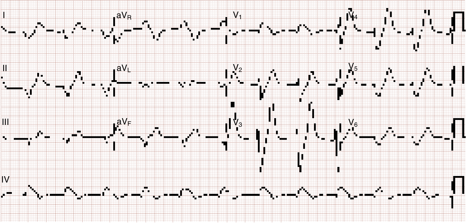
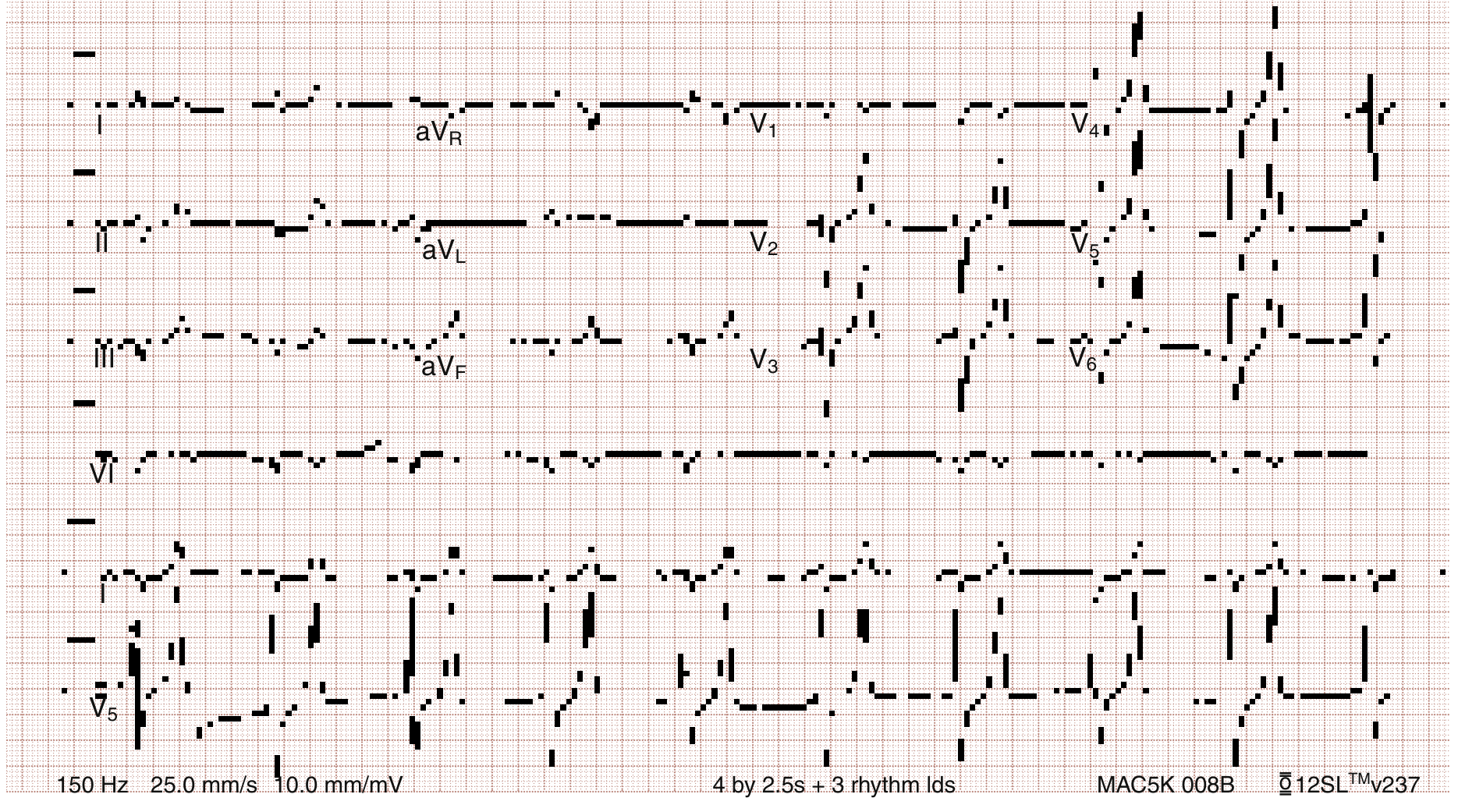
| K+ Level (mEq/L) | ECG Change | Mechanism |
|---|---|---|
| 5.5 - 6.5 | Peaked T waves, short QT | IKr upregulation → faster repolarization |
| 6.5 - 7.5 | P wave flattens/disappears, PR prolongs | Atrial Na+ channel inactivation → slow/absent atrial conduction |
| 7.0 - 8.0 | QRS widens, BBB pattern, ST changes | Ventricular Na+ channel inactivation → slow conduction |
| > 8.0 | Sine wave | QRS and T wave merge |
| > 9-10 | VF / asystole | Total electrical failure |
Why the ECG is Used Clinically (Not Just the Blood Test)
Three important reasons the ECG guides management:
- Speed - ECG result is immediate. A serum K+ lab takes 30-60 minutes. In a crashing patient, you cannot wait.
- Clinical correlation - The same K+ level (e.g., 6.5) may be dangerous in one patient (acute rise) and tolerated in another (chronic CKD). The ECG shows whether the heart is actually being affected.
- Treatment urgency - ECG changes determine the order and choice of interventions:
- ECG changes present → give calcium gluconate first (membrane stabilizer - works in 1-3 min)
- No ECG changes → can proceed with K+ removal at a less urgent pace
A normal ECG does not rule out hyperkalemia - changes may be absent even at severely elevated levels. An ECG showing changes with a plausible clinical history should trigger treatment even before the lab result returns. - Rosen's Emergency Medicine
One-Sentence Summary of the Mechanism
High extracellular K+ reduces the resting membrane potential gradient, partially inactivating voltage-gated Na+ channels - causing faster repolarization early on (peaked T), then slowed conduction in atria (no P) and ventricles (wide QRS), culminating in a sine wave and cardiac arrest.
Sources: Rosen's Emergency Medicine | Medical Physiology (Boron & Boulpaep) | Guyton & Hall | Morgan & Mikhail's Clinical Anesthesiology | Hyperkalemia Revisited, PMC | ESC eJournal - Hyperkalemia & the Heart
Hyperkalemia which condition
Your question "which condition" is a bit broad - let me clarify what you're asking before I dive in.
Asking for Preferences
What do you want to know about hyperkalemia and conditions?
Hyperkalemia is most dangerous when the heart, kidneys, or acid-base balance are already compromised, since these remove the body's safety margins for handling elevated potassium.
Searching sources...
Finding Sources
Searching the Web
hyperkalemia most dangerous conditions high risk patients cardiac renal
Finding Sources
Finding Sources
Finding Sources
Finding Sources
Finding Sources
Finding Sources
Finding Sources
Reading File
I now have enough to give a thorough answer. Here it is:
In Which Conditions Is Hyperkalemia Most Dangerous?
Hyperkalemia is universally dangerous, but certain conditions make it dramatically more lethal - either because they amplify the cardiac toxicity, remove the body's compensatory mechanisms, or cause K+ to rise faster than the heart can adapt.
1. End-Stage Renal Disease (ESRD) / Dialysis Patients
Why most dangerous:
- The kidney is the primary route for K+ excretion. In ESRD, it is completely gone.
- Between dialysis sessions (especially over weekends - 2-3 day gaps), potassium accumulates continuously with no safety valve.
- ESRD patients also carry concurrent LV hypertrophy, heart failure, and valvular disease - a heart already structurally compromised is far more vulnerable to arrhythmias from K+ changes.
- Both hyperkalemia before dialysis AND the rapid K+ drop during dialysis create dangerous swings. Post-dialysis hypokalemia can persist 6-8 hours and trigger sudden cardiac death.
- Uremia, acidosis, and calcium-phosphorus imbalance (all present in ESRD) independently worsen arrhythmia risk on top of hyperkalemia.
"Uremia, hyperkalemia, acidosis, and disorders of calcium-phosphorous balance all link to higher rates of atrial and ventricular arrhythmias." - Braunwald's Heart Disease
Clinical impact: A single high-potassium meal (dried fruits, bananas, potatoes) over a long interdialytic weekend has killed ESRD patients. This is the highest-risk group.
2. Acute Kidney Injury (AKI) with Oliguria or Anuria
Why dangerous:
- Unlike CKD, which develops slowly allowing some adaptation, AKI causes K+ to rise acutely over hours to days.
- The cardiac cells have had no time to adapt to the rising K+, so even a K+ of 6.5 mEq/L can cause fatal arrhythmias in AKI, whereas CKD patients might tolerate 6.8 chronically.
- Rhabdomyolysis and tumor lysis syndrome combine AKI (impaired excretion) with massive K+ release (cell lysis) - a double hit that drives K+ to dangerously high levels very rapidly.
- Rate of rise matters as much as absolute level.
3. Chronic Kidney Disease (CKD) Stage 3-5
Why dangerous:
- Most common risk factor for hyperkalemia overall.
- GFR < 30 mL/min marks the threshold where the kidneys can no longer excrete enough K+.
- CKD patients are typically on ACE inhibitors/ARBs (which raise K+), and have concurrent diabetes, heart failure, and metabolic acidosis - all stacking the risk.
- A study found patients with hyperkalemia in CKD had a 4.4-fold higher risk of all-cause mortality and 5.1-fold higher risk of heart failure compared to normokalemic CKD patients.
4. Heart Failure
Why dangerous:
- The failing heart already has abnormal myocyte electrophysiology, reduced reserve, and susceptibility to arrhythmias at lower K+ thresholds than a normal heart.
- Heart failure patients often have:
- Reduced renal perfusion → impaired K+ excretion
- Diabetes as comorbidity
- They are prescribed spironolactone/eplerenone + ACE inhibitors + ARBs together - a combination that dramatically raises K+
- The RALES trial (spironolactone in heart failure) was associated with a spike in hyperkalemia-related deaths in real-world practice after publication, because physicians combined multiple RAAS-blocking drugs without adequate K+ monitoring.
5. Diabetic Nephropathy / Hyporeninemic Hypoaldosteronism (Type 4 RTA)
Why dangerous:
- Diabetes creates a "perfect storm": insulin deficiency (impairs K+ entry into cells) + hyporeninemic hypoaldosteronism (impairs renal K+ excretion) + nephropathy (reduces GFR).
- These three mechanisms hit simultaneously, making even modest dietary K+ intake dangerous.
- Type 4 RTA is the most common cause of hyperkalemia in outpatients with mild-to-moderate CKD.
- Patients are often on ACE inhibitors for renoprotection - adding another K+-raising mechanism.
6. Metabolic Acidosis (especially non-anion gap)
Why dangerous:
- Acidosis causes transcellular K+ shift OUT of cells (~0.6 mEq/L rise per 0.1 pH unit drop).
- It also directly reduces Na+/K+-ATPase activity.
- When acidosis occurs on top of CKD or AKI, it can rapidly push K+ from a mild level (5.5) into severe territory (7+) within hours.
- Addison disease (adrenal insufficiency) combines acidosis + aldosterone deficiency + often dehydration - hyperkalemia here is a medical emergency.
7. Digitalis Toxicity
Why uniquely dangerous:
- Digoxin inhibits Na+/K+-ATPase → raises serum K+.
- But the simultaneous Na+/K+-ATPase blockade directly impairs myocardial conduction independently, meaning the heart is doubly sensitized.
- Hyperkalemia + digoxin toxicity creates additive cardiotoxicity - the ECG changes and arrhythmia risk occur at lower K+ levels than in other conditions.
- Calcium gluconate (the first-line membrane stabilizer for hyperkalemia) is relatively contraindicated in digoxin toxicity because hypercalcemia worsens digoxin toxicity ("stone heart").
8. Burns, Crush Injury, and Major Trauma
Why dangerous:
- Massive cell lysis releases enormous quantities of intracellular K+ within hours.
- These patients are simultaneously hypovolemic (reduced renal blood flow → impaired excretion) and acidotic.
- Succinylcholine, commonly used for intubation in trauma, can cause a further catastrophic K+ rise (4-8+ mEq/L) in burn and crush patients due to upregulation of extrajunctional acetylcholine receptors - enough to cause cardiac arrest on induction.
9. Tumor Lysis Syndrome (TLS)
Why dangerous:
- Occurs when chemotherapy kills a large tumor burden rapidly - cancer cells lyse and flood the bloodstream with K+, phosphate, and uric acid.
- The simultaneous uric acid and phosphate load causes AKI, which eliminates the renal escape route.
- K+ can rise to lethal levels within 12-24 hours of chemotherapy.
- Common tumors: Burkitt lymphoma, ALL, bulky solid tumors.
Summary Table
| Condition | Why Hyperkalemia is Especially Dangerous |
|---|---|
| ESRD / Dialysis | No renal excretion; structural heart disease; K+ swings from dialysis |
| Acute KI with oliguria | Rapid rise; heart unadapted; often combined with lysis |
| CKD Stage 3-5 | Reduced excretion + multiple drug risk factors + comorbidities |
| Heart failure | Abnormal cardiac substrate; RAAS drug combinations; low renal perfusion |
| Diabetic nephropathy / Type 4 RTA | Triple mechanism: ↓ insulin, ↓ aldosterone, ↓ GFR |
| Metabolic acidosis (non-AG) | Rapid transcellular shift accelerates rise |
| Addison disease | Absent aldosterone → K+ cannot be excreted at all |
| Digitalis toxicity | Double cardiac toxicity; calcium relatively contraindicated |
| Burns / crush injury / rhabdomyolysis | Massive K+ release + succinylcholine risk + AKI |
| Tumor lysis syndrome | Abrupt massive K+ release + AKI |
Key Clinical Rule
The same K+ level is NOT equally dangerous in all patients.
- A K+ of 6.5 mEq/L in an acute MI patient with myocardial ischemia → extremely dangerous (ischemic myocardium is hypersensitive to K+)
- A K+ of 6.8 mEq/L in a stable CKD patient who has been at that level for weeks → lower immediate risk (some adaptation), but still needs treatment
Chronic hyperkalemia is better tolerated than acute hyperkalemia at the same level. The rate of rise is often more important than the absolute value. - Rosen's Emergency Medicine
Sources: Braunwald's Heart Disease | Brenner & Rector's The Kidney | Rosen's Emergency Medicine | AJMC - Burden of Hyperkalemia in CV/Renal Disease | How Dangerous Is Hyperkalemia? PMC
Hyponatremia and hypenatermia
Hyponatremia (Na+ < 135 mEq/L) and hypernatremia (Na+ > 145 mEq/L) are the two disorders of serum sodium, both reflecting abnormalities in water balance rather than sodium content alone. Both can cause serious neurological consequences.
Searching sources...
Finding Sources
Finding Sources
Searching the Web
hyponatremia hypernatremia causes classification management 2024
Your credits are exhausted. You can purchase a top-up or subscribe to a plan to continue using Orris. If you're already subscribed, your daily credits will reset tomorrow.
Create a summary document comparing hyponatremia vs hypernatremia
Your credits are exhausted. You can purchase a top-up or subscribe to a plan to continue using Orris. If you're already subscribed, your daily credits will reset tomorrow.
This is a shared conversation. Sign in to Orris to start your own chat.