
Incisional hernia, definition, classification case performa indetail
"incisional hernia" AND classification
European Hernia Society midline incisional hernia classification definition guidelines
https://pmc.ncbi.nlm.nih.gov/articles/PMC10638550
incisional hernia clinical abdominal wall diagram fascial defect
Incisional hernia
Definition
Bailey and Love's Short Practice of Surgery, 28th ed., p. 1098.
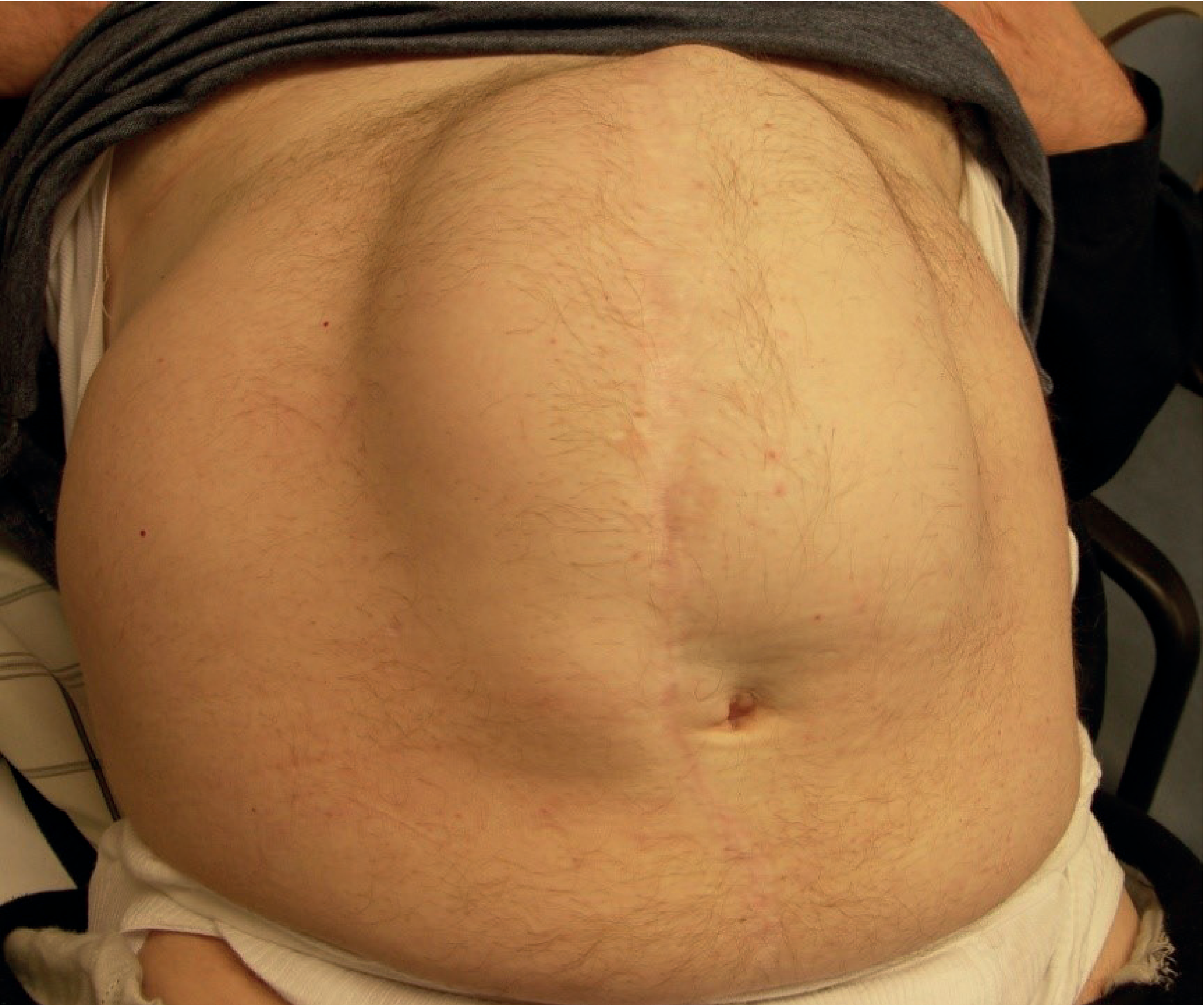
Classification
1. Etiological classification
-
Primary incisional hernia
- First hernia occurring through a previous operative scar.
-
Recurrent incisional hernia
- Hernia recurring after a prior operative repair.
-
Port-site hernia
- Incisional hernia through a laparoscopic trocar site.
-
Parastomal hernia
- Incisional defect adjacent to an intestinal or urinary stoma. Usually classified separately.
2. Classification by anatomical site
A. Midline incisional hernia
- Upper midline
- Epigastric
- Umbilical
- Infraumbilical
- Suprapubic
B. Lateral incisional hernia
- Subcostal
- Flank
- Iliac
- Lumbar
- Oblique scar
- Transverse scar
- Pfannenstiel scar
3. European Hernia Society (EHS) classification of incisional hernia
- Site of defect
- Width of defect
- Recurrence status
A. Midline location: M classification
| Code | Location |
|---|---|
| M1 | Subxiphoidal: xiphoid to 3 cm above umbilicus |
| M2 | Epigastric: 3 cm above umbilicus to 3 cm above umbilicus |
| M3 | Umbilical: 3 cm above to 3 cm below umbilicus |
| M4 | Infraumbilical: 3 cm below umbilicus to 3 cm above pubis |
| M5 | Suprapubic: pubis to 3 cm above pubis |
B. Lateral location: L classification
| Code | Location |
|---|---|
| L1 | Subcostal |
| L2 | Flank |
| L3 | Iliac |
| L4 | Lumbar |
C. Defect width: W classification
| Code | Maximum transverse fascial defect width |
|---|---|
| W1 | < 4 cm |
| W2 | 4-10 cm |
| W3 | > 10 cm |
D. Recurrence: R classification
| Code | Meaning |
|---|---|
| R0 | Primary incisional hernia, no previous repair |
| R1 | First recurrence |
| R2 | Second recurrence |
| R3 | Third or further recurrence |
A first-time infraumbilical midline incisional hernia, 7 cm wide, is documented as:
M4 W2 R0.
Other useful clinical classifications
4. Classification according to reducibility and complications
- Reducible
- Irreducible or incarcerated
- Obstructed
- Strangulated
- Inflamed or ulcerated
5. Classification by contents
- Omentocele
- Enterocele, containing small bowel
- Colocele, containing colon
- Richter-type hernia, where only part of bowel circumference is entrapped
- Sliding hernia, where a viscus forms part of sac wall
6. Classification by size
- Small: < 5 cm
- Medium: 5-10 cm
- Large: > 10 cm
- Giant or loss-of-domain hernia: very large defect with a substantial proportion of viscera permanently residing outside the abdominal cavity.
Incisional Hernia Case Proforma
A. Identification data
- Name:
- Age:
- Sex:
- Occupation:
- Address:
- Hospital number:
- Date of admission:
- Date of examination:
- Informant and reliability:
B. Chief complaints
- Swelling over previous abdominal operative scar for ___ months/years.
- Pain or dragging sensation over swelling for ___ duration.
- Swelling increasing on coughing or straining for ___ duration.
- Difficulty in reducing swelling for ___ duration.
- Vomiting, abdominal distension, constipation, or inability to pass flatus for ___ duration.
- Ulceration, skin discoloration, or discharge over swelling for ___ duration.
C. History of present illness
1. Onset and progression
- When was the swelling first noticed?
- Did it appear soon after surgery or after an interval?
- Was onset sudden or gradual?
- Has it progressively increased in size?
- Is it related to coughing, lifting weights, straining during defecation, micturition, or physical work?
- Does it reduce spontaneously on lying down?
2. Pain
- Site and nature: dull dragging pain, aching, colicky pain, severe continuous pain.
- Relation to exertion, standing, meals, cough, and posture.
- Sudden severe pain may indicate incarceration, obstruction, or strangulation.
3. Reducibility
- Is the swelling completely reducible?
- Does it reduce on lying down?
- Is manual pressure needed?
- Any history of irreducibility?
4. Features of intestinal obstruction
- Colicky abdominal pain
- Vomiting
- Abdominal distension
- Constipation
- Obstipation, inability to pass flatus or stool
5. Features suggesting strangulation
- Sudden severe continuous pain
- Tender irreducible swelling
- Fever
- Tachycardia
- Vomiting
- Skin erythema or discoloration over swelling
- Toxic appearance
6. Previous operation details
- Indication for prior surgery:
- Date of surgery:
- Type of surgery:
- Elective or emergency:
- Open/laparoscopic/robotic procedure:
- Type and site of incision:
- Midline
- Paramedian
- Transverse
- Subcostal
- Pfannenstiel
- Port site
- Stoma site
- Any re-laparotomy?
- Any postoperative wound infection?
- Wound dehiscence or burst abdomen?
- Serosanguineous discharge around postoperative day 5-8?
- Prolonged wound discharge?
- Resuturing or secondary suturing?
- Drain through the main wound?
- Previous hernia repair?
- Type of previous repair: anatomical repair, onlay mesh, sublay/retrorectus mesh, intraperitoneal mesh, laparoscopic IPOM, component separation.
- Number of prior repairs and recurrence interval.
7. Predisposing factors
- Obesity and recent weight gain
- Smoking
- Diabetes mellitus and glycaemic control
- Malnutrition or hypoalbuminaemia
- Anaemia
- Chronic renal failure
- Malignancy
- Steroid use, chemotherapy, immunosuppressive treatment
- Connective-tissue disease or collagen disorder
- Advanced age
- Chronic cough, COPD, tuberculosis, bronchiectasis
- Constipation
- Prostatism or stricture urethra causing straining
- Ascites
- Repeated vomiting
- Heavy manual work
- Pregnancy
- Surgical-site infection
- Poor fascial closure
- Emergency surgery
- Peritonitis or intra-abdominal sepsis
- Repeated abdominal operations
- Tissue ischemia or excessive tension on closure.
D. Past history
- Diabetes mellitus
- Hypertension
- COPD, asthma, tuberculosis
- Chronic cough
- Constipation
- Urinary obstructive symptoms
- Chronic liver disease, ascites
- Renal disease
- Malignancy
- Previous tuberculosis
- Previous abdominal surgeries
- Previous hernia surgeries
- History of wound infection or burst abdomen
E. Personal history
- Appetite and weight loss
- Bowel habits and constipation
- Micturition and straining
- Smoking, tobacco, alcohol
- Occupation involving heavy lifting
- Nutritional history
F. Drug and allergy history
- Corticosteroids
- Immunosuppressive drugs
- Chemotherapy
- Anticoagulants or antiplatelet agents
- Antidiabetic treatment
- Drug allergies
G. Family history
- Hernias in family members
- Known collagen vascular or connective-tissue disorders
Examination
1. General physical examination
- General condition and nutritional status
- Body mass index
- Pallor, icterus, cyanosis, clubbing, lymphadenopathy, edema
- Hydration
- Pulse, blood pressure, temperature, respiratory rate
- Signs of diabetes or chronic steroid use
- Features of COPD or chronic cough
- Evidence of chronic liver disease or ascites
2. Abdominal examination
A. Inspection
- Site of previous scar
- Scar type, length, direction, and quality
- Location of swelling in relation to scar
- Size and shape of swelling
- Single or multiple swellings
- Skin changes:
- Scar
- Dilated veins
- Erythema
- Ulceration
- Excoriation
- Pigmentation
- Thin, shiny, atrophic skin
- Visible peristalsis
- Cough impulse
- Whether swelling becomes prominent on standing, coughing, or raising the head and shoulders from supine position
- Other scars, stomas, drain sites, sinuses, or fistulae
- Divarication of recti
B. Palpation
- Local temperature
- Tenderness
- Consistency
- Surface and margins
- Cough impulse
- Expansile impulse on coughing
- Reducibility:
- Complete or partial
- Spontaneous or manual
- Gurgling on reduction suggests bowel
- Defect:
- Site
- Number
- Shape
- Maximum transverse width in cm
- Vertical length in cm
- Margins, especially lower margin
- Contents:
- Omentum: doughy, non-gurgling
- Bowel: soft, gurgling, may be resonant
- Whether defect is narrow or wide
- Whether there are multiple defects along the scar
C. Percussion
- Resonant: likely bowel-containing sac
- Dull: likely omentum or fluid-containing sac
D. Auscultation
- Bowel sounds over swelling may indicate bowel content.
- High-pitched or exaggerated sounds may suggest intestinal obstruction.
- Absent sounds with severe tenderness is concerning for strangulation or ileus.
E. Tests
- Cough impulse: place fingers over swelling and ask patient to cough.
- Head-raising test: with patient supine, ask them to lift head and shoulders. A hernia becomes more prominent due to contraction of abdominal muscles.
- Reducibility test: gently attempt reduction only if there is no suspicion of strangulation.
- Finger invagination test: not routinely useful in large incisional hernia, but may help identify a small scar defect.
F. Examination of other systems
- Respiratory system for COPD/chronic cough
- Cardiovascular assessment
- Per rectal examination if constipation, bleeding, or obstruction is present
- Digital rectal examination in elderly men with urinary symptoms
- Examine all other hernial sites
Provisional diagnosis
“A ___ year old male/female has a reducible/irreducible, uncomplicated/obstructed/strangulated incisional hernia through a previous ___ incision, with a defect measuring ___ cm × ___ cm, containing probable ___, classified as EHS ___, associated with ___ risk factors.”
“A 58-year-old obese woman with diabetes has a reducible infraumbilical midline incisional hernia following lower midline laparotomy, with a 7 cm transverse and 9 cm vertical fascial defect, containing bowel and omentum, without obstruction, classified as M4 W2 R0.”
Differential diagnosis
- Divarication of recti
- Lipoma of abdominal wall
- Seroma
- Chronic postoperative hematoma
- Abdominal wall abscess
- Stitch granuloma
- Desmoid tumour
- Abdominal wall neoplasm
- Parastomal hernia
- Spigelian hernia, depending on location
Investigations
Routine preoperative tests
- Complete blood count
- Blood group and cross-match if indicated
- Blood glucose and HbA1c
- Renal function tests and electrolytes
- Liver function tests, especially if ascites or malnutrition suspected
- Serum albumin
- Coagulation profile when indicated
- Urine routine and culture where appropriate
- ECG and chest radiograph based on age/comorbidity
- Pulmonary function assessment if COPD is suspected
Hernia-specific investigations
1. Ultrasonography
- Small, clinically equivocal defects
- Dynamic assessment with Valsalva
- Identifying superficial collections
2. CT abdomen and pelvis with Valsalva if feasible
- Fascial defect width and length
- Number of defects
- Hernial contents
- Bowel adhesions or obstruction
- Muscle quality
- Previous mesh position
- Loss of domain
- Operative planning
3. MRI
Brief management outline for case presentation
Initial approach
- Determine whether the hernia is uncomplicated, obstructed, or strangulated.
- Correct dehydration, electrolyte imbalance, hyperglycaemia, anaemia, malnutrition, and respiratory infection.
- Encourage smoking cessation and weight reduction where feasible.
- Treat chronic cough, constipation, urinary obstruction, and ascites.
- An abdominal binder may provide temporary symptomatic support but does not repair the defect.
Indications for urgent operation
- Strangulation
- Intestinal obstruction
- Irreducibility with pain or tenderness
- Peritonitis
- Skin necrosis or impending rupture
Elective repair
- Pain or activity limitation
- Progressive enlargement
- Recurrent obstruction
- Cosmesis affecting quality of life
- Large defect or poor skin condition
Write a history for 46 year old female with swelling on the lumbar region for 6 months not associated with pain and the swelling in more than 5cm, she had her right Kidney removed due to infection which she could not elaborate properly
"lumbar hernia" AND incisional
lumbar incisional hernia nephrectomy flank incision clinical presentation
History
Write it in exam presentation performa
History Case Presentation
Chief complaints
- Swelling in the right lumbar region for 6 months.
History of presenting illness
Past history
- History of right nephrectomy for renal infection, details not available.
- No known history of diabetes mellitus, hypertension, tuberculosis, chronic respiratory illness, or chronic liver disease.
- No previous history of hernia repair.
Personal history
- Appetite: Normal.
- Bowel habits: Regular. No constipation.
- Bladder habits: Normal. No straining during micturition.
- No history of smoking, alcohol intake, or heavy manual work.
Family history
- No similar illness in family members.
Provisional diagnosis
Right lumbar incisional hernia following right nephrectomy, uncomplicated, with a swelling more than 5 cm in size.
Points to confirm on examination
- Presence and site of right flank/lumbar operative scar
- Size and exact location of swelling
- Cough impulse and expansile nature
- Reducibility and contents
- Defect size and margins
- Evidence of obstruction, strangulation, or associated abdominal-wall weakness
- Examination of the left kidney and other hernial sites
Change in details she had surgery 1.5 years back , she is diabetic and uncontrolled, possible constipation problem
incisional hernia risk factors diabetes constipation wound healing
History Case Presentation
Chief complaint
- Swelling in the right lumbar region for 6 months.
History of presenting illness
Past history
- Right nephrectomy 1½ years ago for right renal infection, details not available.
- Diabetes mellitus, poorly controlled.
- Possible constipation.
- No past history of hypertension, chronic respiratory disease, chronic liver disease, ascites, or tuberculosis.
- No history of previous hernia repair.
Personal history
- Appetite: Normal.
- Bowel habits: History suggestive of constipation with occasional straining.
- Bladder habits: No difficulty or straining during micturition.
- Sleep: Normal.
- No history of smoking, alcohol consumption, or heavy manual work.
Family history
- No family history of similar swelling or hernia.
Provisional diagnosis
Right lumbar incisional hernia following right nephrectomy, uncomplicated, in a patient with uncontrolled diabetes mellitus and constipation.
Risk factors in this patient
- Previous right flank/lumbar incision for nephrectomy
- Poorly controlled diabetes mellitus, which can impair wound healing
- Possible chronic straining due to constipation, causing raised intra-abdominal pressure
Points to be elicited/examined further
- Exact site, type, and length of nephrectomy scar
- Whether swelling has an expansile cough impulse
- Reducibility and contents of swelling
- Size of fascial defect and number of defects
- Presence of bowel sounds over the swelling
- Evidence of obstruction or strangulation
- Glycaemic status: random blood sugar, fasting/postprandial glucose, HbA1c
- CT abdomen for confirming defect, contents, and planning repair
History of lifting heavy weights
There is a history of lifting heavy weights regularly as part of her daily household/occupational activities. This may have contributed to increased intra-abdominal pressure and enlargement of the swelling.
There is a possible history of constipation with occasional straining during defecation, and a history of regular heavy weight lifting. There is no history of chronic cough or straining during micturition.
Updated risk factors
- Previous right nephrectomy through a lumbar/flank incision
- Poorly controlled diabetes mellitus
- Constipation with straining
- Regular lifting of heavy weights
Incision in transverse from umbilicus to right flank
The patient underwent a right nephrectomy 1½ years ago for a right renal infection. The surgery was performed through a transverse incision extending from the umbilical region to the right flank. However, she is unable to give further details about the exact renal disease, operative procedure, or postoperative period.
Revised relevant portion of history
Revised provisional diagnosis
Right lateral incisional hernia through the lateral end of a previous transverse umbilico-flank incision, following right nephrectomy, uncomplicated, in a patient with poorly controlled diabetes mellitus, constipation, and a history of heavy weight lifting.
Write the complete final case presentation in one format
Final History Case Presentation
Chief complaint
- Swelling over the right lumbar/flank region for 6 months.
History of presenting illness
Past history
- History of right nephrectomy 1½ years ago for renal infection, with details not available.
- Known case of diabetes mellitus, poorly controlled.
- Possible history of constipation.
- No history suggestive of chronic respiratory disease or chronic cough.
- No history of chronic liver disease or ascites.
- No history of previous hernia repair.
Personal history
- Appetite: To be elicited.
- Bowel habits: Possible constipation with occasional straining.
- Bladder habits: No history of difficulty or straining during micturition.
- Sleep: To be elicited.
- Diet: To be elicited.
- History of heavy weight lifting is present.
- Smoking and alcohol history: To be elicited.
Family history
- No relevant family history of hernia or similar illness is available.
Summary
Provisional diagnosis
Right lateral incisional hernia through the lateral end of a previous transverse umbilico-flank incision following right nephrectomy, uncomplicated, in a patient with poorly controlled diabetes mellitus, constipation, and a history of heavy weight lifting.
Risk factors in this patient
- Previous transverse abdominal incision extending to the right flank
- Poorly controlled diabetes mellitus, with impaired wound healing risk
- Possible constipation and straining
- Regular lifting of heavy weights, causing repeated rises in intra-abdominal pressure
Important points to confirm on examination
- Exact location, length, and condition of the operative scar
- Site and size of swelling and fascial defect
- Cough impulse and expansile nature
- Reducibility and likely contents
- Presence of bowel sounds over the swelling
- Number of defects and status of abdominal wall musculature
- Evidence of obstruction or strangulation
- Examination of other hernial sites
- General assessment, including BMI and diabetic status.
Add the physical examination findings to this case presentation
incisional hernia physical examination cough impulse reducibility clinical examination
"incisional hernia" AND physical examination
Final Case Presentation
Chief complaint
- Swelling over the right flank/lumbar region for 6 months.
History of Presenting Illness
Past History
- History of right nephrectomy 1½ years ago for right renal infection.
- Known case of diabetes mellitus, poorly controlled.
- History suggestive of constipation.
- No history of hypertension, chronic cough, chronic obstructive pulmonary disease, tuberculosis, chronic liver disease, or ascites.
- No history of previous hernia repair.
Personal History
- Appetite: Normal.
- Bowel habits: Occasional constipation with straining during defecation.
- Bladder habits: Normal. No straining during micturition.
- History of regular lifting of heavy weights is present.
- Sleep: Normal.
- No history of smoking or alcohol consumption.
Family History
Physical Examination
The following findings are appropriate for this case presentation. Replace the exact measurements and examination findings with those found in your patient.
General Physical Examination
- No pallor, icterus, cyanosis, clubbing, lymphadenopathy, or pedal edema.
- Pulse: ___/minute, regular.
- Blood pressure: / mmHg.
- Temperature: Afebrile.
- Respiratory rate: ___/minute.
- No clinical features of dehydration or sepsis.
- Body mass index: ___ kg/m².
- Features of poorly controlled diabetes such as skin infection, neuropathy, or diabetic foot should be specifically looked for.
Local Examination of Abdomen and Swelling
Inspection
- Abdomen moves normally with respiration.
- A healed transverse operative scar is present, extending from the umbilical region towards the right flank, consistent with the history of right nephrectomy.
- A visible swelling is noted at the lateral end of this scar in the right flank/lumbar region.
- The swelling is approximately more than 5 cm in maximum dimension. Record exact dimensions as ___ cm × ___ cm.
- The swelling becomes more prominent on standing, coughing, and straining.
- The overlying skin is normal, with no redness, ulceration, dilated veins, sinus, discharge, or visible peristalsis.
- No other abdominal scars, sinuses, or visible swellings are noted.
Palpation
- Local temperature is not raised.
- The swelling is non-tender.
- It is soft in consistency, with smooth surface and indistinct margins.
- An expansile cough impulse is present.
- The swelling is reducible completely or partially on gentle pressure in the supine position. If bowel is present, reduction may produce a gurgling sensation.
- On reduction, a fascial defect is palpable at the lateral aspect of the scar. Its exact transverse and vertical dimensions should be recorded: ___ cm × ___ cm.
- The defect margins are palpable and non-tender.
- There is no local guarding, rigidity, or tenderness.
Percussion
- The swelling is resonant if it contains bowel.
- It may be dull if it predominantly contains omentum.
Auscultation
- Bowel sounds may be audible over the swelling, suggesting bowel as a content.
- No high-pitched bowel sounds are heard.
- There are no findings suggestive of intestinal obstruction.
Rest of the Abdominal Examination
- Abdomen is soft and non-tender.
- No distension, organomegaly, ascites, or free fluid.
- No other palpable abdominal mass.
- All other hernial sites, including inguinal, femoral, umbilical, and previous scar sites, should be examined and documented as normal.
- Per rectal examination may be done to assess constipation and exclude fecal loading, if indicated.
Systemic Examination
- Respiratory system: Normal, with no evidence of chronic cough or COPD.
- Cardiovascular system: Normal.
- Central nervous system: Normal.
- Left renal system: To be assessed clinically and by renal function tests, as the patient has a solitary left kidney.
Case Summary
Provisional Diagnosis
Uncomplicated, reducible right lateral incisional hernia through the lateral end of a previous transverse umbilico-flank incision following right nephrectomy, in a patient with poorly controlled diabetes mellitus, constipation, and history of heavy weight lifting.
Investigations to Plan
- Complete blood count and urine routine examination
- Fasting blood glucose, postprandial blood glucose, and HbA1c
- Renal function tests and serum electrolytes, especially important because she has one remaining kidney
- Serum albumin and nutritional assessment
- Ultrasound abdomen and abdominal-wall defect assessment
- CT abdomen and pelvis for defining the fascial defect, contents, number of defects, and operative planning
- ECG and chest radiograph if required for preoperative assessment
Any other tests and a possible list of differential diagnosis
Clinical tests
clinical examination tests incisional hernia cough impulse reducibility Valsalva
Clinical Tests for Right Lateral Incisional Hernia
1. Cough impulse test
- Place the palm or fingertips over the swelling.
- Ask the patient to cough.
- Positive test: An expansile impulse is felt, supporting a hernia.
- In this case, it is expected to be positive.
2. Valsalva manoeuvre
- Ask the patient to take a deep breath and strain as if passing stool.
- Observe and palpate the swelling.
- Positive test: Swelling becomes more prominent and tense.
- Useful in demonstrating a small or partially reducible hernia.
3. Reducibility test
- Examine with the patient supine and relaxed.
- Gently apply pressure over the swelling towards the defect.
- Positive finding: Swelling reduces completely or partially.
- A gurgling sensation suggests bowel content.
- Smooth, doughy reduction suggests omental content.
- After reduction, palpate the fascial defect and measure its transverse and vertical dimensions.
4. Head-raising or partial sit-up test
- With the patient lying supine, ask her to raise her head and shoulders without using her arms.
- This contracts the abdominal musculature and increases intra-abdominal pressure.
- Positive finding: The swelling becomes more prominent, and the defect may be easier to define.
- It also helps look for associated divarication of recti.
5. Defect palpation after reduction
- Once the swelling is reduced, place fingertips over the scar defect.
- Ask the patient to cough or strain.
- Assess:
- Number of defects
- Size of each defect
- Shape and margins
- Whether margins are firm or tender
- Impulse transmitted through the defect
6. Auscultation over the swelling
- Listen for bowel sounds.
- Presence of bowel sounds supports bowel as a hernial content.
- High-pitched, frequent bowel sounds with distension and vomiting may suggest obstruction.
- Absence of bowel sounds alone does not diagnose strangulation.
7. Percussion
- Resonant note: bowel-containing hernia.
- Dull note: omentum, thick sac, or fluid-containing lesion.
- This is supportive only and does not replace imaging.
8. Examination in different positions
- Examine in:
- Standing position
- Supine position
- During cough and Valsalva
- A hernia generally becomes more evident on standing/straining and decreases or reduces when supine.
Examination Statement for Your Case
On local examination, a swelling of more than 5 cm is present at the lateral end of the healed transverse umbilico-right flank scar. It becomes prominent on standing, coughing, and Valsalva manoeuvre. The swelling is non-tender, has a positive expansile cough impulse, and is reducible on lying down. Following reduction, a fascial defect can be palpated at the lateral end of the scar. Percussion may be resonant and bowel sounds may be audible over the swelling, suggesting bowel content. There are no clinical signs of obstruction or strangulation.
Differential Diagnoses
- Right lateral incisional hernia, most likely
- Primary lumbar hernia
- Postoperative flank bulge due to denervation or muscle paralysis, also called pseudohernia
- Lipoma of the abdominal wall
- Seroma or chronic postoperative collection
- Chronic hematoma
- Cold abscess, including tuberculous abscess
- Abdominal-wall tumour, including desmoid tumour