
Make PPT on complications of trauma according to book neelima malik oral surgery in detail with appropriate pictures and with flow charts also add thank you slide at end
maxillofacial trauma complications facial fracture
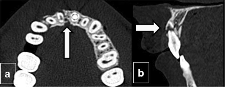
This unenhanced Computed Tomography (CT) study of the facial skeleton demonstrates traumatic injuries to the maxilla and dentition across two planes. Image (a) is an axial section showing a fracture of the upper alveolar process. A dark, linear lucency (indicated by an arrow) signifies a cortical discontinuity and disruption of the alveolar ridge's normal contour near the anterior midline. The fracture exhibits mild comminution and displacement of the surrounding bone fragments. Image (b) is a sagittal reconstruction revealing a dental root fracture of an upper incisor. The fracture is visualized as a transverse radiolucent line (arrow) passing through the root, resulting in a loss of longitudinal structural continuity. These findings are characteristic of maxillofacial trauma, highlighting the clinical significance of CT in assessing alveolar bone integrity and associated dental complications like root fractures or avulsions. The images illustrate key diagnostic criteria for surgeons and radiologists evaluating middle-third facial fractures and the status of the maxillary dental buttress.
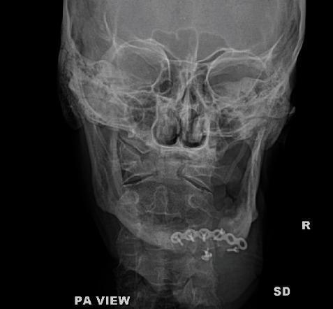
This posteroanterior (PA) radiograph of the skull and facial bones demonstrates internal fixation hardware in the mandible with evidence of mechanical failure. A metallic reconstruction mini-plate is positioned along the parasymphysis and anterior body of the mandible, secured by several cortical screws. A clear discontinuity or fracture is visible within the plate structure at the right parasymphysis region, indicative of hardware failure. There is associated bone lucency around several screw sites, which may suggest screw loosening, focal osteolysis, or localized infection. The underlying mandibular bone shows a persistent fracture line or non-union at the site of the plate break. The surrounding craniofacial structures, including the orbits, nasal cavity, and zygomatic arches, appear grossy intact and symmetric. This image illustrates post-operative complications in maxillofacial trauma management, specifically hardware fatigue and potential secondary infection in a mandibular fracture case.
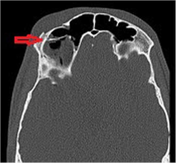
This diagnostic image is an axial non-contrast computerized tomography (CT) scan of the facial bones, utilizing a bone window setting to evaluate traumatic injury. The primary finding is a comminuted and displaced fracture of the right supraorbital rim and the anterior wall of the right frontal sinus, indicated by a red arrow. The fracture disrupts the normal cortical continuity of the bone, with posterior displacement of bony fragments into the sinus cavity. There is associated soft tissue swelling overlying the right supraorbital region. Intracranial air pockets, or pneumocephaly, are visible as hypodense (black) areas within the frontal sinus and extending toward the cranial cavity, suggesting a communication between the sinus and the intracranial space. This image demonstrates key radiological signs of maxillofacial trauma, specifically highlighting the risk of pneumocephaly following frontal sinus wall disruption. It is used in emergency medicine and neurosurgery to assess the severity of skull base or facial fractures and to screen for associated intracranial complications.
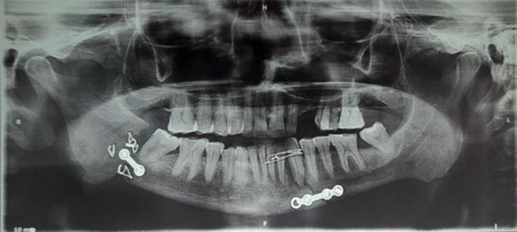
This diagnostic image is a panoramic radiograph (orthopantomogram or OPG) of the human mandible and maxilla. The imaging reveals significant pathology at the right mandibular angle, characterized by a clear cortical discontinuity and radiolucent gap consistent with a non-union of a mandibular fracture. Multiple radiopaque internal fixation devices are visible: a small two-hole metallic plate with screws and several transosseous wires are positioned at the right mandibular angle near the fracture site. Another four-hole metallic plate is fixed at the left mandibular parasymphysis. Additionally, interdental wiring is noted in the anterior maxillary segment, specifically connecting teeth in the upper left quadrant (region of teeth 22 to 24). The radiograph demonstrates clinical findings relevant to oral and maxillofacial surgery, illustrating post-surgical complications and the positioning of osteosynthesis hardware in the treatment of facial trauma.
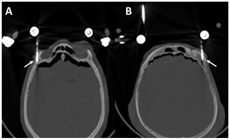
This diagnostic image consists of two axial computerized tomography (CT) scans (labeled A and B) of a human skull, demonstrating the surgical management of complex facial trauma, specifically a Le Fort III fracture. High-density, cylindrical metallic supraorbital pins are visualized (indicated by white arrows) inserted into the lateral third of the upper orbital margin. The pins are placed monocortically, providing external functional fixation (EFF) and stabilization of the fractured craniofacial segments. View A highlights a pin on the patient's right side, while View B demonstrates bilateral pin placement. The scans show the pins are positioned intraosseously, carefully avoiding penetration into the anterior cranial fossa to prevent intracranial complications. Significant bone discontinuity in the frontal and sinus regions is visible, consistent with the described fracture pattern. Streaking artifacts typical of metallic implants are present. This image serves as a clinical reference for optimal drilling angles and anatomical placement of external fixation pins in maxillofacial surgery.
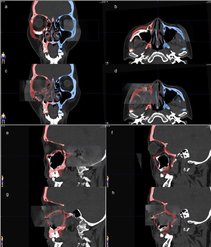
This composite diagnostic image demonstrates the use of surgical navigation and 3D C-arm computed tomography (CT) in the management of complex maxillofacial trauma. Panels a-d show coronal and axial CT views with color-coded overlays from presurgical planning. The blue overlay on the patient's left side represents the unaffected anatomical template, which is mirrored to the right side (red overlay) to guide the reduction of a comminuted zygomatic fracture and reconstruction of a blowout orbital floor fracture. Panels c and d specifically show the intraoperative fusion of 3D C-arm data with the virtual plan, verifying the alignment of bone fragments and the positioning of titanium implants. Panels e-h provide sagittal views comparing the preoperative state (e, f), characterized by orbital floor discontinuity and herniation of contents, with the intraoperative state (g, h). The post-reconstruction images (g, h) visualize a titanium mesh implant restoring the orbital floor contour, achieving high conformity with the mirrored surgical plan. This clinical toolset is essential for ensuring facial symmetry and preventing postoperative complications like enophthalmos in craniomaxillofacial surgery.
diplopia enophthalmos orbital floor fracture
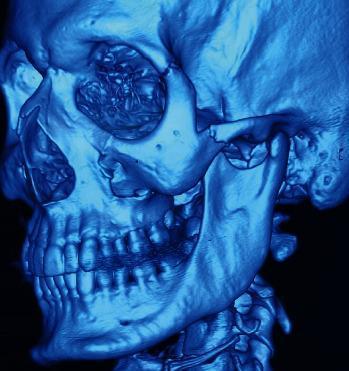
This diagnostic image is a 3D CT reconstruction of a human skull, presented in a monochromatic blue filter, illustrating a complex left zygomatic complex (tripod) fracture. The visualization clearly demonstrates cortical discontinuity and displacement at several key anatomical junctions: the frontozygomatic suture along the lateral orbital rim, the zygomaticomaxillary suture at the inferior orbital rim and anterior maxillary wall, and the zygomaticotemporal suture (zygomatic arch). There is visible disruption of the orbital floor. Adjacent structures including the mandible, maxilla, and dentition appear intact without gross secondary fractures. This image is clinically significant for maxillofacial trauma education, highlighting the structural collapse and multi-point separation characteristic of high-impact zygomatic injuries, often associated with clinical findings like enophthalmos and diplopia.
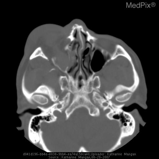
**Imaging Modality:** Non-contrast Computed Tomography (CT) of the head/maxillofacial region, axial view.
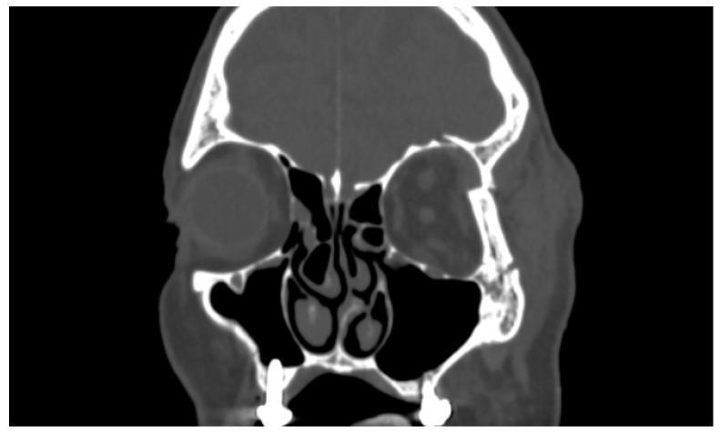
This diagnostic image is a coronal Computed Tomography (CT) scan of the maxillofacial region, focusing on the orbits and paranasal sinuses. The scan reveals a significant fracture of the left zygomaticomaxillary complex, specifically involving the lateral orbital wall and the orbital floor. Key findings include clear cortical discontinuity, fragmentation, and displacement of the lateral bony margin of the left orbit compared to the intact contralateral side. There is associated soft tissue swelling and potential herniation of orbital contents into the adjacent maxillary sinus space. The left globe appears slightly displaced and distorted relative to the normal spherical morphology of the right globe. Anatomical landmarks such as the nasal septum, turbinates, and maxillary sinuses are visible, with the left maxillary sinus demonstrating opacity likely representative of hemorrhage or herniated orbital tissue. This clinical presentation is characteristic of midface trauma and serves as a primary diagnostic tool for evaluating orbital wall integrity and planning surgical reconstruction in patients presenting with symptoms like diplopia or enophthalmos.
TMJ ankylosis condylar fracture
osteomyelitis mandible infection
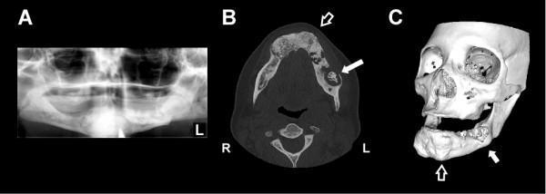
Multi-panel diagnostic imaging of the maxillofacial region demonstrating florid cemento-osseous dysplasia (FCOD) complicated by osteomyelitis. Panel A: A panoramic radiograph showing extensive, well-demarcated mixed radiolucent and radiopaque lesions involving all four quadrants of the mandible and maxilla. The denser opacities present a characteristic 'cotton-wool' appearance surrounded by thin radiolucent rims. Panel B: Axial CT scan highlighting generalized hypertrophic and heterogeneous sclerotic changes within the mandible (open arrow). A significant lytic lesion with cortical loss and central sclerosis is visible in the left mandibular body (solid arrow), indicating secondary infection. Panel C: A 3D reconstructed CT image illustrating global bony expansion and contour irregularities in the maxilla and mandible (open arrow). A focal area of erosion on the left mandible (solid arrow) corresponds to the site of Actinomyces osteomyelitis. This series serves as an educational example of how underlying fibro-osseous dysplasia can predispose patients to secondary infections due to decreased vascularity of the sclerotic bone.
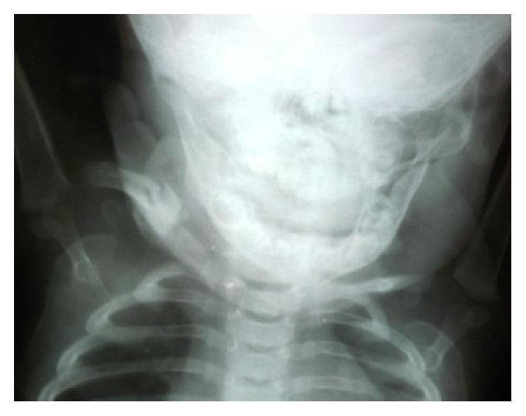
Anterior-posterior (AP) radiograph of a pediatric patient's upper chest and neck region, including the skull, cervical spine, and thorax. The image focuses on the right clavicle, demonstrating the late-stage radiographic appearance of resolved or healing osteomyelitis. The clavicular midshaft shows cortical thickening and mild remodeling, indicative of a resolved infectious process. Surrounding bony structures, including the superior ribs, mandible, and scapulae, are partially visualized. The radiograph illustrates the long-term musculoskeletal outcome and bone regeneration following surgical curettage and antibiotic treatment for a chronic bone infection (osteomyelitis). This diagnostic image is representative of pediatric infectious disease and orthopedic follow-up imaging.
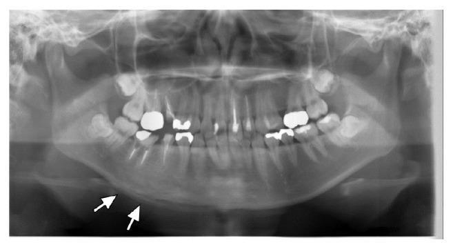
This orthopantomogram (OPG) provides a panoramic diagnostic view of the human maxilla and mandible, including full dentition. The primary pathological finding is localized to the right body of the mandible, where significant sclerotic changes are evident as increased radiopacity compared to the contralateral side. White arrows specifically highlight a periosteal reaction along the inferior border of the right mandible, characterized by a layered or 'onion-skin' appearance of new bone formation. These radiographic features are characteristic of chronic non-suppurative osteomyelitis or sclerosing osteitis. In this clinical context, the absence of obvious odontogenic infection and the presence of multifocal involvement elsewhere suggests SAPHO syndrome (Synovitis, Acne, Pustulosis, Hyperostosis, and Osteitis). The image serves as an educational example of inflammatory bone remodeling and reactive periostitis in the maxillofacial region.
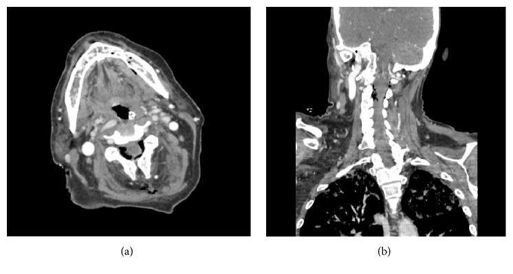
This diagnostic image consists of two Computed Tomography (CT) scans highlighting complications related to maxillofacial and cervical infection. Figure (a) is an axial CT scan at the level of the mandible. It demonstrates the absence of discrete fluid collections or abscesses in the soft tissues, but reveals pathological air within the medullary marrow space of the left posterior mandible, suggesting osteomyelitis with gas-forming organisms or communication with a necrotic track. Figure (b) is a coronal CT reconstruction of the neck and upper thorax. It visualizes the cervical spine, paraspinal soft tissues, and superior mediastinum/lung apices. The significant clinical finding is pneumorrhachis, characterized by the presence of air within the epidural space of the cervical spinal canal. These findings are clinically significant in the context of necrotizing infections, sepsis, and potential spread of air/infection from the oral cavity into deep cervical and spinal spaces.
"maxillofacial injuries" AND complications
facial nerve injury paralysis after trauma
malunion mandible fracture facial asymmetry
/tmp/workspace
~/oral-trauma-ppt/fetch_assets.py
import json, subprocess, base64, os

urls = [
    "https://cdn.orris.care/cdss_images/pmc_clinical_VQA_6eb12d765d8bf9e0d5ae77291e1a0224f06c0bcf6b630e374251b60bc22f8854.jpg",
    "https://cdn.orris.care/cdss_images/pmc_clinical_VQA_5bdab43dca9a79bb8f13222f639c1726ebbb59ac970a8cb58c0420daae18319e.jpg",
    "https://cdn.orris.care/cdss_images/pmc_clinical_VQA_6864f90c722f2ac4abe4354391b632a6979bf8b6a3bdc425b15f7d5b7a8048e5.jpg",
    "https://cdn.orris.care/cdss_images/pmc_clinical_VQA_6f1c0e09fd95ca04b75a929ae83c0719599a7640f6062c02fe713638a62ab1d5.jpg",
    "https://cdn.orris.care/cdss_images/pmc_clinical_VQA_2c0d6e9f709ee3648e8cb9e294ed5a88f049bc1236deeee0f4a1bf091d24eeef.jpg",
    "https://cdn.orris.care/cdss_images/pmc_clinical_VQA_465f94778d8b765adddec301b083554e8de97ac9f037669c0e3e3fe48dae5bb6.jpg",
    "https://cdn.orris.care/cdss_images/pmc_clinical_VQA_5e1d08eac8b9175b1972f558ef306cdbe62a890338bcd1dcb1c5d6547752dd13.jpg",
    "https://cdn.orris.care/cdss_images/pmc_clinical_VQA_38f39f1e7dc0f529c7b2f215c2308a217541735b70b586e054cf95964ae67255.jpg",
    "https://cdn.orris.care/cdss_images/pmc_clinical_VQA_0bbce09bbf6b477382e8e2d532f95b8ad7aa6989bf5673825cd924ecea9bb2d5.jpg",
    "https://cdn.orris.care/cdss_images/pmc_clinical_VQA_375f6f62185a1020d24ec1868e1be1c79dba2ee84fee8a40c009abd9591393fd.jpg",
]

os.makedirs("/tmp/workspace/oral-trauma-ppt/assets", exist_ok=True)

result = json.loads(subprocess.check_output(
    ["python", "/tmp/skills/shared/scripts/fetch_images.py"] + urls
))

names = ["dentoalveolar_fx", "plate_failure_nonunion", "frontal_sinus_pneumocephalus",
         "opg_nonunion_hardware", "lefort3_extfix", "orbital_titanium_recon",
         "zygomatic_3dct", "orbital_floor_coronal", "osteomyelitis_fcod", "osteomyelitis_air"]

for name, img in zip(names, result):
    if img.get("base64"):
        header, b64data = img["base64"].split(",", 1) if "," in img["base64"] else ("", img["base64"])
        ext = "png" if "png" in header else "jpg"
        raw = base64.b64decode(b64data)
        path = f"/tmp/workspace/oral-trauma-ppt/assets/{name}.{ext}"
        with open(path, "wb") as f:
            f.write(raw)
        print("saved", path)
    else:
        print("FAILED", name, img.get("error"))
cd /tmp/workspace/oral-trauma-ppt && python fetch_assets.py
**/fetch_images*.py in /tmp/skills
ls -la /tmp/skills/shared/scripts/ 2>&1 | head -20
cd /tmp/workspace/oral-trauma-ppt && python fetch_assets.py 2>&1 | tail -30
~/oral-trauma-ppt/build.py
from pptx import Presentation
from pptx.util import Inches, Pt, Emu
from pptx.dml.color import RGBColor
from pptx.enum.text import PP_ALIGN, MSO_ANCHOR
from pptx.enum.shapes import MSO_SHAPE, MSO_CONNECTOR
from pptx.oxml.ns import qn
import copy

ASSETS = "/tmp/workspace/oral-trauma-ppt/assets/"

NAVY = RGBColor(0x0B, 0x2E, 0x4D)
TEAL = RGBColor(0x11, 0x7A, 0x8B)
RED = RGBColor(0xB0, 0x2A, 0x2A)
ORANGE = RGBColor(0xD9, 0x7A, 0x1F)
GREEN = RGBColor(0x2E, 0x7D, 0x32)
WHITE = RGBColor(0xFF, 0xFF, 0xFF)
GREY = RGBColor(0x45, 0x45, 0x45)
LIGHT = RGBColor(0xF2, 0xF4, 0xF6)

prs = Presentation()
prs.slide_width = Inches(13.333)
prs.slide_height = Inches(7.5)
BLANK = prs.slide_layouts[6]
SW, SH = prs.slide_width, prs.slide_height


def add_bg(slide, color=WHITE):
    rect = slide.shapes.add_shape(MSO_SHAPE.RECTANGLE, 0, 0, SW, SH)
    rect.fill.solid()
    rect.fill.fore_color.rgb = color
    rect.line.fill.background()
    rect.shadow.inherit = False
    slide.shapes._spTree.remove(rect._element)
    slide.shapes._spTree.insert(2, rect._element)
    return rect


def add_header_bar(slide, title, subtitle=None, bar_color=NAVY):
    bar = slide.shapes.add_shape(MSO_SHAPE.RECTANGLE, 0, 0, SW, Inches(1.15))
    bar.fill.solid()
    bar.fill.fore_color.rgb = bar_color
    bar.line.fill.background()
    bar.shadow.inherit = False
    tb = slide.shapes.add_textbox(Inches(0.5), Inches(0.12), Inches(11.5), Inches(0.7))
    tf = tb.text_frame
    tf.word_wrap = True
    p = tf.paragraphs[0]
    p.text = title
    p.runs[0].font.size = Pt(28)
    p.runs[0].font.bold = True
    p.runs[0].font.color.rgb = WHITE
    p.runs[0].font.name = "Calibri"
    if subtitle:
        tb2 = slide.shapes.add_textbox(Inches(0.5), Inches(0.75), Inches(11.5), Inches(0.35))
        tf2 = tb2.text_frame
        p2 = tf2.paragraphs[0]
        p2.text = subtitle
        p2.runs[0].font.size = Pt(14)
        p2.runs[0].font.italic = True
        p2.runs[0].font.color.rgb = RGBColor(0xCF, 0xE3, 0xEC)
    # accent line
    line = slide.shapes.add_shape(MSO_SHAPE.RECTANGLE, 0, Inches(1.15), SW, Pt(4))
    line.fill.solid()
    line.fill.fore_color.rgb = ORANGE
    line.line.fill.background()
    line.shadow.inherit = False
    return bar


def add_footer(slide, text="Complications of Trauma - Oral & Maxillofacial Surgery"):
    tb = slide.shapes.add_textbox(Inches(0.4), SH - Inches(0.4), Inches(9), Inches(0.3))
    p = tb.text_frame.paragraphs[0]
    p.text = text
    p.runs[0].font.size = Pt(10)
    p.runs[0].font.color.rgb = RGBColor(0xA0, 0xA0, 0xA0)


def bullet_box(slide, x, y, w, h, items, font_size=18, color=GREY, bold_first=False, line_space=1.15):
    tb = slide.shapes.add_textbox(x, y, w, h)
    tf = tb.text_frame
    tf.word_wrap = True
    for i, item in enumerate(items):
        p = tf.paragraphs[0] if i == 0 else tf.add_paragraph()
        if isinstance(item, tuple):
            text, level = item
        else:
            text, level = item, 0
        p.text = ("  " * level) + ("• " if level == 0 else "‑ ") + text
        p.level = 0
        p.space_after = Pt(8)
        p.line_spacing = line_space
        run = p.runs[0]
        run.font.size = Pt(font_size - level * 2)
        run.font.color.rgb = color
        run.font.name = "Calibri"
        if level == 0:
            run.font.bold = True
    return tb


def box(slide, x, y, w, h, text, fill=TEAL, font_color=WHITE, size=13, shape=MSO_SHAPE.ROUNDED_RECTANGLE, bold=True):
    sp = slide.shapes.add_shape(shape, x, y, w, h)
    sp.fill.solid()
    sp.fill.fore_color.rgb = fill
    sp.line.color.rgb = RGBColor(0xFF, 0xFF, 0xFF)
    sp.line.width = Pt(1.5)
    sp.shadow.inherit = False
    tf = sp.text_frame
    tf.word_wrap = True
    tf.margin_left = Pt(4); tf.margin_right = Pt(4); tf.margin_top = Pt(2); tf.margin_bottom = Pt(2)
    tf.vertical_anchor = MSO_ANCHOR.MIDDLE
    p = tf.paragraphs[0]
    p.alignment = PP_ALIGN.CENTER
    p.text = text
    run = p.runs[0]
    run.font.size = Pt(size)
    run.font.bold = bold
    run.font.color.rgb = font_color
    run.font.name = "Calibri"
    return sp


def arrow(slide, x1, y1, x2, y2, color=NAVY, width=2.25):
    conn = slide.shapes.add_connector(MSO_CONNECTOR.STRAIGHT, x1, y1, x2, y2)
    conn.line.color.rgb = color
    conn.line.width = Pt(width)
    ln = conn.line._get_or_add_ln()
    tail = ln.makeelement(qn('a:tailEnd'), {'type': 'triangle', 'w': 'med', 'len': 'med'})
    ln.append(tail)
    return conn


def pic(slide, path, x, y, w=None, h=None):
    return slide.shapes.add_picture(path, x, y, width=w, height=h)


def caption(slide, x, y, w, text, size=11, color=GREY, align=PP_ALIGN.CENTER):
    tb = slide.shapes.add_textbox(x, y, w, Inches(0.4))
    tf = tb.text_frame
    tf.word_wrap = True
    p = tf.paragraphs[0]
    p.alignment = align
    p.text = text
    p.runs[0].font.size = Pt(size)
    p.runs[0].font.italic = True
    p.runs[0].font.color.rgb = color


def picture_frame(slide, path, x, y, w, h, cap_text=None):
    frame = slide.shapes.add_shape(MSO_SHAPE.RECTANGLE, x - Pt(3), y - Pt(3), w + Pt(6), h + Pt(6))
    frame.fill.solid(); frame.fill.fore_color.rgb = RGBColor(0xDD, 0xDD, 0xDD)
    frame.line.color.rgb = NAVY; frame.line.width = Pt(1)
    frame.shadow.inherit = False
    p = pic(slide, path, x, y, w=w, h=h)
    if cap_text:
        caption(slide, x, y + h + Pt(2), w, cap_text)
    return p


# ---------------------------------------------------------------------------
# SLIDE 1 - TITLE
# ---------------------------------------------------------------------------
s = prs.slides.add_slide(BLANK)
add_bg(s, NAVY)
band = s.shapes.add_shape(MSO_SHAPE.RECTANGLE, 0, Inches(4.6), SW, Inches(0.12))
band.fill.solid(); band.fill.fore_color.rgb = ORANGE; band.line.fill.background(); band.shadow.inherit = False

tb = s.shapes.add_textbox(Inches(0.8), Inches(2.5), Inches(11.7), Inches(1.5))
tf = tb.text_frame; tf.word_wrap = True
p = tf.paragraphs[0]; p.alignment = PP_ALIGN.CENTER
p.text = "COMPLICATIONS OF TRAUMA"
p.runs[0].font.size = Pt(48); p.runs[0].font.bold = True; p.runs[0].font.color.rgb = WHITE
p2 = tf.add_paragraph(); p2.alignment = PP_ALIGN.CENTER
p2.text = "Maxillofacial & Oral Trauma - Classification and Management"
p2.runs[0].font.size = Pt(22); p2.runs[0].font.color.rgb = RGBColor(0xCF, 0xE3, 0xEC); p2.runs[0].font.italic = True

tb2 = s.shapes.add_textbox(Inches(0.8), Inches(5.1), Inches(11.7), Inches(0.8))
tf2 = tb2.text_frame; p3 = tf2.paragraphs[0]; p3.alignment = PP_ALIGN.CENTER
p3.text = "Reference framework: Textbook of Oral & Maxillofacial Surgery - Neelima Malik"
p3.runs[0].font.size = Pt(16); p3.runs[0].font.color.rgb = WHITE

tb3 = s.shapes.add_textbox(Inches(0.8), Inches(6.6), Inches(11.7), Inches(0.5))
p4 = tb3.text_frame.paragraphs[0]; p4.alignment = PP_ALIGN.CENTER
p4.text = "Oral & Maxillofacial Surgery | Undergraduate / Postgraduate Seminar"
p4.runs[0].font.size = Pt(13); p4.runs[0].font.color.rgb = RGBColor(0xAF, 0xC7, 0xD6)

# ---------------------------------------------------------------------------
# SLIDE 2 - INTRODUCTION
# ---------------------------------------------------------------------------
s = prs.slides.add_slide(BLANK)
add_bg(s)
add_header_bar(s, "Introduction", "Why complications matter in trauma management")
bullet_box(s, Inches(0.6), Inches(1.5), Inches(7.6), Inches(5.3), [
    "Trauma to the maxillofacial region can injure bone, soft tissue, teeth, nerves, vessels and adjacent structures (orbit, cranial base, airway).",
    "A 'complication' is any adverse event that arises as a direct or indirect consequence of the injury or its treatment.",
    "Complications may occur despite optimal treatment because of the severity of injury, delay in reporting, associated systemic injury, or patient factors (age, nutrition, comorbidities, infection).",
    "Neelima Malik's textbook classifies complications of trauma on a TIME-based framework, which guides emergency priorities:",
    ("Immediate - occurring at the time of injury / within hours", 1),
    ("Intermediate (Early) - occurring within days", 1),
    ("Late (Delayed) - occurring weeks to months/years later", 1),
], font_size=17)

picture_frame(s, ASSETS + "dentoalveolar_fx.jpg", Inches(8.5), Inches(1.6), Inches(4.2), Inches(3.4),
              "CT: alveolar & dental root fracture following maxillofacial trauma")
add_footer(s)

# ---------------------------------------------------------------------------
# SLIDE 3 - MASTER FLOWCHART / CLASSIFICATION
# ---------------------------------------------------------------------------
s = prs.slides.add_slide(BLANK)
add_bg(s)
add_header_bar(s, "Classification of Complications of Trauma", "Time-based framework")

box(s, Inches(4.9), Inches(1.5), Inches(3.5), Inches(0.7), "TRAUMA (Facial / Oral)", fill=NAVY, size=18)
arrow(s, Inches(6.65), Inches(2.2), Inches(6.65), Inches(2.55))

box(s, Inches(4.9), Inches(2.55), Inches(3.5), Inches(0.6), "COMPLICATIONS", fill=ORANGE, size=16)

y0 = Inches(3.5)
cols = [
    (Inches(0.6), "IMMEDIATE", RED, ["Haemorrhage & shock", "Airway obstruction / asphyxia", "Aspiration (blood/tooth/FB)",
                                       "Air embolism", "Surgical emphysema", "Associated head/spine/chest injury"]),
    (Inches(4.85), "INTERMEDIATE (Early)", ORANGE, ["Wound / bone infection", "Delayed haemorrhage", "Fat embolism",
                                        "Tetanus", "CSF leak, meningitis", "Cavernous sinus thrombosis"]),
    (Inches(9.1), "LATE (Delayed)", GREEN, ["Mal-/non-/delayed union", "Chronic osteomyelitis", "Malocclusion, deformity",
                                  "TMJ ankylosis", "Nerve injury, diplopia", "Scarring, psychological"]),
]
for x, title, color, items in cols:
    arrow(s, Inches(6.65), Inches(3.15), x + Inches(1.7), y0, color=NAVY)
    box(s, x, y0, Inches(3.5), Inches(0.55), title, fill=color, size=16)
    tb = s.shapes.add_textbox(x, y0 + Inches(0.65), Inches(3.5), Inches(3.2))
    tf = tb.text_frame; tf.word_wrap = True
    for i, it in enumerate(items):
        p = tf.paragraphs[0] if i == 0 else tf.add_paragraph()
        p.text = "• " + it
        p.space_after = Pt(6)
        p.runs[0].font.size = Pt(14)
        p.runs[0].font.color.rgb = GREY
add_footer(s)

# ---------------------------------------------------------------------------
# SLIDE 4 - IMMEDIATE: HAEMORRHAGE & SHOCK + AIRWAY (flow)
# ---------------------------------------------------------------------------
s = prs.slides.add_slide(BLANK)
add_bg(s)
add_header_bar(s, "Immediate Complications (1): Haemorrhage, Shock & Airway", "Occur at the time of injury - life-threatening, need instant action")

bullet_box(s, Inches(0.6), Inches(1.55), Inches(6.6), Inches(5.4), [
    "Haemorrhage - from lacerated facial vessels (facial, maxillary, lingual artery), fracture surfaces, or scalp; may be brisk in Le Fort/panfacial fractures.",
    "Haemorrhagic shock - hypovolaemia from unrecognised blood loss; treat with airway-breathing-circulation (ABC) protocol, fluid resuscitation, local pressure/pack.",
    "Airway obstruction / asphyxia - most common cause of early death in maxillofacial trauma:",
    ("Posterior displacement of tongue (bilateral mandible fracture)", 1),
    ("Blood clots, avulsed teeth, dentures, bone fragments", 1),
    ("Soft-tissue oedema / haematoma of floor of mouth", 1),
    "Aspiration of blood, tooth fragments or vomitus - risk of asphyxia or aspiration pneumonia.",
    "Air embolism - rare, via open venous channels in facial/skull fractures.",
    "Surgical (subcutaneous) emphysema - air trapped in soft tissue, often from paranasal sinus fractures.",
], font_size=15.5)

# mini flow chart airway
fx = Inches(7.5)
box(s, fx, Inches(1.6), Inches(4.8), Inches(0.5), "Facial trauma", fill=RED, size=14)
arrow(s, fx+Inches(2.4), Inches(2.1), fx+Inches(2.4), Inches(2.35))
box(s, fx, Inches(2.35), Inches(4.8), Inches(0.6), "Tongue fall-back / clot / oedema / FB", fill=ORANGE, size=13)
arrow(s, fx+Inches(2.4), Inches(2.95), fx+Inches(2.4), Inches(3.2))
box(s, fx, Inches(3.2), Inches(4.8), Inches(0.5), "Airway obstruction", fill=RED, size=14)
arrow(s, fx+Inches(2.4), Inches(3.7), fx+Inches(2.4), Inches(3.95))
box(s, fx, Inches(3.95), Inches(4.8), Inches(0.5), "Hypoxia -> Shock -> Death (if untreated)", fill=NAVY, size=13)
arrow(s, fx+Inches(2.4), Inches(4.45), fx+Inches(2.4), Inches(4.7))
box(s, fx, Inches(4.7), Inches(4.8), Inches(0.6), "Emergency: positioning, suction,\njaw thrust, airway adjunct / tracheostomy", fill=GREEN, size=12.5)

caption(s, fx, Inches(5.45), Inches(4.8), "Emergency airway management flow in facial trauma")
add_footer(s)

# ---------------------------------------------------------------------------
# SLIDE 5 - ASSOCIATED / SEVERE COMPLEX TRAUMA
# ---------------------------------------------------------------------------
s = prs.slides.add_slide(BLANK)
add_bg(s)
add_header_bar(s, "Immediate Complications (2): Associated Injuries", "Complex/panfacial trauma requires whole-body assessment")
bullet_box(s, Inches(0.6), Inches(1.55), Inches(7.6), Inches(5.2), [
    "High-energy facial trauma (RTA, assault, fall from height) is frequently associated with:",
    ("Traumatic brain injury / cervical spine injury - always assess before manipulating the neck", 1),
    ("Ocular injury - globe rupture, orbital fracture", 1),
    ("Chest and abdominal injury in polytrauma patients", 1),
    "Cervical spine must be immobilised until cleared radiographically, especially in unconscious patients.",
    "Severe panfacial/Le Fort III fractures may need external fixation (halo frame / pins) for temporary stabilisation before definitive ORIF.",
    "A recent systematic review/meta-analysis found a strong association between maxillofacial fractures and traumatic brain injury, reinforcing the need for combined neurosurgical-maxillofacial assessment (Othman et al. 2023).",
], font_size=16)
picture_frame(s, ASSETS + "lefort3_extfix.jpg", Inches(8.5), Inches(1.6), Inches(4.2), Inches(3.6),
              "CT: supraorbital external fixation pins for Le Fort III fracture")
add_footer(s)

# ---------------------------------------------------------------------------
# SLIDE 6 - INTERMEDIATE FLOWCHART: INFECTION PATHWAY
# ---------------------------------------------------------------------------
s = prs.slides.add_slide(BLANK)
add_bg(s)
add_header_bar(s, "Intermediate (Early) Complications (1): Infection Pathway", "Days after injury - contamination and delayed treatment are key risk factors")

fx = Inches(1.2)
steps = [
    ("Contaminated wound / compound fracture / delayed treatment / poor oral hygiene", RED),
    ("Bacterial colonisation of wound / fracture haematoma", ORANGE),
    ("Wound infection / cellulitis", ORANGE),
    ("Osteomyelitis of jaw (if bone involved, esp. mandible)", RED),
    ("Chronic osteomyelitis, sequestrum, sinus tract, non-union", NAVY),
]
yy = Inches(1.55)
for i, (txt, col) in enumerate(steps):
    box(s, fx, yy, Inches(6.6), Inches(0.65), txt, fill=col, size=14)
    if i < len(steps)-1:
        arrow(s, fx+Inches(3.3), yy+Inches(0.65), fx+Inches(3.3), yy+Inches(0.9))
    yy += Inches(0.9)

bullet_box(s, Inches(8.1), Inches(1.55), Inches(4.6), Inches(4.3), [
    "Predisposing factors:",
    ("Delay in treatment/reduction", 1),
    ("Retained tooth/root/FB in fracture line", 1),
    ("Poor blood supply, comminution", 1),
    ("Systemic disease, immunosuppression", 1),
    "Other early complications:",
    ("Delayed/secondary haemorrhage", 1),
    ("Fat embolism (long bone + facial trauma)", 1),
    ("Tetanus (contaminated wounds)", 1),
], font_size=14.5)
add_footer(s)

# ---------------------------------------------------------------------------
# SLIDE 7 - INFECTION IMAGES
# ---------------------------------------------------------------------------
s = prs.slides.add_slide(BLANK)
add_bg(s)
add_header_bar(s, "Intermediate Complications (2): Osteomyelitis & Wound Infection", "Radiological features")
picture_frame(s, ASSETS + "osteomyelitis_fcod.jpg", Inches(0.6), Inches(1.6), Inches(5.9), Inches(4.1),
              "Panoramic/CT/3D-CT: sclerotic, mixed radiolucent-radiopaque changes of osteomyelitis of the mandible")
picture_frame(s, ASSETS + "osteomyelitis_air.jpg", Inches(6.9), Inches(1.6), Inches(5.9), Inches(4.1),
              "CT: intramedullary gas in mandible with spread of infection - necrotising infection")
bullet_box(s, Inches(0.6), Inches(5.9), Inches(12.1), Inches(1.3), [
    "Clinical features: persistent pain, swelling, pus discharge, sequestrum formation, pathological fracture, trismus, sinus tract.",
    "Management: culture-guided antibiotics, drainage, sequestrectomy/saucerisation, removal of infected hardware, nutritional support.",
], font_size=15)
add_footer(s)

# ---------------------------------------------------------------------------
# SLIDE 8 - INTERMEDIATE: CRANIAL BASE COMPLICATIONS
# ---------------------------------------------------------------------------
s = prs.slides.add_slide(BLANK)
add_bg(s)
add_header_bar(s, "Intermediate Complications (3): CSF Leak & Intracranial Spread", "Anterior skull base / frontal sinus involvement")
bullet_box(s, Inches(0.6), Inches(1.55), Inches(6.6), Inches(5.2), [
    "Fractures involving the frontal sinus, cribriform plate, or orbital roof can tear the dura.",
    "CSF rhinorrhoea/otorrhoea - clear watery discharge, 'halo/ring sign' on filter paper, positive beta-2 transferrin.",
    "Complications if untreated:",
    ("Ascending meningitis", 1),
    ("Brain abscess", 1),
    ("Pneumocephalus (air in cranial cavity)", 1),
    ("Cavernous sinus thrombosis - spread of infection via valveless facial veins (especially from midface/'danger triangle' infections)", 1),
    "Management: neurosurgical consultation, prophylactic broad-spectrum antibiotics debate, bed rest with head elevation, dural repair if leak persists >7-10 days.",
], font_size=15.5)
picture_frame(s, ASSETS + "frontal_sinus_pneumocephalus.jpg", Inches(7.5), Inches(1.6), Inches(5.2), Inches(4.0),
              "CT: frontal sinus/supraorbital fracture with pneumocephalus")
add_footer(s)

# ---------------------------------------------------------------------------
# SLIDE 9 - LATE: BONE HEALING FLOWCHART
# ---------------------------------------------------------------------------
s = prs.slides.add_slide(BLANK)
add_bg(s)
add_header_bar(s, "Late Complications (1): Disorders of Bone Healing", "Malunion, non-union and delayed union of jaw fractures")

box(s, Inches(4.9), Inches(1.5), Inches(3.5), Inches(0.6), "Fracture of jaw", fill=NAVY, size=16)
arrow(s, Inches(6.65), Inches(2.1), Inches(6.65), Inches(2.35))
box(s, Inches(4.9), Inches(2.35), Inches(3.5), Inches(0.6), "Reduction & Fixation", fill=TEAL, size=15)

y0 = Inches(3.35)
outs = [
    (Inches(0.7), "Normal Union", GREEN, "Anatomic healing, normal occlusion & function"),
    (Inches(4.9), "Delayed Union", ORANGE, "Healing slower than expected; bone ends mobile but viable"),
    (Inches(9.1), "Mal-union", ORANGE, "Healing in abnormal position -> facial asymmetry, malocclusion"),
]
for x, title, color, desc in outs:
    arrow(s, Inches(6.65), Inches(2.95), x+Inches(1.6), y0, color=NAVY)
    box(s, x, y0, Inches(3.4), Inches(0.55), title, fill=color, size=15)
    tb = s.shapes.add_textbox(x, y0+Inches(0.65), Inches(3.4), Inches(1.3))
    p = tb.text_frame.paragraphs[0]; tb.text_frame.word_wrap=True
    p.text = desc; p.runs[0].font.size = Pt(13); p.runs[0].font.color.rgb = GREY

box(s, Inches(4.9), Inches(5.5), Inches(3.5), Inches(0.55), "Non-union", fill=RED, size=15)
arrow(s, Inches(6.65), Inches(2.95), Inches(6.65), Inches(5.5), color=RED)
tb = s.shapes.add_textbox(Inches(4.9), Inches(6.1), Inches(3.5), Inches(1.0))
p = tb.text_frame.paragraphs[0]; tb.text_frame.word_wrap = True
p.text = "Failure of union - persistent mobility, fibrous tissue/pseudarthrosis at fracture site; needs re-fixation +/- bone grafting"
p.runs[0].font.size = Pt(13); p.runs[0].font.color.rgb = GREY
add_footer(s)

# ---------------------------------------------------------------------------
# SLIDE 10 - BONE HEALING IMAGES
# ---------------------------------------------------------------------------
s = prs.slides.add_slide(BLANK)
add_bg(s)
add_header_bar(s, "Late Complications (2): Radiographic Evidence of Non-union", "Hardware failure and persistent fracture gap")
picture_frame(s, ASSETS + "plate_failure_nonunion.jpg", Inches(0.6), Inches(1.6), Inches(5.9), Inches(4.3),
              "PA radiograph: mini-plate fracture/hardware failure with non-union at mandible")
picture_frame(s, ASSETS + "opg_nonunion_hardware.jpg", Inches(6.9), Inches(1.6), Inches(5.9), Inches(4.3),
              "OPG: radiolucent gap at mandibular angle - non-union despite plate/wire fixation")
bullet_box(s, Inches(0.6), Inches(6.1), Inches(12.1), Inches(1.1), [
    "Risk factors: infection, inadequate fixation/mobility, poor patient compliance, comminution, avascular fragments, smoking, systemic disease.",
], font_size=15)
add_footer(s)

# ---------------------------------------------------------------------------
# SLIDE 11 - LATE: FUNCTIONAL / TMJ / GROWTH
# ---------------------------------------------------------------------------
s = prs.slides.add_slide(BLANK)
add_bg(s)
add_header_bar(s, "Late Complications (3): Functional & Occlusal Sequelae", "TMJ, occlusion and growth disturbance")
bullet_box(s, Inches(0.6), Inches(1.55), Inches(12.1), Inches(5.3), [
    "Malocclusion - due to mal-union, condylar displacement, loss of vertical/horizontal ramus height, or premature contacts.",
    "TMJ ankylosis - fibrous or bony fusion of the joint, most often following intracapsular condylar fractures with haemarthrosis, especially in children; leads to restricted mouth opening and facial asymmetry.",
    "Facial asymmetry / deformity - from mal-union, growth disturbance or soft-tissue scarring.",
    "Growth disturbance in children - condylar (growth centre) injury can cause hemifacial micrognathia/asymmetry that becomes evident with growth.",
    "Trismus - from muscle splinting, fibrosis, or joint pathology; needs jaw physiotherapy.",
    "Persistent pain / myofascial dysfunction of the masticatory muscles.",
], font_size=17)
add_footer(s)

# ---------------------------------------------------------------------------
# SLIDE 12 - LATE: NERVE & OCULAR
# ---------------------------------------------------------------------------
s = prs.slides.add_slide(BLANK)
add_bg(s)
add_header_bar(s, "Late Complications (4): Nerve Injury & Ocular Complications", "Sensory, motor and visual sequelae")
bullet_box(s, Inches(0.6), Inches(1.55), Inches(6.6), Inches(5.3), [
    "Nerve injury:",
    ("Inferior alveolar / mental nerve - paraesthesia/anaesthesia of lip and chin after mandible fracture", 1),
    ("Infraorbital nerve - numbness of cheek/upper lip after zygomatico-maxillary or orbital floor fracture", 1),
    ("Facial nerve - rare, from condylar/temporal bone involvement or surgical approach", 1),
    "Ocular / orbital complications (orbital floor & zygomatic complex fractures):",
    ("Diplopia - from entrapment of inferior rectus/oblique or oedema", 1),
    ("Enophthalmos - increased orbital volume from blow-out fracture", 1),
    ("Restricted globe movement, traumatic mydriasis, rarely blindness (retrobulbar haemorrhage/optic nerve injury)", 1),
], font_size=15.5)
picture_frame(s, ASSETS + "orbital_floor_coronal.jpg", Inches(7.5), Inches(1.55), Inches(5.2), Inches(2.75),
              "Coronal CT: orbital floor/zygomaticomaxillary fracture with herniation")
picture_frame(s, ASSETS + "zygomatic_3dct.jpg", Inches(7.5), Inches(4.5), Inches(5.2), Inches(2.4),
              "3D CT: zygomatic complex (tripod) fracture")
add_footer(s)

# ---------------------------------------------------------------------------
# SLIDE 13 - LATE: SCARRING, PSYCHOLOGICAL, ORBITAL RECON IMAGE
# ---------------------------------------------------------------------------
s = prs.slides.add_slide(BLANK)
add_bg(s)
add_header_bar(s, "Late Complications (5): Soft-tissue, Psychological & Reconstructive Aspects")
bullet_box(s, Inches(0.6), Inches(1.55), Inches(6.6), Inches(5.3), [
    "Scarring and soft-tissue contracture - hypertrophic scar, keloid, contour deformity, ectropion/eclabium near eyelid or lip wounds.",
    "Salivary/duct injury - parotid duct fistula or sialocele if soft tissue laceration crosses the duct.",
    "Psychological complications - anxiety, depression, and post-traumatic stress disorder (PTSD) are increasingly recognised after facial trauma because of visible disfigurement.",
    ("A 2024 systematic review/meta-analysis (Lal et al.) confirmed a substantial burden of PTSD among maxillofacial trauma victims, supporting routine psychological screening.", 1),
    "Reconstructive complications - implant/mesh extrusion, infection, malposition of titanium plates/mesh used for orbital or midface reconstruction.",
], font_size=15.5)
picture_frame(s, ASSETS + "orbital_titanium_recon.jpg", Inches(7.5), Inches(1.6), Inches(5.2), Inches(4.6),
              "Navigation-guided titanium mesh reconstruction of orbital floor/zygomatic fracture")
add_footer(s)

# ---------------------------------------------------------------------------
# SLIDE 14 - SUMMARY FLOWCHART
# ---------------------------------------------------------------------------
s = prs.slides.add_slide(BLANK)
add_bg(s)
add_header_bar(s, "Summary Flowchart: Complications of Trauma", "Quick revision map")

box(s, Inches(5.15), Inches(1.4), Inches(3.0), Inches(0.55), "TRAUMA", fill=NAVY, size=16)
arrow(s, Inches(6.65), Inches(1.95), Inches(6.65), Inches(2.2))
box(s, Inches(4.9), Inches(2.2), Inches(3.5), Inches(0.5), "COMPLICATIONS", fill=ORANGE, size=15)

data = [
    ("IMMEDIATE\n(minutes-hours)", RED, "Haemorrhage/Shock  •  Airway obstruction\nAspiration  •  Air embolism  •  Associated injury"),
    ("INTERMEDIATE\n(days)", ORANGE, "Infection/Osteomyelitis  •  Fat embolism\nTetanus  •  CSF leak/Meningitis  •  Cavernous sinus thrombosis"),
    ("LATE\n(weeks-years)", GREEN, "Mal/non/delayed union  •  Malocclusion  •  TMJ ankylosis\nNerve injury  •  Diplopia/Enophthalmos  •  Scar/PTSD"),
]
yy = Inches(3.0)
for title, color, desc in data:
    arrow(s, Inches(6.65), Inches(2.7), Inches(6.65), yy, color=NAVY)
    box(s, Inches(1.3), yy, Inches(2.6), Inches(0.9), title, fill=color, size=14)
    tb = s.shapes.add_textbox(Inches(4.1), yy, Inches(8.4), Inches(0.9))
    tf = tb.text_frame; tf.word_wrap = True; tf.vertical_anchor = MSO_ANCHOR.MIDDLE
    p = tf.paragraphs[0]; p.text = desc
    p.runs[0].font.size = Pt(14); p.runs[0].font.color.rgb = GREY
    yy += Inches(1.35)
add_footer(s)

# ---------------------------------------------------------------------------
# SLIDE 15 - KEY TAKEAWAYS
# ---------------------------------------------------------------------------
s = prs.slides.add_slide(BLANK)
add_bg(s)
add_header_bar(s, "Key Takeaways", "Principles to minimise complications")
bullet_box(s, Inches(0.7), Inches(1.6), Inches(11.9), Inches(5.3), [
    "Follow ABC (Airway, Breathing, Circulation) priority before definitive facial fracture management - immediate complications kill fastest.",
    "Early, atraumatic reduction and stable fixation reduce the incidence of infection, mal-union and non-union.",
    "Always rule out associated head, cervical spine, ocular, chest and abdominal injury in high-energy trauma.",
    "Anticipate and monitor for intermediate complications (infection, CSF leak) in comminuted or open/compound fractures.",
    "Long-term follow-up is essential to detect and manage late complications - occlusion, TMJ function, nerve sensation, facial growth (children) and psychological wellbeing.",
    "A multidisciplinary approach (oral & maxillofacial surgery, neurosurgery, ophthalmology, psychiatry/psychology) gives the best functional and aesthetic outcome.",
], font_size=18)
add_footer(s)

# ---------------------------------------------------------------------------
# SLIDE 16 - THANK YOU
# ---------------------------------------------------------------------------
s = prs.slides.add_slide(BLANK)
add_bg(s, NAVY)
band = s.shapes.add_shape(MSO_SHAPE.RECTANGLE, 0, Inches(4.6), SW, Inches(0.12))
band.fill.solid(); band.fill.fore_color.rgb = ORANGE; band.line.fill.background(); band.shadow.inherit = False
tb = s.shapes.add_textbox(Inches(1), Inches(2.8), Inches(11.3), Inches(1.5))
tf = tb.text_frame; p = tf.paragraphs[0]; p.alignment = PP_ALIGN.CENTER
p.text = "THANK YOU"
p.runs[0].font.size = Pt(54); p.runs[0].font.bold = True; p.runs[0].font.color.rgb = WHITE
tb2 = s.shapes.add_textbox(Inches(1), Inches(4.3), Inches(11.3), Inches(0.6))
p2 = tb2.text_frame.paragraphs[0]; p2.alignment = PP_ALIGN.CENTER
p2.text = "Questions & Discussion"
p2.runs[0].font.size = Pt(20); p2.runs[0].font.color.rgb = RGBColor(0xCF, 0xE3, 0xEC)

out = "/tmp/workspace/oral-trauma-ppt/Complications_of_Trauma_Oral_Surgery.pptx"
prs.save(out)
print("SAVED", out, "slides:", len(prs.slides.__iter__.__self__._sldIdLst))
print("total slides:", len(prs.slides._sldIdLst))
cd /tmp/workspace/oral-trauma-ppt && python build.py 2>&1 | tail -40
ls -la /tmp/workspace/oral-trauma-ppt/*.pptx
Complications of Trauma - Oral & Maxillofacial Surgery
- Title slide
- Introduction
- Master classification flowchart (Trauma → Immediate/Intermediate/Late)
- Immediate complications: haemorrhage, shock, airway obstruction (with emergency-airway flowchart)
- Immediate complications: associated injuries (TBI, C-spine, ocular, chest/abdomen) + Le Fort III image
- Intermediate complications: infection pathway flowchart
- Osteomyelitis/wound infection with radiographic images
- Intermediate complications: CSF leak, meningitis, cavernous sinus thrombosis + frontal sinus fracture image
- Late complications: bone-healing flowchart (union/delayed union/mal-union/non-union)
- Radiographic evidence of non-union (plate failure, OPG)
- Late complications: TMJ ankylosis, malocclusion, growth disturbance
- Late complications: nerve injury, diplopia/enophthalmos + orbital CT images
- Late complications: scarring, PTSD, reconstruction images
- Summary flowchart (full revision map)
- Key takeaways
- Thank you slide
Complications of Trauma Oral Surgery
Presentation · PPTX