
Mechanism of invasion of malignant tumors according to Ramdas nayak in detail
Reading File
Now let me search for the relevant content in the medical library.
Finding Sources
Finding Sources
Finding Sources
Finding Sources
Ramdas Nayak is not in the library, but Robbins is. Let me search for the invasion mechanism content in Robbins and also look at specific headings.
Finding Sources
Finding Sources
Now let me read the detailed content on invasion from Robbins Pathologic Basis of Disease (the larger, more detailed edition):
Reading File
Reading File
I have great content. Let me now read the continuation of the invasion section plus the Local Invasion section and also look at images:
Reading File
Excellent content. Let me also fetch the key diagram showing the invasion mechanism:
Loading Image
Loading Image
I now have everything needed to write a comprehensive answer. Note: Ramdas Nayak's "Essentials of Pathology" is not in the library, but the content it covers on tumor invasion follows the same framework as Robbins (which Nayak's book is based upon). The steps described are identical. Let me now compile the detailed answer.
Mechanism of Invasion of Malignant Tumors (Ramdas Nayak / Robbins Pathology)
Note: Ramdas Nayak's Essentials of Pathology is not directly available in the library, but its content on tumor invasion is drawn from the same authoritative framework as Robbins & Cotran. The explanation below follows that framework in detail.
Invasion is a defining hallmark of malignant tumors that distinguishes them from benign neoplasms. It is an active, multi-step process by which tumor cells breach normal tissue barriers and spread locally. The full cascade then enables distant metastasis.
Contrast: Benign vs. Malignant Growth
| Feature | Benign | Malignant |
|---|---|---|
| Growth pattern | Expansile, cohesive | Infiltrative, destructive |
| Capsule | Well-formed fibrous capsule | Pseudocapsule at best; no true capsule |
| Tissue planes | Clearly defined, easily excised | Poorly defined, no cleavage plane |
| Invasive capacity | Absent | Present |
Slowly growing malignant tumors may develop an apparent fibrous "pseudocapsule," but histologic sections always reveal tongues of tumor cells extending beyond it into adjacent tissue.
Mechanism of Invasion of the Extracellular Matrix (ECM)
Invasion is the first phase of the metastatic cascade. Normal tissues are separated by two types of ECM: basement membrane and interstitial connective tissue (collagens, glycoproteins, proteoglycans). Carcinoma cells must:
- Breach the underlying basement membrane
- Traverse the interstitial connective tissue
- Penetrate the vascular basement membrane (for hematogenous spread)
This process is repeated in reverse at the distant site during extravasation.
The invasion of ECM is resolved into four sequential steps:
Step 1 - Loosening of Tumor Cell-to-Tumor Cell Interactions (Detachment)
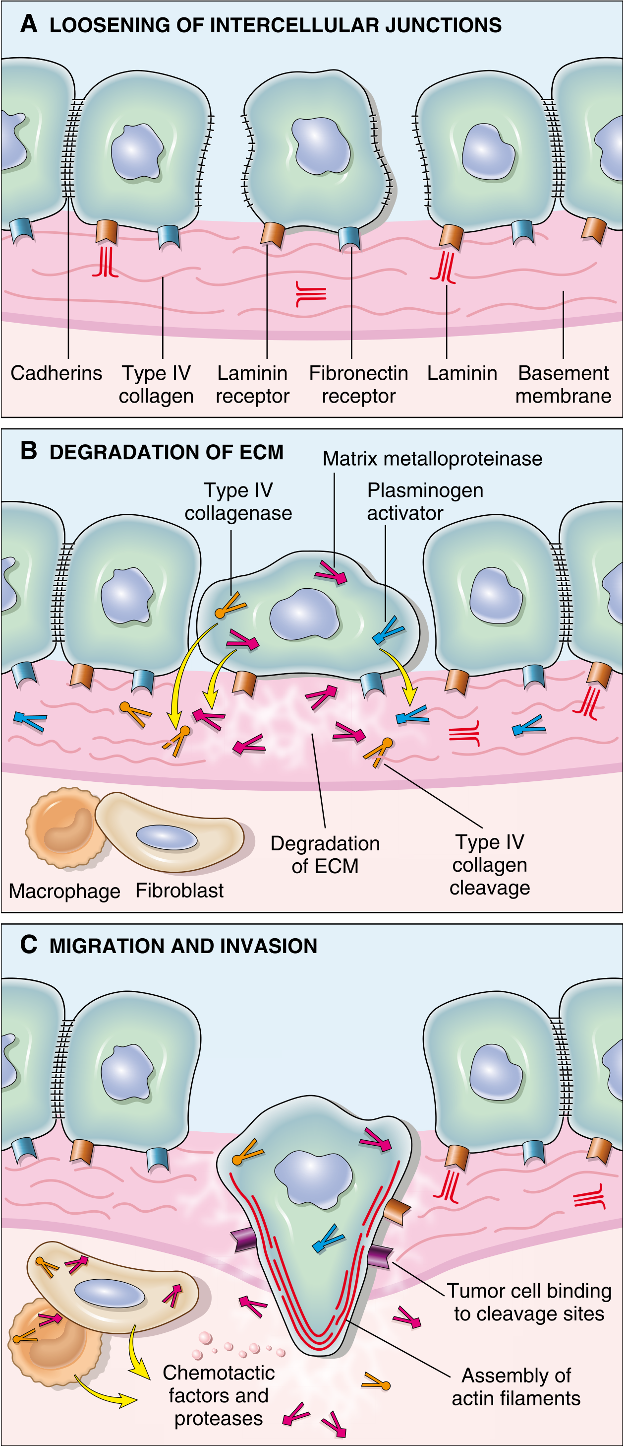
- Normal epithelial cells are tightly held together by E-cadherins - transmembrane glycoproteins that mediate homotypic adhesion and relay intercellular signals.
- In malignant tumors, E-cadherin function is lost due to:
- Mutations in the E-cadherin gene (CDH1) - seen in gastric and breast adenocarcinomas
- Transcriptional silencing through Epithelial-Mesenchymal Transition (EMT)
- EMT is a key process controlled by the transcription factors SNAIL and TWIST, which:
- Downregulate epithelial markers (E-cadherin)
- Upregulate mesenchymal markers (vimentin, smooth muscle actin)
- Produce a pro-migratory phenotype that facilitates invasion and metastasis
- Loss of E-cadherin removes the "glue" that holds cells together, enabling individual tumor cells to detach and become mobile.
Step 2 - Degradation of ECM (Enzymatic Destruction of Basement Membrane and Connective Tissue)
- Tumor cells degrade the ECM by secreting proteolytic enzymes or by inducing stromal cells (fibroblasts, macrophages) to do so.
- Key enzymes involved:
- Matrix metalloproteinases (MMPs) - the most important group. Particularly MMP-9 (gelatinase), which cleaves type IV collagen found in the epithelial and vascular basement membrane. MMP-9 also:
- Releases VEGF from ECM-sequestered pools (promoting angiogenesis)
- Generates collagen and proteoglycan cleavage products with chemotactic, angiogenic, and growth-promoting effects
- Cathepsin D - a lysosomal protease
- Urokinase plasminogen activator (uPA) - activates plasmin, which further degrades ECM
- Matrix metalloproteinases (MMPs) - the most important group. Particularly MMP-9 (gelatinase), which cleaves type IV collagen found in the epithelial and vascular basement membrane. MMP-9 also:
- Benign tumors (breast, colon, stomach) show little MMP-9 activity, while malignant counterparts overexpress it.
- Simultaneously, the concentrations of metalloproteinase inhibitors (TIMPs) are reduced in many cancers, tilting the balance further toward degradation.
Step 3 - Altered Cell Attachment to ECM (Changes in Integrin Expression)
- Integrins are transmembrane proteins that mediate cell-to-cell and cell-to-ECM adhesion.
- In normal epithelial cells, integrins that bind basement membrane laminin and collagens are restricted to the basal aspect - maintaining a resting, polarized state.
- Loss of adhesion in normal cells triggers anoikis (apoptosis induced by detachment).
- In tumor cells, however:
- The profile of integrins expressed is altered
- There is a shift toward integrins that bind degraded ECM components (fibronectin fragments, osteopontin, etc.) and provide traction for migration
- Tumor cells are able to migrate along cleavage sites created by MMP-mediated ECM destruction
- Tumor cells become resistant to anoikis, allowing survival after detachment
Step 4 - Migration and Invasion of Tumor Cells
- This is the final active phase in which tumor cells use the degraded ECM as a scaffold to migrate through tissues.
- Migration is driven by:
- Chemotactic factors - produced by inflammatory cells (macrophages) and stromal cells (fibroblasts). These include:
- Autocrine motility factors secreted by tumor cells themselves
- Cleavage products of ECM components (e.g., collagen fragments) acting as chemoattractants
- Growth factors such as IGF-I and IGF-II that stimulate cell locomotion
- Assembly of actin filaments at the leading edge of the cell - providing the cytoskeletal force for locomotion
- Tumor cells bind to newly exposed cleavage sites in the ECM and use them as "traction points" for forward movement
- Chemotactic factors - produced by inflammatory cells (macrophages) and stromal cells (fibroblasts). These include:
The Full Metastatic Cascade (Overview)
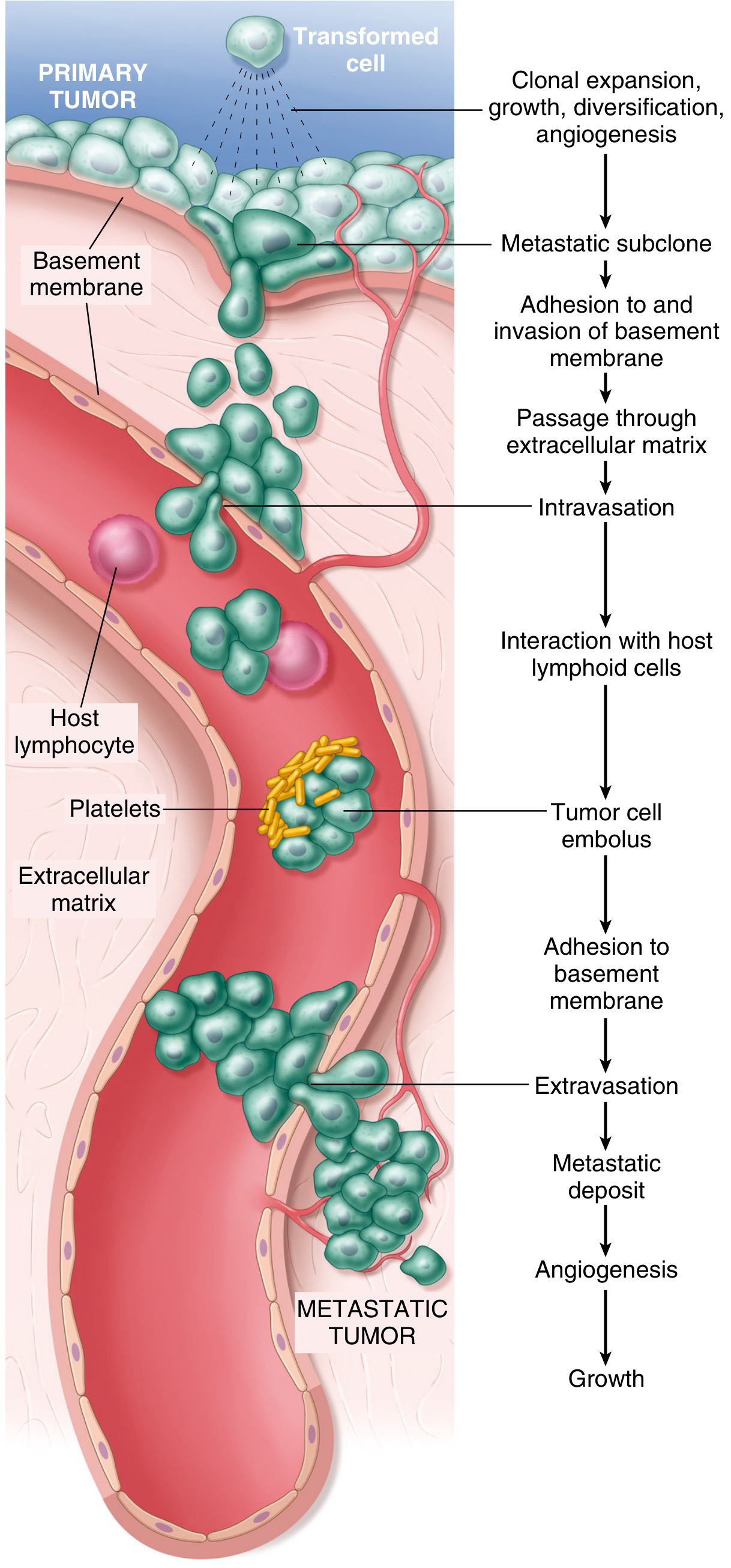
Once local invasion is complete, the metastatic cascade proceeds:
- Clonal expansion and angiogenesis in primary tumor
- Development of a metastatic subclone with additional mutations
- Adhesion to and invasion of basement membrane (as above)
- Passage through the ECM
- Intravasation - entry into blood vessels or lymphatics
- Interaction with host lymphoid cells; formation of tumor cell embolus (with platelets)
- Adhesion to vascular basement membrane at distant site
- Extravasation into the distant tissue
- Formation of metastatic deposit with new angiogenesis and growth
Key Molecular Players - Summary Table
| Molecule | Role in Invasion |
|---|---|
| E-cadherin | Cell-cell adhesion; loss allows detachment |
| SNAIL / TWIST | EMT transcription factors; suppress E-cadherin |
| Vimentin, SMA | Mesenchymal markers upregulated in EMT |
| MMP-9 | Cleaves type IV collagen in basement membrane |
| Cathepsin D | Lysosomal protease, degrades ECM |
| Urokinase (uPA) | Activates plasmin; ECM degradation |
| TIMPs | Inhibitors of MMPs; reduced in cancer |
| Integrins | Altered expression; traction for migration |
| VEGF | Released from ECM by MMP-9; promotes angiogenesis |
| Autocrine motility factors | Promote tumor cell locomotion |
Why Metastasis is Inefficient
Although many locally invasive cells enter the bloodstream each day, very few produce metastases. This is because the process requires:
- Overcoming immune defenses at every step
- Adapting to a completely different microenvironment at the distant site (e.g., bone marrow, brain, liver)
- Individual "metastasis genes" have not been identified because the metastatic phenotype likely requires the accumulation of multiple complementary genetic and epigenetic alterations
There is also evidence that cohesive groups of cells (not single cells) may migrate together and that successful metastases may arise from collaborating subclones, each contributing different functions to the process.
Content based on Robbins, Cotran & Kumar Pathologic Basis of Disease, which forms the foundational source for Ramdas Nayak's Essentials of Pathology on this topic.
This is a shared conversation. Sign in to Orris to start your own chat.