
Write notes on vitreous substitutes- types and indications
Reading File
Now let me search the medical library for vitreous substitutes content in parallel.
Finding Sources
Finding Sources
Reading File
Finding Sources
Finding Sources
Good. Now let me read the full vitreous substitutes section from Goodman & Gilman and also search for more ophthalmology-specific content.
Reading File
Finding Sources
Finding Sources
Finding Sources
Searching PubMed
vitreous substitutes tamponade agents retinal detachment
I have comprehensive information. Let me also check the Wills Eye Manual for more clinical detail on indications.
Finding Sources
I now have sufficient information to write comprehensive notes. Let me compile everything.
Vitreous Substitutes - Types and Indications
What is the Vitreous?
The vitreous humor is a clear, gel-like substance filling the posterior segment of the eye (~4 mL). It is composed primarily of water (~98%), collagen fibrils, and hyaluronic acid. During vitreoretinal surgery (vitrectomy), the vitreous is partially or completely removed and must be replaced with a substance that maintains the eye's shape, provides internal tamponade of retinal breaks, and prevents re-detachment.
Classification of Vitreous Substitutes
Vitreous substitutes fall into three main categories:
- Gases (air and expansile gases)
- Perfluorocarbon liquids (PFCLs)
- Silicone oils (including combinations)
1. GASES
Gases act as temporary vitreous substitutes. They provide internal tamponade by creating a surface-tension force at the gas-retina interface that seals retinal breaks.
A. Air
| Property | Detail |
|---|---|
| Duration | 3-7 days |
| Expansion | Non-expansile |
Properties:
- Least effective tamponade due to short duration
- Non-toxic, absorbed rapidly
- No special precautions for anesthesia
Indications:
- Fluid-air exchange at the end of vitrectomy
- Macular hole surgery (small holes)
- Short-term tamponade after simple retinal detachment repair
- Descemet's membrane detachment (anterior segment use)
B. Sulfur Hexafluoride (SF₆)
| Property | Detail |
|---|---|
| Duration | 10-14 days |
| Expansion | ~2x original volume |
Properties:
- Expansile gas - expands to approximately twice its injected volume by absorbing surrounding gases
- Typically used as a 20-25% concentration (non-expansile) or pure (expansile)
- In the anterior segment: used in non-expansile concentration after DSAEK/endothelial keratoplasty to press the lamellar graft against the posterior cornea; also used to treat Descemet's membrane detachments after cataract surgery
Indications (posterior segment):
- Rhegmatogenous retinal detachment (pneumatic retinopexy) - superior breaks
- Primary vitrectomy for uncomplicated retinal detachment
- Macular hole repair
Anterior segment indications:
- Post-DSEK/DSAEK to assist graft adherence (non-expansile concentration)
- Descemet's membrane detachment
C. Perfluoropropane (C₃F₈)
| Property | Detail |
|---|---|
| Duration | 55-65 days |
| Expansion | ~4x original volume |
Properties:
- Longest-lasting of the commonly used gases
- Expands to ~4 times injected volume
- Available in concentrations of 12-16% (non-expansile)
Indications:
- Giant retinal tears (GRT)
- Complex retinal detachment with PVR (proliferative vitreoretinopathy) - as an alternative to silicone oil for moderately severe cases
- Macular hole surgery (especially large holes, high myopia)
- Inferior retinal breaks (when patient can maintain prone positioning)
- Cases requiring longer tamponade than SF₆ provides but shorter than silicone oil
Critical anesthesia point: Patients with any intraocular gas (SF₆ or C₃F₈) must NOT receive nitrous oxide (N₂O) during general anesthesia. N₂O rapidly diffuses into the gas bubble, causing dangerous expansion and acute elevation of IOP, which can result in vision loss. This restriction continues for the duration of the gas bubble (up to 2 months for C₃F₈).
Complications of Gases (All Types)
- Elevated intraocular pressure (IOP)
- Corneal edema
- Posterior subcapsular cataract
- Subretinal gas if injected under the retina
- Optic nerve damage from IOP spikes
- Positional requirements (face-down positioning) can be burdensome
2. PERFLUOROCARBON LIQUIDS (PFCLs)
PFCLs are heavy liquids with a specific gravity of 1.76-1.94 (denser than vitreous at 1.005), which causes them to sink to the posterior pole and flatten the retina.
Common agents:
- Perfluoro-n-octane (C₈F₁₈)
- Perfluorodecalin
- Perfluoroperhydrophenanthrene
Properties:
- Immiscible with water; clear, colorless
- High density - sink posteriorly, push detached retina back
- Chemically inert but toxic to the retina with prolonged exposure
- Strictly intraoperative use only - must be removed at the end of surgery
Intraoperative Indications:
- Giant retinal tears (GRT) - excellent for unfolding the inverted retina; considered gold standard intraoperatively for GRT
- Posterior dislocated lens fragments or IOL - to float the nucleus/IOL forward
- Subretinal proliferation removal
- Stabilizing the posterior pole during complex membrane peeling
- Reattaching the posterior retina while working anteriorly
- Combined traction-rhegmatogenous retinal detachments
Why not permanent? Retained PFCL causes sterile inflammation, photoreceptor toxicity, and foreign body reactions - so it must be completely removed and replaced with a permanent substitute (silicone oil or gas) at the end of surgery.
3. SILICONE OIL (SO)
Silicone oil is the only long-term liquid vitreous substitute. It is composed of polydimethylsiloxane polymers.
Standard Silicone Oil (Lighter than Water)
| Property | Detail |
|---|---|
| Viscosity | 1000-5000 centistokes (cs) |
| Specific gravity | ~0.97 (less dense than water) |
| Refractive index | ~1.404 |
Properties:
- Floats on aqueous - provides tamponade of superior retinal breaks
- 1000 cs (lower viscosity): easier to inject/remove, slightly higher emulsification risk
- 5000 cs (higher viscosity): more resistant to emulsification, harder to remove
- Must be surgically removed (usually after 3-6 months)
Indications:
- Complex retinal detachment with severe PVR (grade C or higher) - the most common indication
- Giant retinal tears (GRT) especially when the patient cannot maintain face-down positioning
- Tractional retinal detachment (e.g., from proliferative diabetic retinopathy)
- Pediatric retinal detachments (unable to cooperate with positioning requirements of gas)
- HIV retinopathy / CMV retinitis with associated retinal detachment
- Ocular trauma with retinal detachment
- Cases where re-operation is anticipated
- Phthisis-prone eyes
- Inferior retinal breaks that cannot be treated adequately with gas
- When the patient cannot fly (gas bubbles expand at altitude)
Complications:
- Glaucoma (most common serious complication): emulsified droplets block trabecular meshwork
- Cataract (posterior subcapsular and nuclear)
- Corneal edema and band keratopathy (from emulsified oil in the anterior chamber, especially in aphakic eyes)
- Emulsification of oil over time
- Retinal toxicity (long-term)
- Hypotony after removal
- Inferior oil keratopathy in aphakes
Heavy Silicone Oil (HSO) / SO-PFA Combinations
| Property | Detail |
|---|---|
| Composition | Silicone oil + partially fluorinated alkanes (PFA) e.g., F4H5, F6H8 |
| Specific gravity | 1.02-1.06 (denser than water) |
Rationale: Standard silicone oil floats and is suboptimal for inferior retinal breaks. Heavy silicone oil sinks to the inferior pole and provides tamponade of inferior retinal breaks.
Properties:
- Density slightly greater than vitreous/aqueous
- FDA/CE approved combinations (e.g., Oxane HD - silicone oil + perfluorohexyloctane)
- Different refractive index than standard SO
Indications:
- Inferior retinal detachment with PVR
- Inferior giant retinal tears
- Cases where inferior tamponade is needed but patient cannot position face-down
- Lower retinal breaks not amenable to conventional SO or gas
Limitations:
- Risk of superior oil keratopathy
- Different emulsification profile
- Less clinical data compared to standard SO
COMPARISON TABLE
| Agent | Duration | Density | Position Required | Tamponades | Removal Needed |
|---|---|---|---|---|---|
| Air | 3-7 days | < water | Face down | Superior | No |
| SF₆ | 10-14 days | < water | Face down | Superior | No |
| C₃F₈ | 55-65 days | < water | Face down | Superior | No |
| PFCL | Hours (intraop) | > water | - | Posterior pole intraop | Yes (mandatory) |
| SO (standard) | Months | < water | Face up | Superior | Yes (3-6 mo) |
| Heavy SO | Months | > water | Face down / lateral | Inferior | Yes |
SPECIAL CONSIDERATIONS
Posturing Requirements
- Gas (air, SF₆, C₃F₈): Patients need face-down positioning to bring the gas bubble to tamponade superior or posterior breaks. Duration depends on the gas used.
- Standard SO: Patients should lie on their back (face up) so oil floats to tamponade superior breaks.
- Heavy SO: Patients lie face down to bring the heavy oil to the inferior retina.
Contraindications to Gas Tamponade
- Inability to maintain required positioning
- Contralateral poor vision (risk of temporary visual loss from the gas)
- Anticipated need for general anesthesia soon (N₂O risk)
- Air travel within the gas resorption period (expansion at altitude)
The "Ideal" Vitreous Substitute
No currently available substitute is ideal. The ideal vitreous substitute would be:
- Optically clear, non-toxic, biocompatible
- Stable over the long term without emulsification
- Correct density and viscosity to maintain retinal tamponade
- Self-resorbing with no surgical removal needed
- Able to serve as a drug delivery vehicle
Active research areas include hydrogels, synthetic polymers, and drug-eluting vitreous substitutes (PMID: 36125790, 33987966).
SOURCES
- Goodman & Gilman's The Pharmacological Basis of Therapeutics (Table 74-14, Vitreous Substitutes section)
- Shettigar MP et al. (2024). Vitreous substitutes and tamponades - A review. Indian Journal of Ophthalmology [PMID: 39078953]
- Siwik P et al. (2025). Retinal displacement following vitrectomy for RRD. J Clin Med [PMID: 39797332]
- Mondelo-Garcia C et al. (2021). Current Situation and Challenges in Vitreous Substitutes. Macromol Biosci [PMID: 33987966]
Short note on impression cytology
Finding Sources
Finding Sources
Finding Sources
Finding Sources
Searching PubMed
impression cytology ocular surface conjunctiva
Finding Sources
Finding Sources
Searching the Web
impression cytology ocular surface technique indications grading ophthalmology
Finding Sources
Reading Web Page
https://eyewiki.org/Impression_Cytology
Reading Web Page
https://pmc.ncbi.nlm.nih.gov/articles/PMC1772976
I now have comprehensive information to write detailed notes on impression cytology.
Impression Cytology - Short Note
Definition and Introduction
Impression cytology (IC) is a minimally invasive technique for sampling the superficial layers of the ocular surface epithelium. It was first described by Egbert et al. in 1977 for studying conjunctival goblet cells and has since become a valuable diagnostic and research tool in ocular surface disease.
Basic principle: A cellulose acetate filter paper (or other membrane) is applied to the ocular surface under gentle pressure. The adhesive affinity of the filter paper removes the topmost 2-3 layers of epithelial cells. These cells are then subjected to histological, immunohistochemical, or molecular analysis.
Relevant Anatomy
The conjunctiva is a translucent mucous membrane covering:
- The anterior surface of the globe (bulbar conjunctiva)
- The posterior surface of the eyelids (palpebral conjunctiva)
Goblet cells are mucin-secreting cells interspersed among the conjunctival epithelial cells. They:
- Arise from the basal layer of the epithelium
- Are most concentrated in the inferonasal quadrant
- Are responsible for the mucin layer of the tear film
- Are destroyed by chronic inflammation and severe dry eye
- Are a marker of conjunctival health - their density reflects ocular surface status
In limbal stem cell deficiency, conjunctivalization of the cornea occurs, and goblet cells appear on the corneal surface (normally goblet cell-free).
Technique
1. Patient Preparation
- Topical anesthesia is applied (e.g., proparacaine 0.5%), though the procedure can be done without it
- The ocular surface is dried carefully - excessive tears or medications are wiped away to maximize cell yield
- Patient looks in the direction opposite to the area being sampled
2. Filter Paper Preparation
- Standard filter: Cellulose acetate filter paper (Millipore, pore size 0.025-0.45 µm), cut into strips approximately 5 × 10 mm
- Newer alternatives: pure nitrocellulose membranes (better for immunocytochemistry)
- The filter is applied with firm, even pressure to the area of interest for 2-5 seconds
- Generally 2-3 layers of cells are removed per application
- Deeper layers can be sampled by repeat application at the same site
3. Sites of Sampling
- Bulbar conjunctiva (nasal and temporal quadrants most common)
- Tarsal conjunctiva
- Limbal area
- Corneal surface (when conjunctivalization is suspected)
4. Staining Methods
| Stain | Purpose |
|---|---|
| PAS (Periodic Acid-Schiff) | Stains goblet cells magenta; standard stain for goblet cell assessment |
| Haematoxylin and eosin (H&E) | General morphology |
| Papanicolaou (Pap) | Cytoplasmic and nuclear detail; used for OSSN (dysplasia) |
| Alcian blue | Mucin staining |
| Immunocytochemistry | CK4, CK13, CK7, MUC5AC, HLA-DR, p53, Ki-67 |
| Electron microscopy | Ultrastructural analysis (specialized fixatives required) |
For immunocytochemistry on cellulose acetate, xylene clearing must be avoided as it destroys cell surface antigens. Nitrocellulose membranes with spray fixative are preferred.
Grading Systems
Nelson's Classification (for squamous metaplasia)
The most widely used grading system, based on goblet cell density and epithelial cell morphology:
| Grade | Goblet Cell Density | Epithelial Cell Features | Interpretation |
|---|---|---|---|
| Grade 0 | > 500 cells/mm² | Small, round cells; large nuclei; high N:C ratio | Normal |
| Grade 1 | 100-500 cells/mm² | Intermediate morphology | Mild metaplasia |
| Grade 2 | < 100 cells/mm² | Large polygonal cells; small nuclei; low N:C ratio | Moderate metaplasia |
| Grade 3 | Absent | Large, polygonal, keratinized cells; pyknotic nuclei | Severe metaplasia |
Grade 2 or higher = Abnormal (pathological squamous metaplasia)
Tseng's Classification
Tseng classified conjunctival squamous metaplasia into 6 stages based on:
- Presence/absence and density of goblet cells
- Nuclear morphology and nucleus-to-cytoplasm (N:C) ratio
- Metachromatic changes in cytoplasmic color
- Emergence of keratinization
Indications
1. Dry Eye Disorders
- Keratoconjunctivitis sicca (KCS) / Sjögren's syndrome - assess goblet cell loss and squamous metaplasia
- Vitamin A deficiency - one of the earliest and most sensitive markers; xerophthalmia staging
- Superior limbic keratoconjunctivitis (SLK)
2. Cicatricial / Inflammatory Conditions
- Cicatricial ocular pemphigoid (MMP) - staging fibrosis and metaplasia
- Stevens-Johnson syndrome / Toxic epidermal necrolysis
- Trachoma
- Vernal keratoconjunctivitis (VKC) - eosinophils and goblet cell changes
- Atopic keratoconjunctivitis
- Graft-versus-host disease (GVHD) affecting the ocular surface
3. Limbal Stem Cell Deficiency (LSCD)
- Diagnosis of conjunctivalization of the cornea
- Monitoring after limbal stem cell transplantation
- Chemical/thermal burns
- Aniridia
4. Neoplastic / Pre-neoplastic Conditions
- Ocular Surface Squamous Neoplasia (OSSN) - dysplasia, CIN, SCC
- Sensitivity: ~80% accuracy for predicting histological diagnosis
- 77% predictability for moderate dysplasia
- Conjunctival melanoma / primary acquired melanosis (PAM)
- Differentiating benign vs. malignant lesions
5. Infections
- Acanthamoeba keratitis - IC has a yield as high as 94.6%, higher than corneal scraping in some reports
- Viral conjunctivitis (inclusion bodies)
- Chlamydial conjunctivitis
- Fungal keratitis
6. Systemic Disease Monitoring
- Mucopolysaccharidoses
- Epidermolysis bullosa
- Ichthyosis
- Psoriasis affecting the ocular surface
7. Treatment Monitoring
- Assessing response to topical cyclosporine, vitamin A drops, or anti-inflammatory therapy
- Post-limbal stem cell transplantation
- Documenting sequential changes over time (serial IC)
Advantages
- Non-invasive / minimally invasive - no surgical excision required
- Easy to perform in the outpatient clinic
- Minimal patient discomfort
- Can be repeated serially to monitor disease progression or treatment response
- Allows multiple types of analysis (histology, immunocytochemistry, molecular)
- Does not disturb the deeper epithelial layers
- No need for local injection or surgical setup
Limitations
- Only the superficial 2-3 epithelial layers are sampled - subepithelial pathology is not accessible
- Sampling variability - cell yield can vary considerably with technique
- Drying of the ocular surface is essential but technically challenging
- False negatives can occur in patchy lesions
- Cannot replace histopathological biopsy for definitive diagnosis of malignancy
- Immunocytochemistry on cellulose acetate is technically difficult (background staining, antigen destruction with xylene)
- Requires an experienced cytopathologist for interpretation
Summary
| Feature | Detail |
|---|---|
| First described | Egbert et al., 1977 |
| Material used | Cellulose acetate filter paper (or nitrocellulose) |
| Cells sampled | Superficial 2-3 layers of conjunctival/corneal epithelium |
| Primary stain | PAS (goblet cells), Pap smear (cytology/OSSN) |
| Key grading | Nelson's classification (Grade 0-3) |
| Main uses | Dry eye, OSSN, LSCD, infections, cicatricial disease |
| Key advantage | Non-invasive, repeatable |
| Key limitation | Superficial sampling only |
Sources:
- Singh R et al. (2005). Impression cytology of the ocular surface. Br J Ophthalmol [PMID: 15834085] - foundational review, 344 citations
- EyeWiki: Impression Cytology - American Academy of Ophthalmology
- Laver NMV (2021). Ocular cytology: Diagnostic features and ongoing practices. Cancer Cytopathology [PMID: 33136340]
Short note on choroidal effusion- clinical features and management
Finding Sources
Finding Sources
Excellent - two strong sources found. Let me read both in parallel.
Reading File
Reading File
Reading File
Finding Sources
Reading File
Searching Images
choroidal detachment effusion fundus orange brown elevation
I have all the information needed. Let me also fetch the textbook image.
Loading Image
I have all the information needed for comprehensive notes. Here they are:
Choroidal Effusion (Choroidal Detachment) - Short Note
Definition
Choroidal effusion (also called ciliochoroidal detachment or choroidal detachment) refers to the accumulation of fluid - either serous transudate or blood - in the suprachoroidal space (between the choroid and sclera). It is not a true detachment in the rhegmatogenous sense; there is no break in the choroid.
Types
| Type | Contents | IOP | Transillumination |
|---|---|---|---|
| Serous (Exudative) | Protein-rich fluid | Low (often < 6 mmHg) | Positive |
| Hemorrhagic | Blood | High (if large) | Negative |
Etiology
Serous Choroidal Effusion
1. Postoperative / Intraoperative (most common)
- Wound leak or filtering bleb over-filtration (after trabeculectomy / glaucoma drainage surgery)
- Perforation of the sclera from a superior rectus bridle suture
- After retinal detachment repair (scleral buckling or vitrectomy)
- After laser photocoagulation or cryotherapy
- Cyclodialysis cleft (traumatic or surgical)
- Post-cataract surgery
2. Traumatic
- Ruptured globe
- Blunt ocular trauma
3. Inflammatory
- Posterior uveitis
- Posterior scleritis
- Sympathetic ophthalmia
4. Drug-induced
- Sulfonamide drugs: acetazolamide, topiramate (ciliary body edema causing anterior rotation and angle closure)
5. Other
- Nanophthalmos / uveal effusion syndrome
- Carotid-cavernous fistula
- Primary or metastatic choroidal tumor
- Hypotony from any cause
- Chronic rhegmatogenous retinal detachment
Hemorrhagic Choroidal Detachment
- Intraoperative (expulsive hemorrhage): Rupture of short posterior ciliary arteries during anterior displacement of ocular contents - the most feared intraoperative complication
- Postoperative
- Spontaneous (especially in patients on anticoagulants)
- Perforation of a corneal ulcer
Clinical Features
Fundus Appearance
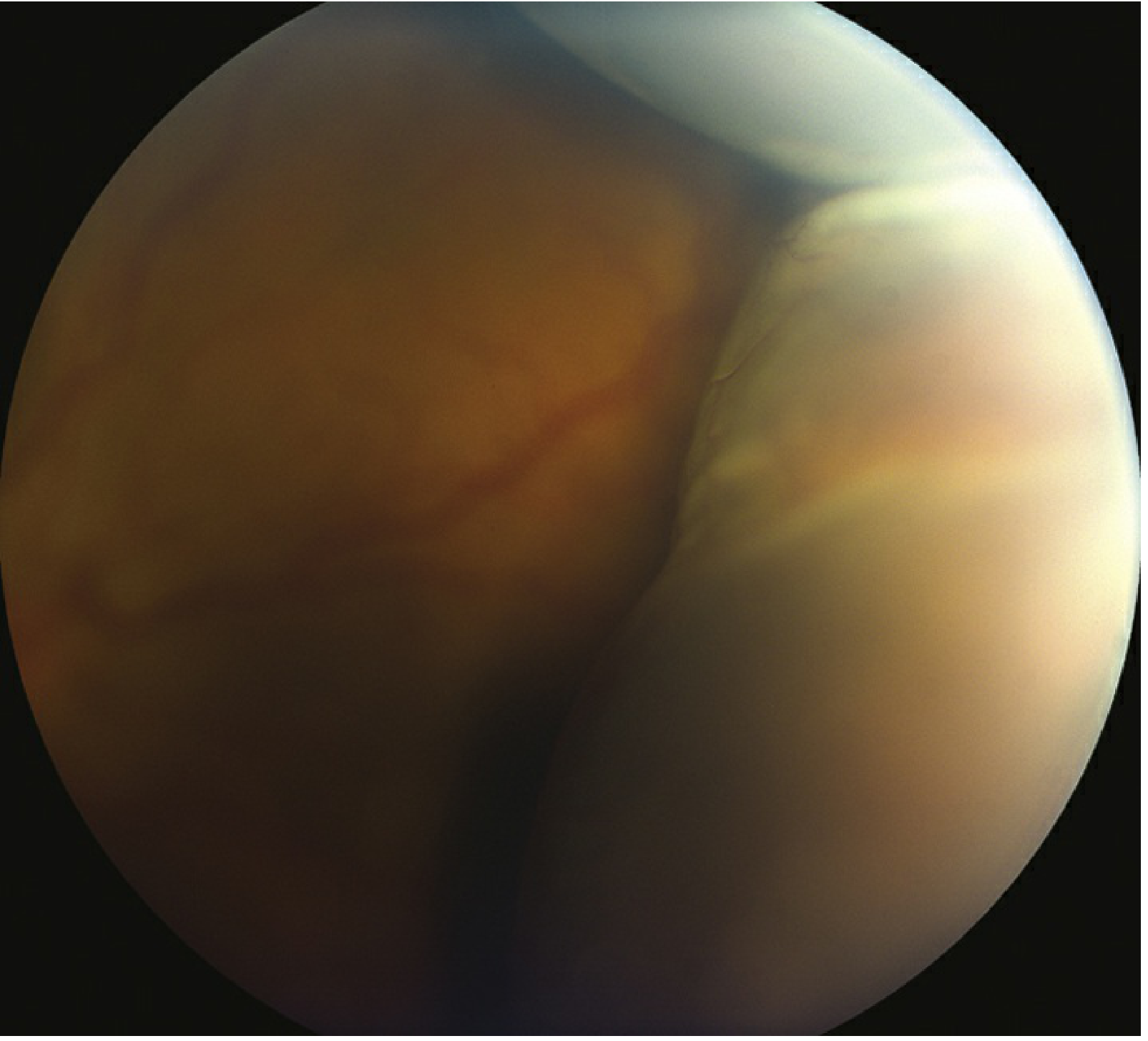
Critical finding: Smooth, bullous, orange-brown elevation of the retina and choroid, usually extending 360 degrees around the periphery in a lobular/multilobular configuration
- Four lobes typically present, separated by the vortex veins
- Temporal and nasal bullae tend to be most prominent
- Elevations are convex, smooth, and relatively immobile (unlike RD which undulates with eye movement)
- The ora serrata can be seen without scleral depression (elevations extend beyond the ora serrata anteriorly)
- Elevations do not extend to the posterior pole - limited by the vortex veins entering their scleral canals
- "Kissing choroidals" - when two large lobules of detached choroid appose each other in the central vitreous cavity, obscuring the view of the fundus
Symptoms
Serous:
- Decreased vision (if extends posteriorly or involves the macula)
- Visual field defect (peripheral shadow) if extensive
- No photopsia or floaters (no vitreoretinal traction)
- Often asymptomatic if peripheral
Hemorrhagic:
- Moderate to severe pain
- Red eye
- Sudden marked visual loss
Slit Lamp / Anterior Segment
| Finding | Serous | Hemorrhagic |
|---|---|---|
| IOP | Low (often < 6 mmHg) | High (if large) |
| Anterior chamber | Shallow | Shallow / flat |
| Cell and flare | Mild | Mild |
| Transillumination | Positive (fluid transmits light) | Negative (blood blocks light) |
Differential Diagnosis
| Condition | Distinguishing Features |
|---|---|
| Rhegmatogenous RD | White, undulates with eye movement; retinal break seen; pigment cells in vitreous |
| Melanoma of ciliary body | Not multilobular; no transillumination in pigmented tumors; B-scan distinguishes |
| Suprachoroidal hemorrhage | Negative transillumination; high IOP; B-scan shows echogenic contents |
| Exudative RD | Shifts with position; no retinal break |
Investigations (Workup)
- History - Recent ocular surgery, trauma, medication use (topiramate, acetazolamide), systemic disease
- Slit lamp examination - Anterior chamber depth, Seidel test (to rule out wound leak / filtering bleb leak)
- Gonioscopy - Look for cyclodialysis cleft
- Dilated fundus examination - Extent and location of choroidal elevation; assess for concomitant RD; examine the contralateral eye
- B-scan ultrasonography - Most useful investigation:
- Distinguishes serous (optically empty, low reflectivity) from hemorrhagic (echogenic)
- Determines if hemorrhage is mobile (fresh) or coagulated (organized, solid)
- Shows characteristic V-shaped or dome-shaped elevation
- Rules out intraocular tumor
- Transillumination of globe - Serous transilluminates; hemorrhagic does not
Management
General Treatment (Both Types)
- Cycloplegic - Atropine 1% three times daily (deepens anterior chamber, reduces ciliary spasm, prevents posterior synechiae)
- Topical corticosteroids - Prednisolone acetate 1% four to six times per day (reduces inflammation)
- Oral corticosteroids - Consider in inflammatory cases
Surgical Drainage - Indications
Drainage of suprachoroidal fluid is indicated for:
- Flat anterior chamber - especially with inflammation (risk of peripheral anterior synechiae, PAS)
- Lens-corneal touch (Grade III flat chamber) - surgical emergency; prompt correction needed to prevent corneal decompensation and lens opacification
- "Kissing" choroidals with:
- Intractable pain, or
- IOP elevation
- (Note: kissing choroidals alone can be tolerated if no pain or IOP rise)
- Progressive shallowing of anterior chamber unresponsive to medical treatment
Drainage technique: Partial-thickness sclerectomy (radial or posteriorly placed incisions over the suprachoroidal space, typically 4-5 mm behind the limbus) to allow egress of the fluid.
For hemorrhagic choroidal detachment, drainage is generally delayed 7-14 days to allow the clot to liquefy before attempting drainage.
Specific Treatment - Directed at Underlying Cause
| Cause | Specific Treatment |
|---|---|
| Wound / bleb leak | Patch 24h; reduce steroids; add aqueous suppressants; suture/glue the leak; bandage contact lens |
| Cyclodialysis cleft | Laser therapy, diathermy, cryotherapy, or surgical suturing of cleft |
| Uveitis | Topical cycloplegic + steroid; systemic immunosuppression |
| Drug-induced (topiramate/acetazolamide) | Stop offending drug |
| RD | Surgical retinal detachment repair |
| Hemorrhagic | Drainage after clot liquefaction (7-14 days) |
Uveal Effusion Syndrome (Special Entity)
A rare, idiopathic condition considered a subtype of serous choroidal effusion. Features:
- Affects middle-aged hypermetropic men predominantly
- Often bilateral
- Associated with nanophthalmos (abnormally small eye) or abnormal scleral thickness/composition
- Pathogenesis: impaired fluid drainage from choroid via sclera or vortex veins
- Fundus: ciliochoroidal detachment + exudative RD; on resolution, characteristic "leopard spot" RPE mottling due to protein-rich SRF
- Treatment: Full-thickness sclerectomy, particularly in nanophthalmos
Summary
| Feature | Serous | Hemorrhagic |
|---|---|---|
| Common cause | Post-glaucoma surgery, uveitis | Intraoperative (expulsive), anticoagulants |
| Symptom | Asymptomatic / mild | Pain, red eye, sudden visual loss |
| IOP | Low | High |
| Transillumination | + | - |
| B-scan | Optically empty | Echogenic |
| Medical Rx | Atropine + topical steroid | Same + stop anticoagulants |
| Surgery timing | When indicated | Delay 7-14 days for clot lysis |
Sources:
- The Wills Eye Manual, 8th ed. - §11.27 Choroidal Effusion/Detachment
- Kanski's Clinical Ophthalmology, 10th ed. - Choroidal Detachment & Uveal Effusion Syndrome
Long question- metabolic diseases involving ocular structure
Finding Sources
Finding Sources
Finding Sources
Finding Sources
Reading File
Finding Sources
Finding Sources
Finding Sources
Reading File
Finding Sources
Finding Sources
Reading File
Finding Sources
Metabolic Diseases Involving Ocular Structures
Introduction
The eye, as a metabolically active organ with diverse tissue types, is a window into systemic metabolic disease. Inborn errors of metabolism, endocrine disorders, storage diseases, and acquired metabolic disturbances all produce characteristic ocular signs - often pathognomonic for a specific disease. These are organized below by the primary ocular structure involved, then by disease category.
I. DISORDERS OF CARBOHYDRATE METABOLISM
1. Diabetes Mellitus
The most common metabolic disease with ocular manifestations. The eye is affected at nearly every level.
A. Diabetic Retinopathy (DR)
The leading cause of blindness in working-age adults.
Pathogenesis: Chronic hyperglycemia → pericyte loss → capillary leakage and occlusion → ischemia → VEGF-driven neovascularization
Classification:
| Stage | Features |
|---|---|
| Non-Proliferative DR (NPDR) | |
| Mild | Microaneurysms only |
| Moderate | Microaneurysms + dot/blot hemorrhages, hard exudates, cotton-wool spots |
| Severe (4-2-1 rule) | ≥20 hemorrhages in all 4 quadrants, OR venous beading in ≥2 quadrants, OR IRMA in ≥1 quadrant |
| Proliferative DR (PDR) | New vessels on disc (NVD) or elsewhere (NVE); vitreous hemorrhage; tractional RD |
| Diabetic Macular Edema | Clinically significant macular edema (CSME) - thickening within 500 µm of fovea |
B. Diabetic Cataract
- "Snowflake" or "metabolic" cataract in type 1 DM - bilateral, rapid subcapsular opacification from sorbitol accumulation
- Nuclear sclerosis accelerated in type 2 DM
- Mechanism: Aldose reductase converts glucose → sorbitol → osmotic damage to lens fibers
C. Rubeosis Iridis (Neovascular Glaucoma)
- New vessels on iris (rubeosis) from VEGF secretion in ischemic retina
- Progress to neovascular glaucoma with angle closure
D. Diabetic Corneal Changes
- Reduced corneal sensitivity (peripheral neuropathy)
- Persistent epithelial defects (diabetic keratopathy)
- Susceptibility to recurrent erosions
E. Refractive Changes
- Acute hyperglycemia → lens swelling → myopia (from increased refractive index)
- Glucose-induced osmotic shifts in lens
F. Cranial Nerve Palsies
- Ischemic mononeuropathy affects CN III, IV, VI
- Pupil-sparing CN III palsy is characteristic (ischemia spares outer pupillomotor fibers)
G. Optic Neuropathy
- Non-arteritic anterior ischemic optic neuropathy (NAION) - more common in diabetics
2. Galactosemia
Defect: Deficiency of galactose-1-phosphate uridyl transferase (classic) or galactokinase
Ocular manifestations:
- Cataract - the hallmark; may be the sole manifestation in galactokinase deficiency
- Characteristic "oil-droplet" opacity - seen with retroillumination, a central nuclear opacity resembling an oil droplet in water
- In classic galactosemia: associated with mental retardation, cirrhosis, sepsis
- May be reversible with early dietary galactose restriction (neonatal screening is key)
- Mechanism: galactose → galactitol accumulation in lens → osmotic damage
II. DISORDERS OF AMINO ACID METABOLISM
3. Homocystinuria
Defect: Cystathionine β-synthase deficiency → accumulation of homocysteine
Ocular manifestations:
- Ectopia lentis (lens subluxation) - most characteristic; typically bilateral, inferior and medial subluxation (downward and inward) - contrast with Marfan's where subluxation is superior and temporal
- Lens dislocation can be complete, causing acute angle-closure glaucoma
- Myopia (due to axial elongation and lens changes)
- Retinal detachment (secondary to lens dislocation)
- Optic atrophy (from vascular occlusions)
- Thromboembolic tendency → central retinal artery/vein occlusion
Systemic: Marfanoid habitus, intellectual disability, osteoporosis, thromboembolism
4. Alkaptonuria (Ochronosis)
Defect: Homogentisate oxidase deficiency → accumulation of homogentisic acid → polymerizes to ochronotic pigment
Ocular manifestations:
- Scleral ochronosis - brownish-black pigmentation of the sclera, most prominent in the horizontal palpebral fissure (anterior to the insertions of the lateral and medial rectus muscles) - pathognomonic
- Conjunctival pigmentation
- Corneal involvement (rare)
5. Tyrosinemia (Type II - Richner-Hanhart Syndrome)
Defect: Tyrosine aminotransferase deficiency → tyrosine crystal deposits
Ocular manifestations:
- Dendritic corneal ulcers (may be mistaken for herpetic keratitis)
- Corneal crystals
- Photophobia and lacrimation
- Associated with palmoplantar keratoderma and intellectual disability
III. DISORDERS OF LIPID / SPHINGOLIPID METABOLISM (STORAGE DISEASES)
6. Tay-Sachs Disease (GM₂ Gangliosidosis)
Defect: Hexosaminidase A deficiency → GM₂ ganglioside accumulation in neurons
Ocular manifestation:
- Cherry-red spot at the macula - the single most important sign
- Pathogenesis: ganglioside accumulation in all ganglion cells of the retina → retina appears white/opaque; however, the foveola has no ganglion cells → underlying choroidal circulation shows through as a red spot against the white surrounding retina
- Progressive optic atrophy and blindness
- Also seen in: GM₁ gangliosidosis, Niemann-Pick disease, Sandhoff disease, metachromatic leukodystrophy (rarely), Farber disease, sialidosis
7. Niemann-Pick Disease
Defect: Sphingomyelinase deficiency (Type A/B) or NPC1/NPC2 (Type C)
Ocular manifestations:
- Cherry-red spot (Types A and B)
- Ocular motor apraxia (inability to initiate voluntary saccades) - Types C and D
- Impairment of vertical gaze (Type C - supranuclear vertical gaze palsy)
- Macular degeneration
8. Gaucher Disease
Defect: Glucocerebrosidase deficiency
Ocular manifestations:
- Pinguecula-like conjunctival changes (wedge-shaped yellow-brown triangular deposits near limbus)
- Ocular motility defects - jerky eye movements, limited abduction (late infantile form)
- Macular degeneration (occasionally)
9. Fabry Disease (X-Linked Lysosomal Storage Disorder)
Defect: Alpha-galactosidase A deficiency → glycosphingolipid accumulation
Ocular manifestations:
- Cornea verticillata (vortex keratopathy) - bilateral whorl-like subepithelial opacities radiating from a central point, visible on slit-lamp; does not affect vision but is virtually pathognomonic
- Spoke-wheel lens opacities - anterior and posterior subcapsular cataracts in a spoke-wheel pattern
- Dilated tortuous conjunctival and retinal vessels ("Fabry vessels")
- Retinal vascular occlusions
Note: Cornea verticillata is also caused by amiodarone, chloroquine, indomethacin - drug-induced keratopathy (important differential)
10. Mucopolysaccharidoses (MPS)
Common feature: Glycosaminoglycan (GAG) accumulation in tissues
| MPS Type | Enzyme Deficiency | Corneal Clouding | Retinal Degeneration | Other Ocular |
|---|---|---|---|---|
| I-H (Hurler) | α-L-iduronidase | Severe | + | Optic atrophy, glaucoma |
| I-S (Scheie) | α-L-iduronidase | Severe | + | Normal intelligence |
| II (Hunter) | Iduronate-2-sulfatase | Absent | + | Papilledema |
| III (Sanfilippo) | Heparan sulfatase | Mild/absent | + | Mainly CNS |
| IV (Morquio) | N-acetylgalactosamine sulfatase | Present | - | |
| VI (Maroteaux-Lamy) | Arylsulfatase B | Severe | - | Optic atrophy |
Corneal clouding mechanism: GAG deposits (dermatan sulfate, heparan sulfate) accumulate in corneal stroma → diffuse bilateral haziness
IV. DISORDERS OF COPPER METABOLISM
11. Wilson's Disease (Hepatolenticular Degeneration)
Defect: ATP7B mutation → impaired hepatic copper excretion → copper accumulates in liver, brain, cornea, kidney
Ocular manifestations:
A. Kayser-Fleischer (KF) Ring - PATHOGNOMONIC
- Golden-to-greenish-brown ring in the peripheral cornea (Descemet's membrane)
- Copper deposits in Descemet's membrane; fluid streaming favors accumulation near the limbus
- Starts at the superior and inferior poles (12 and 6 o'clock), then progresses circumferentially
- Best seen with slit-lamp examination; may not be visible to the naked eye in early stages
- Present in virtually 100% of patients with neurological Wilson's disease
- May disappear with successful chelation therapy (penicillamine, trientine)
B. Sunflower Cataract
- A disc-shaped, greenish-brown posterior subcapsular cataract with petal-like projections resembling a sunflower
- Due to copper deposition in the lens
- Rare but specific for Wilson's disease
- Does not impair vision significantly
- Also resolves with copper chelation
V. DISORDERS OF PURINE METABOLISM
12. Gout (Hyperuricemia)
Defect: Uric acid accumulation → monosodium urate crystal deposition
Ocular manifestations:
- Episcleritis and scleritis - urate deposits in episcleral/scleral tissue causing inflammation
- Band keratopathy - calcium deposits (secondary to chronic uveitis from gout)
- Urate crystals in conjunctiva and cornea - rare direct deposits
- Acute anterior uveitis - urate crystal-induced inflammation
- Corneal urate crystalline keratopathy - rare, subepithelial yellow-white deposits
VI. ENDOCRINE METABOLIC DISORDERS
13. Thyroid Eye Disease (Graves' Ophthalmopathy / TED)
The most common cause of bilateral and unilateral proptosis in adults
Pathogenesis: TSH-receptor antibodies → fibroblast activation in the orbit → GAG deposition → orbital fat and extraocular muscle enlargement → exophthalmos
Ocular manifestations:
A. Lid Signs (due to sympathetic overactivity and lid retractor involvement)
- Lid retraction - most common sign; Dalrymple's sign (upper lid retraction - sclera visible above limbus)
- Lid lag - von Graefe's sign (upper lid lags behind globe on downgaze)
- Stellwag's sign (infrequent blinking)
- Joffroy's sign (absent forehead wrinkling on upgaze)
B. Proptosis (Exophthalmos)
- Usually bilateral but often asymmetric
- Hertel exophthalmometry measures the degree
C. Extraocular Muscle Involvement
- Restrictive myopathy from muscle fibrosis
- Order of frequency: Inferior rectus > Medial rectus > Superior rectus > Lateral rectus (mnemonic: I'M SLow or I'M a Slow Learner)
- Inferior rectus restriction → limitation of upgaze (most common motility complaint)
- Diplopia on extremes of gaze
D. Optic Neuropathy (Dysthyroid Optic Neuropathy)
- From compression of optic nerve at orbital apex by enlarged extraocular muscles
- Presents with decreased vision, color vision defect, RAPD
- Most serious complication of TED
E. Corneal/Conjunctival Complications
- Exposure keratopathy from incomplete lid closure (lagophthalmos)
- Conjunctival chemosis and injection
- Superior limbic keratoconjunctivitis (SLK)
Clinical Activity Score (CAS): Used to assess activity - pain, redness, swelling, impaired function (score ≥3/7 = active disease)
14. Hypothyroidism
Ocular manifestations:
- Periorbital puffiness/myxedema (mucopolysaccharide infiltration of dermis)
- Loss of outer third of eyebrows (Hertoghe's sign)
- Dry eye
- Delayed relaxation of extraocular movements
VII. DISORDERS OF CALCIUM METABOLISM
15. Hypercalcemia (Hyperparathyroidism, Sarcoidosis, Vitamin D toxicity)
Ocular manifestations:
- Band keratopathy - calcium (calcium hydroxyapatite) deposits in Bowman's layer, initially at the 3 and 9 o'clock positions (within the palpebral aperture), eventually forming a band across the cornea with characteristic "Swiss cheese" holes (from corneal nerve perforations)
- Conjunctival calcium deposits ("conjunctival calcification")
- Calcium deposits in eyelid skin (metastatic calcification)
VIII. LIPID DISORDERS
16. Hyperlipidemias
Ocular manifestations:
- Arcus senilis (Corneal arcus) - bilateral white-grey lipid (cholesterol ester) arc/ring in the peripheral stroma with a clear zone between it and the limbus; normal in elderly but arcus in patients < 45 years suggests hyperlipidemia (Type II)
- Xanthelasma - yellowish cholesterol deposits in the nasal eyelid skin (particularly type IIa and III hyperlipoproteinemia)
- Xanthomas of eyelids - also represent lipid deposits
- Lipemia retinalis - triglycerides > 2000 mg/dL → creamy-white retinal vessels ("salmon-colored" arteries and veins) from chylomicrons; typically in type I or V hyperlipoproteinemia
IX. DISORDERS OF AMINO ACID TRANSPORT
17. Cystinosis
Defect: Lysosomal cystine transport defect → cystine crystal deposition throughout the body
Ocular manifestations:
- Corneal cystine crystals - polychromatic, needle-shaped crystals distributed throughout the full-thickness corneal stroma; visible on slit-lamp; cause photophobia, blepharospasm, and eventually corneal erosions
- The most characteristic ocular sign; corneal crystals develop in the first year of life
- Retinal pigment epithelial changes ("salt-and-pepper" retinopathy)
- Photophobia is often the presenting complaint
- Treatment: Topical cysteamine drops dissolve corneal crystals
18. Marfan Syndrome (Fibrillin-1 Defect / Connective Tissue - Associated)
Ocular manifestations:
- Ectopia lentis - bilateral, typically superior and temporal subluxation of the lens (zonular dehiscence); in ~60% of patients
- Iridodonesis (trembling of the iris with eye movement - loss of zonular support)
- High myopia (axial elongation)
- Flat cornea (decreased keratometry)
- Increased risk of rhegmatogenous retinal detachment
- Megalocornea, glaucoma
Comparison of ectopia lentis:
- Marfan syndrome: superotemporal subluxation
- Homocystinuria: inferonasal subluxation
- Weill-Marchesani: microspherophakia, anteroinferior subluxation
X. LYSOSOMAL ENZYME DEFICIENCY
19. Metachromatic Leukodystrophy (MLD)
- Optic atrophy and blindness (progressive)
- Brown-colored macula (granular pigmentation)
- Nystagmus
- Absent foveal reflex
SUMMARY TABLE: Key Ocular Signs in Metabolic Diseases
| Ocular Sign | Disease |
|---|---|
| Cherry-red spot | Tay-Sachs, GM₁ gangliosidosis, Niemann-Pick, Sandhoff, sialidosis, CRAO |
| Corneal clouding | Hurler/Scheie/Morquio/Maroteaux-Lamy MPS, mucolipidosis, cystinosis, tyrosinemia |
| Corneal crystals | Cystinosis, gout (rare), hyperuricemia |
| Cornea verticillata | Fabry disease, amiodarone, chloroquine |
| Kayser-Fleischer ring | Wilson's disease (pathognomonic) |
| Sunflower cataract | Wilson's disease |
| Oil-droplet cataract | Galactosemia |
| Posterior subcapsular cataract | Diabetes, corticosteroids, irradiation |
| Ectopia lentis (superior) | Marfan syndrome |
| Ectopia lentis (inferior) | Homocystinuria |
| Band keratopathy | Hypercalcemia, gout, chronic uveitis, sarcoidosis |
| Arcus senilis in young | Hyperlipidemia (Type II) |
| Xanthelasma | Hyperlipidemia |
| Lipemia retinalis | Severe hypertriglyceridemia (Types I, V) |
| Proptosis (bilateral) | Thyroid eye disease (Graves') |
| Rubeosis iridis | Diabetes, CRVO, ocular ischemic syndrome |
| Scleral ochronosis | Alkaptonuria |
| Retinal pigmentary degeneration | MPS, neuronal ceroid lipofuscinosis |
| Spoke-wheel lens | Fabry disease |
| Optic atrophy | MLD, neuroaxonal dystrophy, Wilson's disease |
Sources:
- Kanski's Clinical Ophthalmology, 10th ed. - Thyroid Eye Disease, Ectopia Lentis, Uveal conditions
- The Wills Eye Manual, 8th ed. - Cataract Etiology, Metabolic Cataracts
- Adams & Victor's Principles of Neurology, 12th ed. - Ophthalmic Signs in Metabolic/Storage Diseases
- Harrison's Principles of Internal Medicine, 22nd ed. - Wilson's Disease, Graves' Ophthalmopathy
- Goldman-Cecil Medicine - Wilson's Disease
- Sleisenger & Fordtran's GI and Liver Disease - Kayser-Fleischer ring
This is a shared conversation. Sign in to Orris to start your own chat.