
Give me the abdominal blood supply and its branches
abdominal aorta branches diagram anatomy
Give me the image abdominal blood supply and its branches
abdominal aorta branches blood supply diagram anatomy
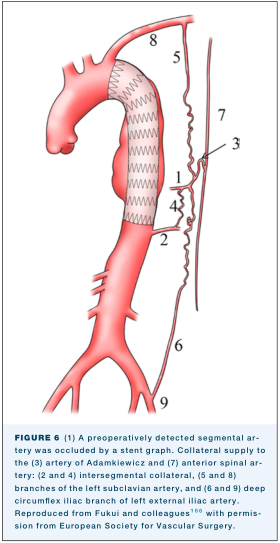
Summary : This is a medical illustration showing the anatomy of the thoracic and abdominal aorta, with a stent graft in place, and the collateral arterial supply to the spinal cord. The diagram is annotated with numbered arteries and collateral pathways, referencing their anatomical names and clinical significance. illustration: # Scene Overview : • Main subject: The thoracic and abdominal aorta, with a stent graft placed in the descending thoracic aorta. • Perspective: Sagittal (side) view of the aorta and its major branches. • Composition: The aorta is centrally depicted, with collateral arteries and spinal cord shown to the right. • Colour palette: Red for arteries, grey for stent graft, black for annotation numbers and lines. # Technical Details : • Stent graft: Shown as a grey, zigzag-patterned tube within the descending thoracic aorta. • Numbered arteries: – 1: Preoperatively detected segmental artery (occluded by stent graft). – 2, 4: Intersegmental collateral arteries. – 3: Artery of Adamkiewicz. – 5, 8: Branches of the left subclavian artery. – 6, 9: Deep circumflex iliac branch of left external iliac artery. – 7: Anterior spinal artery. • Collateral pathways: Wavy lines connecting the aorta to the spinal cord, indicating alternate blood supply routes. # Spatial Relationships : • The stent graft is placed in the upper descending thoracic aorta, occluding the segmental artery (1). • Collateral arteries (2, 4, 5, 6, 8, 9) are shown branching from the aorta and iliac arteries, connecting to the spinal cord. • The artery of Adamkiewicz (3) and anterior spinal artery (7) are highlighted as key vessels for spinal cord perfusion. # Analysis : • The illustration emphasizes the importance of collateral circulation to the spinal cord when a segmental artery is occluded by a stent graft. • Multiple collateral pathways are depicted, suggesting redundancy in spinal cord blood supply. • The diagram is useful for understanding vascular anatomy relevant to aortic stent grafting and spinal cord protection.
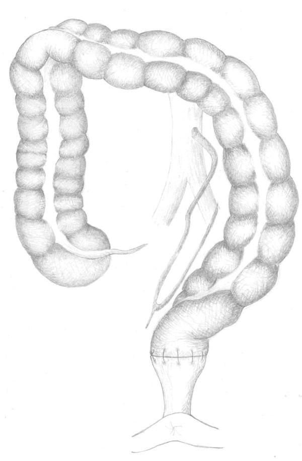
This medical anatomical illustration depicts the surgical anatomy of the colon and its arterial supply following a 'low tie' ligation of the inferior mesenteric artery (IMA). The diagram shows the colon with characteristic sacculated haustra, looping through the abdominal cavity. A significant portion of the distal colon is shown undergoing an anastomosis, indicated by visible surgical sutures at the junction with the rectum. The vascular network illustrates the preservation of the left colic artery while showing ligation of the IMA branches distal to its origin from the aorta. The arterial branches are shown traveling through the mesentery to reach the mesenteric border of the colon, providing collateral circulation to the splenic flexure and descending colon. This illustration serves as an educational resource for colorectal surgery, specifically demonstrating oncological resection techniques and the preservation of blood supply to the conduit during low anterior resections.
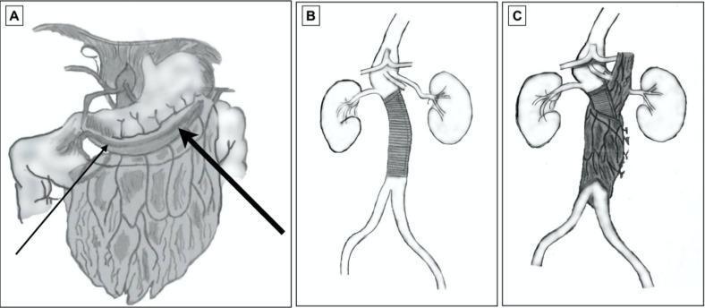
A three-part anatomical diagram (A-C) illustrating the surgical technique of omental wrapping for an abdominal aortic graft. Panel A shows the preparation of the greater omentum, highlighting its vascular supply from the gastroepiploic arteries; a thin arrow indicates the right gastroepiploic artery and a thick arrow indicates the left gastroepiploic artery along the greater curvature of the stomach. Panel B depicts the abdominal aorta following the implantation of a tubular Dacron graft in the infrarenal position, situated between the renal arteries and the iliac bifurcation. Panel C demonstrates the completed procedure where a vascularized pedicle of the greater omentum is circumferentially wrapped around the Dacron graft. This technique is typically used in the management of infected aortic aneurysms or complex reconstructions to provide a viable blood supply, facilitate infection control, and protect the prosthetic material. The diagram serves as an educational tool for vascular surgery, focusing on surgical anatomy and tissue flap transposition.
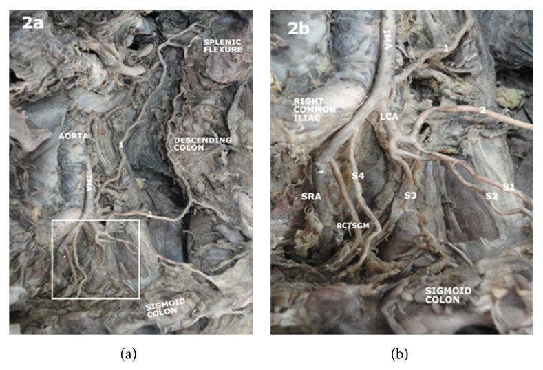
This clinical photograph of a gross anatomical dissection (Figures 2a and 2b) displays the posterior abdominal wall, focusing on the arterial supply to the distal colon. The abdominal aorta is visible, giving rise to the inferior mesenteric artery (IMA). The IMA originates and subsequently branches into the left colic artery (LCA), which provides a transverse branch to the descending colon near the splenic flexure. The dissection illustrates a specific anatomical variation where the LCA continues inferiorly to give rise to multiple sigmoid arteries (labeled S1 through S4) directed toward the sigmoid colon. Further distal branching includes the rectosigmoid branch (RCTSGM) and the terminal superior rectal artery (SRA). Figure 2b provides a magnified view of the vascular bifurcation, highlighting the spatial relationship between the IMA, LCA, and the right common iliac artery. This educational material is designed to demonstrate mesenteric vascular anatomy and potential variations in the blood supply to the hindgut for surgical and anatomical study.
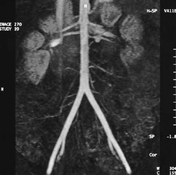
This diagnostic image is a Magnetic Resonance Angiography (MRA) or Angio-MRI of the abdominal aorta and its major branches, presented in a coronal Maximum Intensity Projection (MIP). The central vascular structure is the abdominal aorta, which descends vertically with a straight course. It shows bilateral branching into the renal arteries, which supply the kidneys visualized as paired, mottled, bean-shaped lateral masses. Inferiorly, the aorta exhibits a clear bifurcation at the level of the aortic bifurcation into the left and right common iliac arteries. Smaller mesenteric and lumbar arterial branches are visible along the aortic trunk. The image serves as an educational tool for studying abdominal vascular anatomy, the renal blood supply, and the primary arterial distribution in the retroperitoneum. Clinical relevance includes the assessment of vascular patency, stenosis, or aneurysmal changes in the aorta and iliac system.
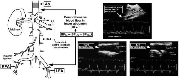
This composite educational graphic combines an anatomical line diagram and duplex ultrasound imaging to illustrate the methodology for calculating comprehensive lower abdominal blood flow (BFAb). The anatomical schematic depicts the abdominal aorta (Ao) and its primary branches, including the coeliac artery (CA), superior mesenteric artery (SMA), renal artery (RA), and inferior mesenteric artery (IMA). Shaded regions indicate Doppler measurement sites on the supra-coeliac aorta and the bilateral right and left femoral arteries (RFA, LFA) distal to the inguinal ligament. Three ultrasound panels provide B-mode anatomical views and pulsed-wave Doppler spectrograms. The abdominal aorta panel shows a sharp, pulsatile waveform with minimal reverse flow. In contrast, the RFA and LFA panels display classic triphasic arterial waveforms, characterized by a rapid systolic peak, early diastolic flow reversal, and a small late diastolic forward component. A mathematical formula defines BFAb as the difference between aortic blood flow and the sum of bilateral femoral arterial flow, representing total perfusion to the splanchnic, renal, and pelvic regions.
celiac trunk superior mesenteric inferior mesenteric artery anatomy
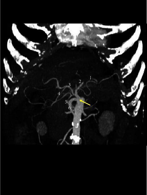
This diagnostic image is a 3D volume rendering CT scan illustrating a rare vascular anomaly known as a complete celiac-mesenteric trunk (CMT), indicated by a yellow arrow. The image demonstrates a single arterial trunk originating from the abdominal aorta, giving rise to both celiac and superior mesenteric arterial branches. Labeled anatomical structures include the splenic artery (1) coursing toward the left, the left gastric artery (2) ascending superiorly, and the common hepatic artery (3) extending toward the right, which further branches into the gastroduodenal artery (4). Mesenteric components are represented by the inferior pancreaticoduodenal artery (5) and superior mesenteric ramifications (6) descending inferiorly. This clinical imaging is essential for surgical planning and understanding congenital variations in abdominal arterial anatomy. The visualization uses high-contrast reconstruction to delineate the complex branching patterns and spatial relationships of the visceral vasculature relative to the vertebral column and ribs.
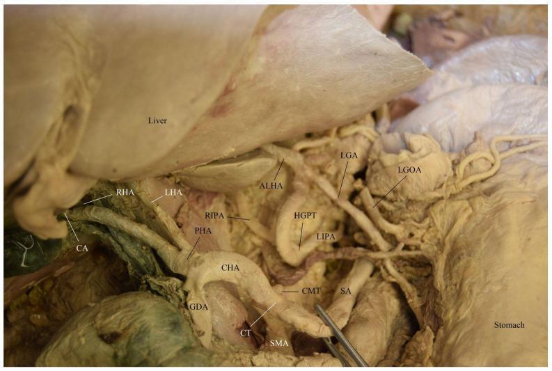
This clinical photograph displays a detailed cadaveric dissection of the upper abdominal vasculature, focusing on rare anatomical variations of the celiac region. The primary feature is a celiacomesenteric trunk (CMT), where the celiac trunk (CT) and superior mesenteric artery (SMA) share a common origin. The CT further bifurcates into the splenic artery (SA) and the common hepatic artery (CHA). Adjacent to this, a distinct hepatogastrophrenic trunk (HGPT) is visible, giving rise to the left gastric artery (LGA), left inferior phrenic artery (LIPA), and an accessory left hepatic artery (ALHA). Further branching of the hepatic system shows the CHA dividing into the gastroduodenal artery (GDA) and proper hepatic artery (PHA), with the latter branching into the right hepatic artery (RHA), left hepatic artery (LHA), and right inferior phrenic artery (RIPA). The cystic artery (CA) is seen near the hepatic hilum. Key landmarks include the liver (superiorly retracted) and the stomach. This image serves as an advanced educational resource for surgical anatomy, illustrating complex vascular anomalies critical for hepatobiliary and gastric surgical planning.
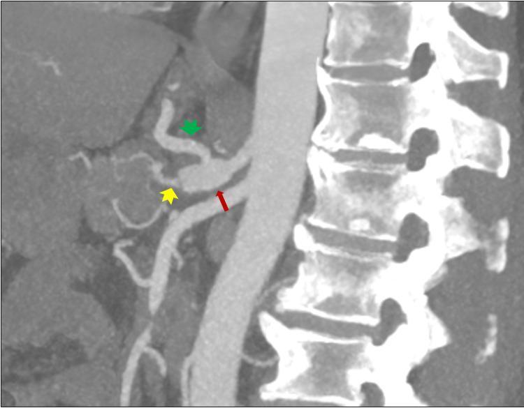
This diagnostic image is a sagittal Maximum Intensity Projection (MIP) from a CT Angiography (CTA) of the upper abdomen, demonstrating the vascular anatomy of the celiac axis. The image shows the abdominal aorta running anterior to the lumbar vertebral bodies. A red arrow highlights a focal dilatation of the celiac trunk at its origin. Branching from this trunk, a green arrowhead identifies a patent and prominent left gastric artery, while a yellow arrowhead indicates the splenic artery, which appears patent but exhibits a thick-walled morphology. The superior mesenteric artery is also visible inferior to the celiac trunk. This clinical imaging is essential for evaluating vascular pathologies such as aneurysmal dilatation, stenosis, or occlusion in patients presenting with acute abdominal symptoms. The anatomical relationship between the major visceral arteries and the spine is clearly depicted, serving as an educational tool for radiology and vascular surgery specialties.
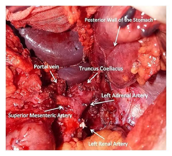
This clinical photograph provides an intraoperative view of the retroperitoneum and upper abdominal cavity following an extensive en bloc tumor resection. The image displays critical vascular anatomy and organ structures with white labels and arrows. Centrally, the celiac trunk (truncus coeliacus) is shown after resection of its primary branches. Positioned superiorly is the portal vein, visible as a large caliber vessel. The superior mesenteric artery is identified inferior to the celiac origin. In the lower portion of the surgical field, the left renal artery and a small left adrenal artery are visualized. To the right of the vessel complex, the posterior wall of the stomach is visible after partial gastrectomy. The surgical field exhibits evidence of multi-organ resection, including distal pancreatectomy and splenectomy, with extensive mobilization of the retroperitoneal tissue. This image is a high-level educational resource for oncological surgery, demonstrating the complex relationships of the mesenteric and celiac vasculature during major abdominal tumor debulking.
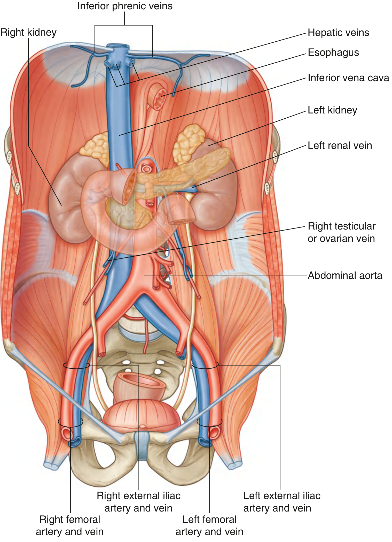
"Fig. 4.167" in /textbooks/9780323934237
Abdominal Blood Supply - The Abdominal Aorta and Its Branches
Overview
MRA showing the abdominal aorta and its bifurcation:
Anatomical overview - posterior abdominal wall (Gray's Anatomy, Fig. 4.169):
Branches - Classified into 3 Groups
1. VISCERAL BRANCHES
| Artery | Level | Supplies |
|---|---|---|
| Celiac trunk | T12 - just below aortic hiatus | Abdominal foregut (stomach, liver, gallbladder, spleen, upper duodenum, pancreas) |
| Superior mesenteric artery (SMA) | L1 - just below celiac | Abdominal midgut (lower duodenum to 2/3 transverse colon) |
| Inferior mesenteric artery (IMA) | L3 - below renal arteries | Abdominal hindgut (left 1/3 transverse to upper rectum) |
| Artery | Level | Supplies |
|---|---|---|
| Middle suprarenal arteries | Just above renal arteries | Suprarenal (adrenal) glands |
| Renal arteries | L1-L2, below SMA | Kidneys |
| Testicular / Ovarian arteries | Below renal arteries | Testes (male) / Ovaries (female) |
2. POSTERIOR BRANCHES (Parietal - supply body wall & diaphragm)
| Artery | Origin | Supplies |
|---|---|---|
| Inferior phrenic arteries | Just below aortic hiatus (or from celiac trunk) | Inferior diaphragm + suprarenal glands |
| Lumbar arteries (4 pairs) | Posterior surface of aorta | Posterior abdominal wall + spinal cord (segmental) |
| Median sacral artery | Just above bifurcation (L4) | Sacrum, coccyx |
3. TERMINAL BRANCHES
| Artery | Notes |
|---|---|
| Right common iliac artery | Divides into right internal + external iliac |
| Left common iliac artery | Divides into left internal + external iliac |
Celiac Trunk - 3D CT showing branches:
Key Relations of the Abdominal Aorta
- Anterior: Pancreas and splenic vein, left renal vein, inferior duodenum, prevertebral nerve plexus
- Right: IVC, cisterna chyli, thoracic duct, azygos vein, right crus of diaphragm
- Left: Left crus of diaphragm
Quick Memory Aid
"I Love Sipping Really Cold Margaritas"
- Inferior phrenic
- Lumbar (x4 pairs)
- Suprarenal (middle)
- Renal
- Celiac trunk
- Mesenteric (superior then inferior)
- Testicular/Ovarian → then bifurcation at L4