
Surface marking of lung
Reading File
Searching sources...
Finding Sources
Finding Sources
Finding Sources
Finding Sources
Finding Sources
Finding Sources
Reading File
Finding Sources
Reading File
Reading File
Excellent content found. Let me fetch the key diagram images.
Loading Image
Loading Image
Reading File
I now have excellent content and diagrams. Let me compile the comprehensive answer.
Surface Markings of the Lung
Surface markings describe how the lungs and their key features (apex, borders, fissures, lobes) project onto the body surface.
1. Apex of the Lung
- The apex projects 2.5 cm above the medial one-third of the clavicle anteriorly.
- Posteriorly, it is related to the neck of rib I and lies approximately 3.5 cm above the costal cartilage of rib I.
- This is clinically significant: the apex is vulnerable in a Pancoast (superior sulcus) tumor, subclavian vessel injury, and during central line placement.
2. Anterior Border
- Both lungs approach the midline behind the upper part of the sternum.
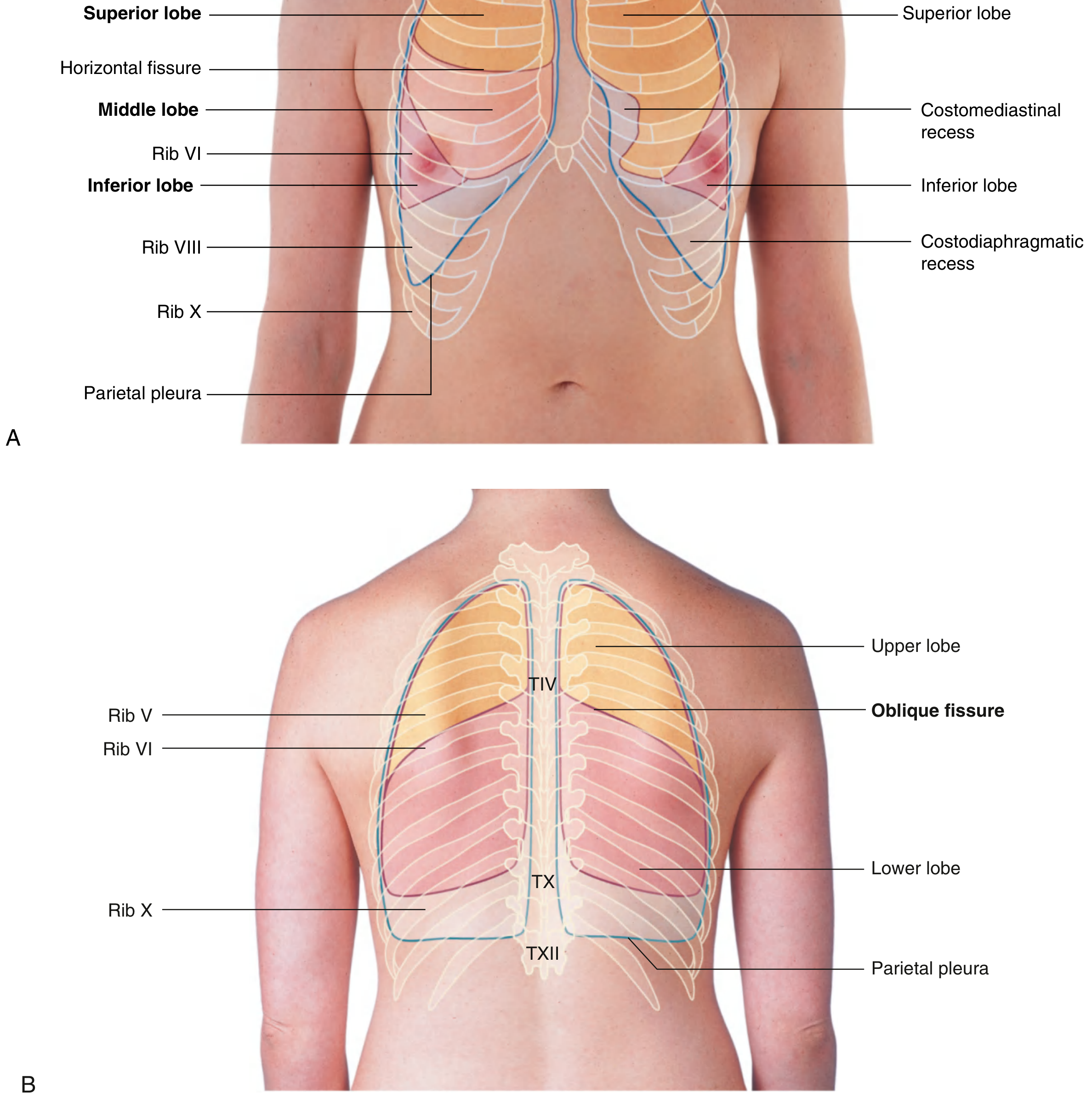
- On the right, the anterior border runs vertically close to the midline from the sternoclavicular joint down to the 6th costal cartilage.
- On the left, the anterior border diverges laterally from the level of the 4th costal cartilage (cardiac notch), creating the costomediastinal recess on the left.
3. Inferior Border (Lung Margin)
During quiet respiration, the inferior margin of the lung follows the contour:
| Reference Line | Rib Level |
|---|---|
| Midclavicular line | Rib VI |
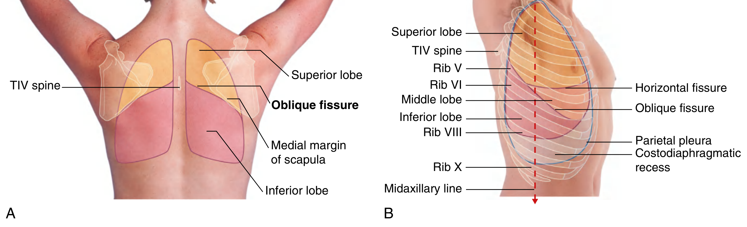
| Midaxillary line | Rib VIII |
| Posteriorly (paravertebral) | Vertebra TX (T10) |
This is the "VI, VIII, X" rule for the lung margin.
4. Inferior Border of the Pleura (Costodiaphragmatic Recess)
The parietal pleura extends lower than the lung edge, creating the costodiaphragmatic recess:
| Reference Line | Rib Level |
|---|---|
| Midclavicular line | Rib VIII |
| Midaxillary line | Rib X |
| Posteriorly | Vertebra TXII (T12) |
This is the "VIII, X, XII" rule for the pleura.
The gap between the lung border (VI, VIII, X) and the pleural border (VIII, X, XII) represents the costodiaphragmatic recess - a potential space that fills with fluid in pleural effusion.
5. Fissures and Lobes
Oblique Fissure (both lungs)
- Posteriorly: begins at the spine of vertebra TIV (T4), near the midline.
- Runs laterally and downward, crossing the 4th and 5th intercostal spaces.
- Reaches rib VI laterally and anteriorly, where it meets the costal cartilage of rib VI.
- A practical landmark: with the arm raised, the medial border of the scapula parallels the oblique fissure.
Horizontal Fissure (right lung only)
- Anteriorly: follows the line of rib IV and its costal cartilage, extending from the oblique fissure laterally to the anterior border of the lung.
Summary of Lobes (anterior view)
| Right Lung | Left Lung |
|---|---|
| Superior, Middle, Inferior lobes | Superior, Inferior lobes |
| Superior and Middle divided by Horizontal Fissure (Rib IV) | No horizontal fissure |
| Middle and Inferior divided by Oblique Fissure (Rib VI) | Superior and Inferior divided by Oblique Fissure (Rib VI) |
Diagrams
Anterior and Posterior views - Surface projections of lobes and fissures:
Posterior view and lateral view showing oblique fissure, horizontal fissure, and pleural borders:
Quick Memory Aid
| Structure | Landmark |
|---|---|
| Apex | 2.5 cm above medial clavicle |
| Lung inferior border | VI (MCL) - VIII (MAL) - X (posterior) |
| Pleura inferior border | VIII (MCL) - X (MAL) - XII (posterior) |
| Oblique fissure | TIV posteriorly → rib VI anteriorly |
| Horizontal fissure (R) | Along rib IV |
| Oblique fissure landmark | Medial border of raised scapula |
Sources: Gray's Anatomy for Students, pp. 277-278; Color Atlas of Human Anatomy Vol. 2 (Thieme), pp. 209-210
This is a shared conversation. Sign in to Orris to start your own chat.