
Areas more prone for neuropathic and ischemic ulcers and cause
neuropathic ulcer vs ischemic ulcer foot comparison sites
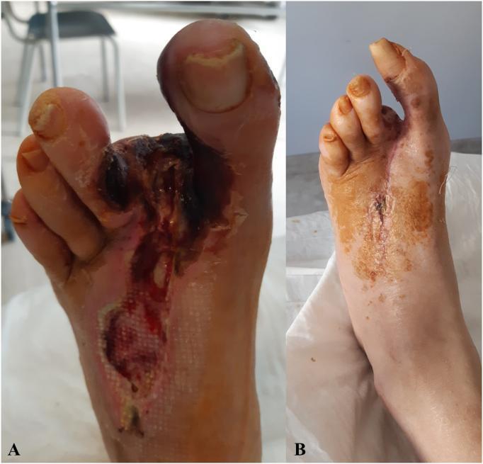
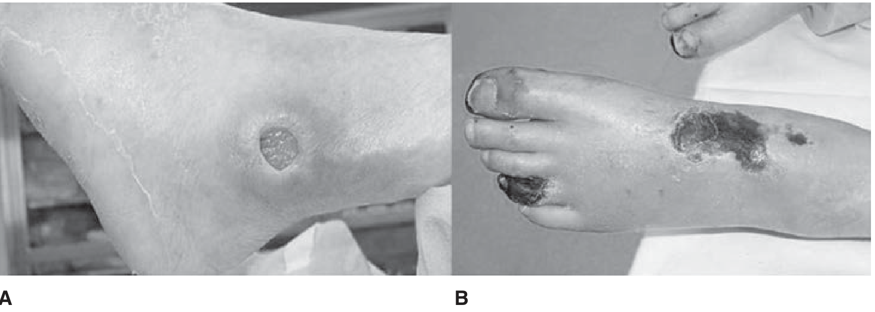
This clinical photograph set provides a side-by-side comparison of a patient's left foot before (A) and three weeks after (B) vascular intervention for peripheral artery disease. Panel A illustrates a severe, longitudinally-oriented ischemic ulcer on the dorsal aspect of the foot, extending into the first interdigital space. The wound displays dark necrotic tissue (eschar), exposed subcutaneous layers, and signs of secondary infection with surrounding erythema and inflammatory changes. A mesh-patterned wound dressing is visible within the proximal ulcer bed. Panel B shows the same foot following successful revascularization and wound management. The primary ulcer has been surgically closed, evidenced by a midline incision secured with sutures or staples. The necrotic tissue is resolved, and there is a significant reduction in peripheral edema and inflammation. Residual hyperpigmentation and staining from topical antiseptic use are visible on the distal skin. This comparison highlights the clinical progression from critical limb-threatening ischemia (CLTI) to successful wound healing and limb salvage.
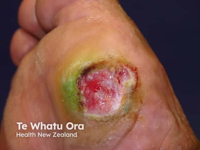
Clinical photography of a neuropathic foot ulcer. Modality: color digital photograph capturing dorsal aspect of the foot. The lesion is a round to irregular shallow ulcer located on the dorsum near the digits, displaying a moist, pink-red wound bed with areas of yellowish slough at the center and surrounding periwound erythema. The wound margins are largely defined with a surrounding greenish-yellow discoloration perhaps indicative of slough or necrotic tissue, and peripheral edema. The periwound tissue shows mild erythema and induration; the surrounding skin may be callused. The imaging reveals features typical of a neuropathic ulcer in the setting of diabetes mellitus: loss of protective sensation with impaired healing capacity; reduced perfusion may contribute to wound chronicity. The central wound bed lacks a robust epithelial rim, indicating ongoing tissue breakdown and delayed healing. Clinically, this image would be used for wound assessment, staging, and monitoring response to therapy, including debridement, offloading, advanced dressings, infection surveillance, and consideration of vascular assessment. Differential diagnoses include diabetic (neuropathic) foot ulcer, ischemic or mixed arterial insufficiency ulcers, pressure ulcers, and venous stasis ulcers, but the profile strongly supports neuropathic etiology. This image is relevant for medical education, wound care, dermatology, endocrinology, podiatry, and research on diabetic complications.
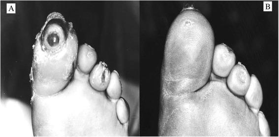
This clinical comparison photograph demonstrates the progression of diabetic foot ulcer healing on the distal aspect of the toes. Image A (baseline) shows a large, deep circular ulcer on the tip of the hallux (great toe) with exposed underlying tissue and irregular, macerated edges. Smaller, superficial ulcers or skin erosions are also visible on the tips of the second and third digits. The nail plate of the hallux appears absent or significantly degraded. Image B (follow-up) illustrates complete re-epithelialization and wound closure across all affected areas. The hallux now displays intact skin with restored dermatoglyphic patterns and a smooth texture, indicating successful tissue repair. The adjacent toes also show resolved skin lesions. This comparison is used to illustrate the efficacy of wound care interventions in managing neuropathic or ischemic diabetic foot ulcers, highlighting the transition from an open, Wagner-graded wound to a fully healed state with restored skin integrity.
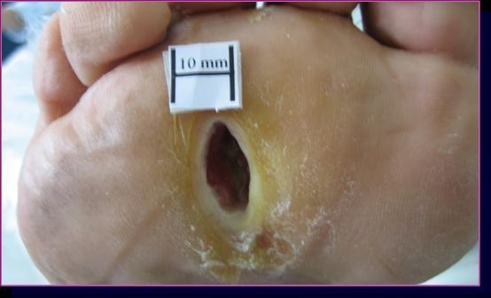
This clinical photograph displays a diabetic neuropathic foot ulcer located on the plantar surface of the third intermetatarsal space. The lesion is oval-shaped with a deep, crater-like morphology. The ulcer bed exhibits dark red granulation tissue, while the surrounding wound margins show a thick ring of yellowish, hyperkeratotic callous tissue typical of neuropathic pressure ulcers. The surrounding skin appears dry with evidence of scaling, consistent with diabetic xerosis. A white 10 mm scale marker is positioned superior to the lesion to provide a quantitative measurement reference. This image is used to demonstrate the baseline state of a chronic, non-ischemic diabetic foot ulcer prior to therapeutic intervention, such as extracorporeal shock wave therapy (ESWT). Key educational concepts include wound morphology, the role of pressure-induced hyperkeratosis in neuropathic ulcers, and clinical documentation of diabetic foot complications.
Neuropathic vs. Ischemic Ulcers: Sites and Causes
Sites (Areas More Prone)
Neuropathic Ulcers
- Plantar surface of the foot - the classic and most common location
- Pressure points under the metatarsal heads (especially 1st and 5th)
- Heel
- Areas overlying bony prominences or deformities (Charcot foot)
- The ulcer forms wherever repetitive, unperceived pressure accumulates
Ischemic Ulcers
- Toes - most commonly affected (any toe can be involved)
- Lateral surface of the ankle
- Distal digits and tips of toes
- Dorsum of the foot (occasionally)
- Glabrous (hairless) skin margins
- Atypical positions are possible from friction (e.g., poorly fitting shoes)
Causes
Neuropathic Ulcers - Cause
- Loss of protective sensation means the patient cannot feel repetitive trauma, pressure, or injury
- Continued weight-bearing on insensate foot causes tissue breakdown
- Autonomic neuropathy leads to decreased sweating → dry, cracked skin → entry point for infection
- Motor neuropathy causes intrinsic muscle wasting → foot deformities (clawing of toes, prominent metatarsal heads) → altered pressure distribution
- Arteriovenous shunting increases blood flow locally → warm foot with dilated veins and bounding pulses
- Callus formation over pressure areas precedes breakdown
Ischemic Ulcers - Cause
- Reduced arterial blood flow → tissue hypoxia → skin and tissue breakdown
- Any minor trauma (friction from shoes, minor injury) can trigger ulceration when perfusion is borderline
- Risk factors: long-standing hypertension, smoking, diabetes mellitus, hyperlipidemia
- In progressive disease, consider Thromboangiitis obliterans (Buerger disease)
- Cholesterol emboli are another arterial cause of lower limb ulceration
- The initial lesion is a red, painful plaque that breaks down into a superficial ulcer with purpuric erythema
Comparative Table
| Feature | Neuropathic Ulcer | Ischemic Ulcer |
|---|---|---|
| Pain | Painless | Painful |
| Pulses | Normal | Absent |
| Margins | Regular, punched-out | Irregular |
| Location | Plantar surface of foot | Toes, lateral ankle, glabrous margins |
| Calluses | Present | Absent or infrequent |
| Sensation | Lost (sensation, reflexes, vibration) | Variable |
| Blood flow | Increased (AV shunting) | Decreased |
| Veins | Dilated | Collapsed |
| Foot temperature | Dry, warm | Cold |
| Bony deformities | Present | Absent |
| Appearance | Red/hyperemic | Pale and cyanotic |
Key Clinical Memory Points
- Neuropathic = plantar + painless + punched-out + pulses present + warm + callused → caused by loss of sensation (diabetic neuropathy)
- Ischemic = toes/lateral ankle + painful + pale/gangrenous + pulseless + cold → caused by arterial occlusion (atherosclerosis/PAD)
- A mixed ulcer (neuro-ischemic) is common in diabetic patients - neuropathy + PAD coexist
- ABI < 0.75 confirms arterial insufficiency; ABI < 0.5 indicates substantial ischemia