
Discuss stages of pregnancy changes that occur in various during pregnancy
Reading File
Now I'll search the medical library for comprehensive content on physiological changes during pregnancy.
Finding Sources
Finding Sources
Finding Sources
Reading File
Excellent content found. Let me read more of this section and search for additional obstetrics content.
Reading File
Finding Sources
Reading File
Finding Sources
Reading File
Reading File
I now have comprehensive content to compile an in-depth answer. Let me also quickly fetch the uteroplacental circulation diagram.
Loading Image
I now have all the content needed for a thorough, well-organized response.
Physiological Changes During Pregnancy
Pregnancy affects virtually every organ system in the body. These changes are primarily adaptive, helping the mother tolerate the stresses of pregnancy, labor, and delivery while supporting fetal growth. The changes below are organized by system and, where relevant, by trimester.
Overview: Maximum Physiological Changes at Term
| System/Parameter | Change |
|---|---|
| Neurological - MAC | -40% |
| Respiratory - O₂ consumption | +20 to +50% |
| Minute ventilation | +50% |
| Tidal volume | +40% |
| FRC | -20% |
| PaCO₂ | -15% |
| HCO₃ | -15% |
| Cardiovascular - Blood volume | +35% |
| Plasma volume | +55% |
| Cardiac output | +40% |
| Stroke volume | +30% |
| Heart rate | +20% |
| Systolic BP | -5% |
| Diastolic BP | -15% |
| Peripheral resistance | -15% |
| Hematological - Hemoglobin | -20% |
| Clotting factors | +30 to +250% |
| Renal - GFR | +50% |
(Morgan and Mikhail's Clinical Anesthesiology, 7e, Table 40-1)
1. Central Nervous System (CNS)
- Minimum alveolar concentration (MAC) for all inhalational anesthetics progressively decreases, reaching as much as -40% at term; returns to normal by the 3rd day postpartum.
- Progesterone, which rises up to 20 times normal at term, is sedating and partly responsible for this MAC reduction.
- A surge in β-endorphin levels during labor also contributes.
- Pregnant patients show enhanced sensitivity to local anesthetics during regional anesthesia, with epidural dose requirements reduced by as much as 30%.
- Obstruction of the inferior vena cava (IVC) by the enlarging uterus causes engorgement of the epidural venous plexus, reducing the CSF and epidural space volumes, thereby enhancing cephalad spread of local anesthetics.
2. Respiratory System
First and Second Trimesters
- Rising progesterone stimulates the respiratory center, driving up minute ventilation (up to +50% by term) primarily through increased tidal volume (+40%) and to a lesser extent respiratory rate (+15%).
Third Trimester
- The growing uterus elevates the diaphragm, but diaphragmatic motion is preserved; the chest compensates by increasing its anteroposterior diameter.
- Functional residual capacity (FRC) falls by up to 20% due to reduced expiratory reserve volume; FRC returns to normal within 48 hours of delivery.
- Vital capacity and closing capacity are minimally affected; airway resistance decreases (-35%).
- Intrapulmonary shunting increases toward term.
- PaCO₂ falls to 28-32 mm Hg (respiratory alkalosis), which is compensated by a decrease in plasma bicarbonate to prevent significant pH change.
- PaO₂ rises slightly (+10%), and the P50 for hemoglobin increases from 27 to 30 mm Hg (rightward shift), enhancing O₂ delivery.
- Oxygen consumption rises up to 50% at term.
- Decreased FRC + increased O₂ consumption = rapid desaturation during apnea - this is why preoxygenation before general anesthesia is mandatory in pregnancy.
- Mucosal engorgement of the upper airways predisposes to trauma, bleeding, and obstruction during laryngoscopy.
3. Cardiovascular System
First Trimester
- A substantial fall in peripheral vascular resistance begins early (nadir in mid-second trimester).
- Heart rate and stroke volume begin to increase.
Second Trimester
- Peripheral resistance reaches its nadir.
- Blood volume and plasma volume continue to rise rapidly.
Third Trimester
- Cardiac output increases 40% at term via +20% heart rate and +30% stroke volume.
- Cardiac chambers enlarge and mild myocardial hypertrophy may be seen on echocardiography.
- The heart is displaced upward and leftward by the elevated diaphragm, causing apparent cardiomegaly on chest X-ray, left axis deviation, and T-wave changes on ECG.
- Blood volume increases 1000-1500 mL, enabling women to tolerate delivery blood loss (200-500 mL vaginal; 800-1000 mL for caesarean section). Blood volume normalizes 1-2 weeks postpartum.
- Plasma volume increases 55%, exceeding red cell mass increase (45%), producing dilutional anemia (hemoglobin usually stays above 11 g/dL).
- Supine hypotension syndrome (aortocaval compression): After week 20, the gravid uterus can compress the IVC when supine, reducing venous return and causing hypotension, pallor, sweating, nausea/vomiting in ~5% of women at term. This is corrected by placing a wedge (>15°) under the right hip.
- Chronic partial caval obstruction in the third trimester predisposes to venous stasis, phlebitis, and lower extremity edema.
- Blood pressure falls slightly (systolic -5%, diastolic -15%) due to vasodilation.
4. Uteroplacental Circulation
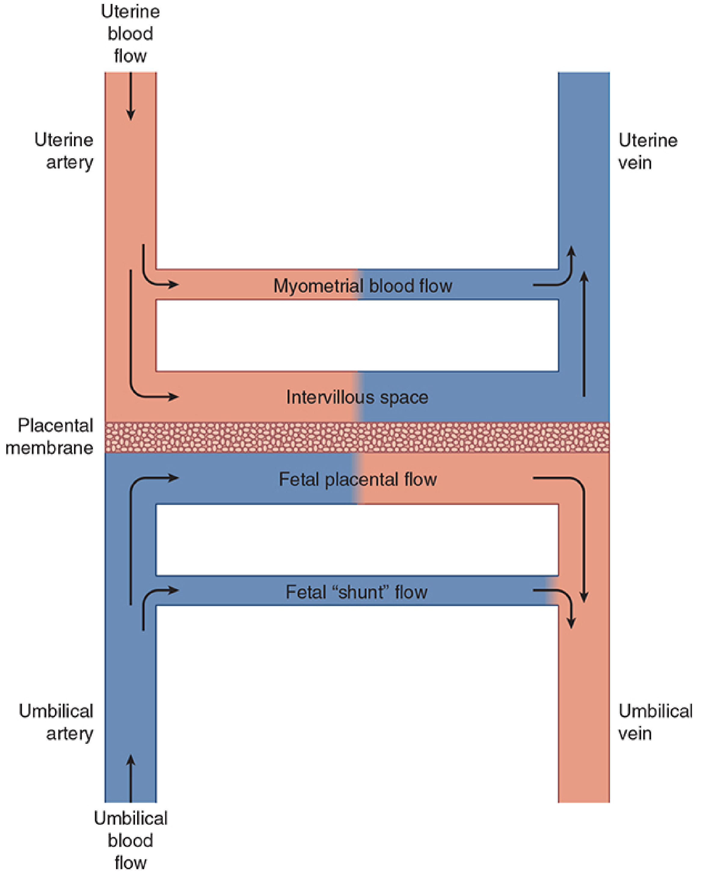
- At term, uterine blood flow = ~10% of cardiac output (600-700 mL/min), compared to just 50 mL/min in the non-pregnant state.
- 80% of uterine blood flow goes to the placenta; 20% to the myometrium.
- The uterine vasculature is maximally dilated, so autoregulation is absent but it remains sensitive to α-adrenergic agonists.
- Three major factors reduce uterine blood flow: (1) systemic hypotension, (2) uterine vasoconstriction, and (3) uterine contractions.
- Placental gas exchange occurs by passive diffusion across a large surface area.
5. Renal System
- Renal plasma flow and GFR increase by ~50% throughout pregnancy.
- Serum creatinine may fall to as low as 0.5 mg/dL; BUN to ~9 mg/dL.
- Glycosuria (1-10 g/day) and mild proteinuria (<300 mg/day) are common due to reduced tubular threshold - not necessarily pathological.
- Plasma osmolality decreases by 8-10 mOsm/kg.
6. Gastrointestinal System
- Gastroesophageal reflux and esophagitis are common due to:
- Reduced gastric motility
- Upward/anterior displacement of the stomach by the uterus
- Incompetence of the gastroesophageal sphincter
- This places pregnant patients at high risk for regurgitation and pulmonary aspiration (Mendelson's syndrome risk).
- Gastric acidity and volume do not change significantly.
- High progesterone inhibits cholecystokinin release, causing incomplete gallbladder emptying and predisposing to cholesterol gallstones.
7. Hepatic System
- Overall hepatic function and blood flow remain unchanged.
- Minor elevations in serum transaminases and LDH may occur in the third trimester.
- Serum alkaline phosphatase is mildly elevated (placental secretion).
- Serum albumin decreases mildly (dilution effect), reducing colloid oncotic pressure.
- Pseudocholinesterase activity decreases 25-30% at term (rarely clinically significant with succinylcholine); normalizes by 6 weeks postpartum.
8. Hematological System
Pregnancy creates a hypercoagulable state to limit blood loss at delivery:
- Fibrinogen increases significantly
- Factors VII, VIII, IX, X, XII all increase (+30-250%)
- Factor XI may decrease slightly
- Platelet count decreases ~10% in the third trimester
- Leukocytosis (up to 21,000/μL) is normal
- Accelerated fibrinolysis can be observed late in the third trimester
- Iron and folate deficiencies develop readily if supplements are not taken (fetal utilization)
9. Metabolic and Endocrine System
- Complex metabolic changes favor fetal growth:
- Low blood glucose and amino acids
- High free fatty acids, ketones, and triglycerides
- Resembles starvation biochemically, yet is a diabetogenic state
- Insulin levels rise steadily; pancreatic beta-cell hyperplasia occurs to meet demand.
- Human placental lactogen (hCS) secreted by the placenta causes relative insulin resistance.
- Thyroid gland hypertrophies: HCG and estrogens stimulate it and increase thyroid-binding globulin; total T4 is elevated, but free T4 remains normal (euthyroid state).
- Progesterone rises 20-fold by term, contributing to CNS sedation, smooth muscle relaxation (GI tract, uterus in early pregnancy).
Summary by Trimester
| Feature | 1st Trimester | 2nd Trimester | 3rd Trimester |
|---|---|---|---|
| Cardiac output | Begins rising | Further rise | Plateau; spikes in labor |
| Blood volume | Starts expanding | Maximal expansion begins | Fully expanded |
| Peripheral resistance | Begins to fall | Nadir | Slight recovery |
| FRC | Minimally affected | Starts to fall | Falls up to 20% |
| Aortocaval compression | Absent | Risk begins >20 weeks | Most significant |
| GI symptoms | Nausea/vomiting peak | Improves | Reflux/heartburn |
| Glycosuria | May begin | Present | Present |
(Source: Morgan and Mikhail's Clinical Anesthesiology, 7e, Chapter 40)
These physiological adaptations are largely driven by hormonal changes (progesterone, estrogen, HCG, human placental lactogen) and the mechanical effects of the growing uterus. Understanding them is essential for clinical management, particularly in anesthesia, obstetric emergencies, and drug prescribing during pregnancy.
This is a shared conversation. Sign in to Orris to start your own chat.